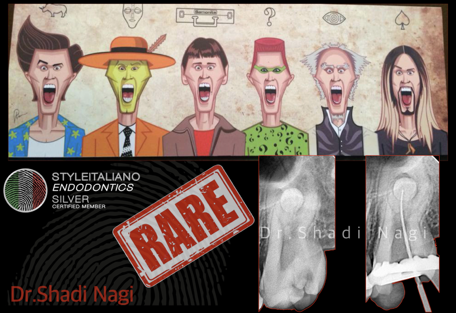
This article demonstrates the protocols followed on two teeth with unique anatomical features in a conservative, feasible, teachable and repeatable way.

 Dealing with hard canals, the easy way.
This article demonstrates the protocols followed on two teeth with unique anatomical features in a conservative, feasible, teachable and repeatable way.
 The probability of two fives
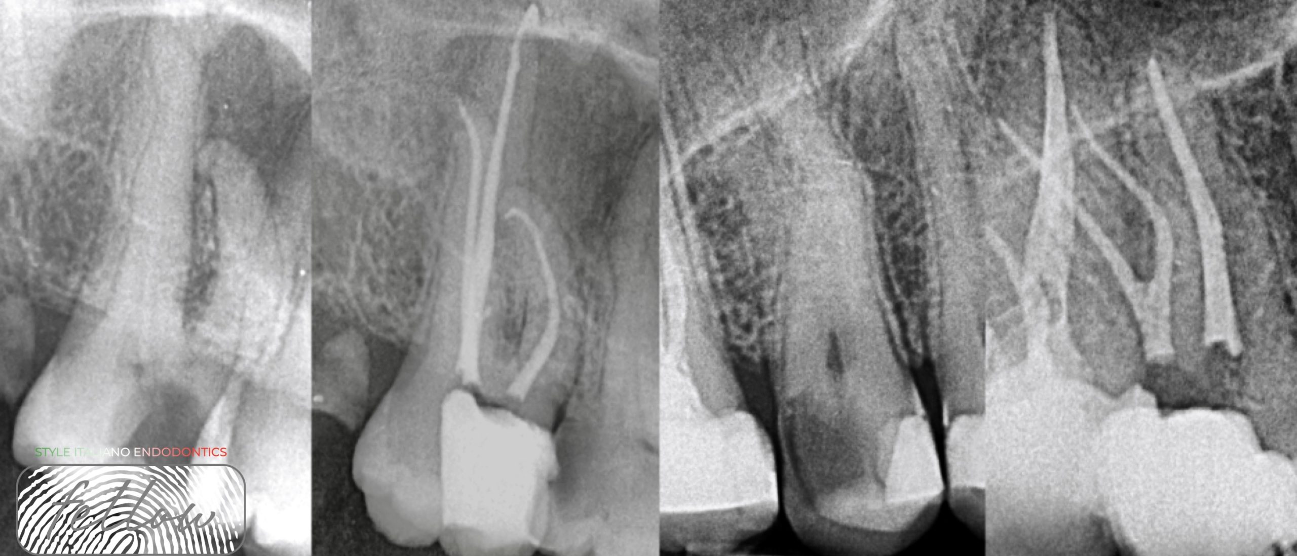
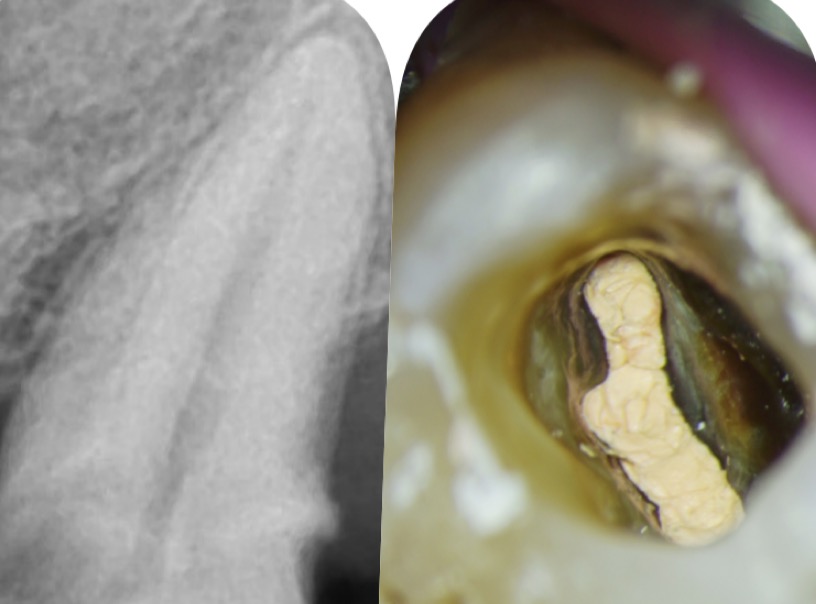
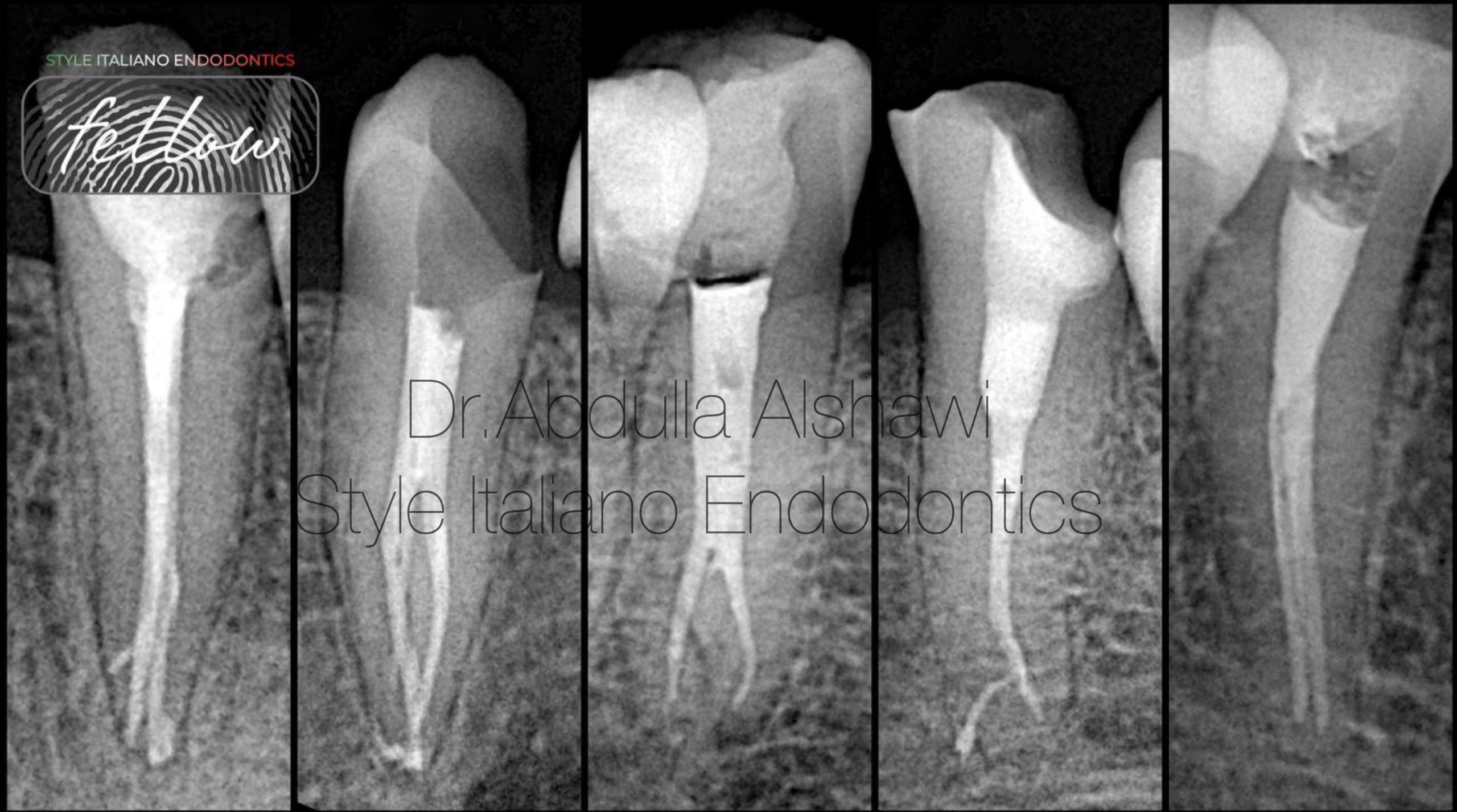
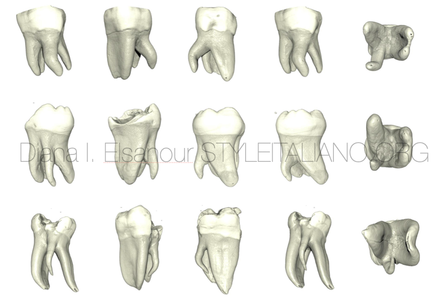
With increasing number of reports and studies of unusual root canal morphology, It is of utmost importance that the clinician must have detailed knowledge of the pulp canal anatomy to achieve effectively proper cleaning and shaping of the root canal system. Failure to recognize the aberrant root canal anatomy will lead to an unsuccessful treatment and thus failure of the endodontic therapy.
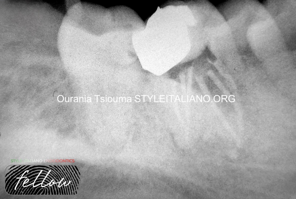
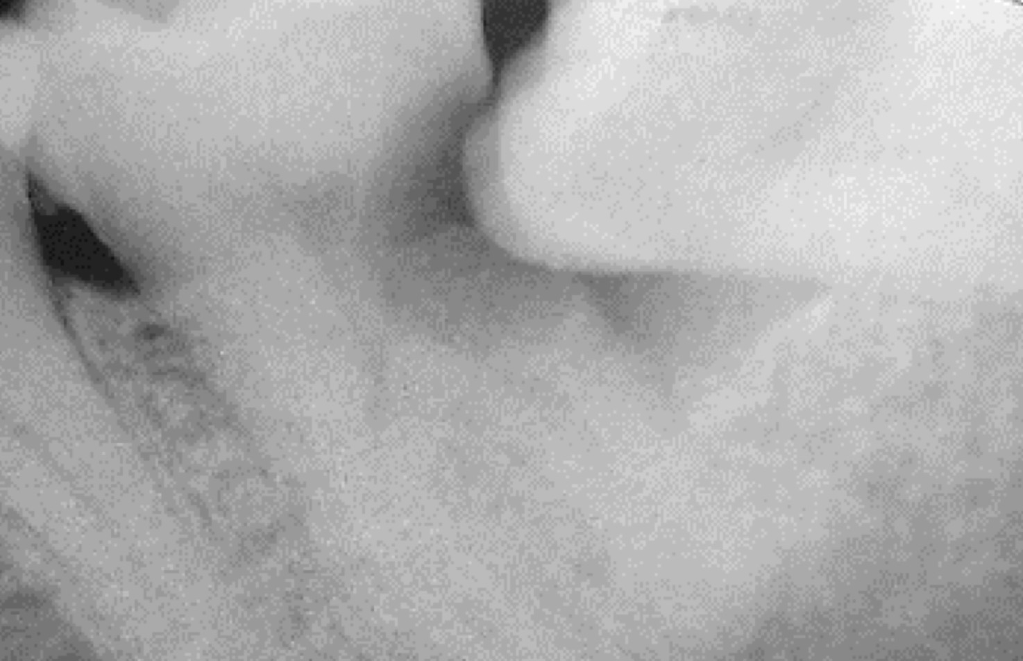
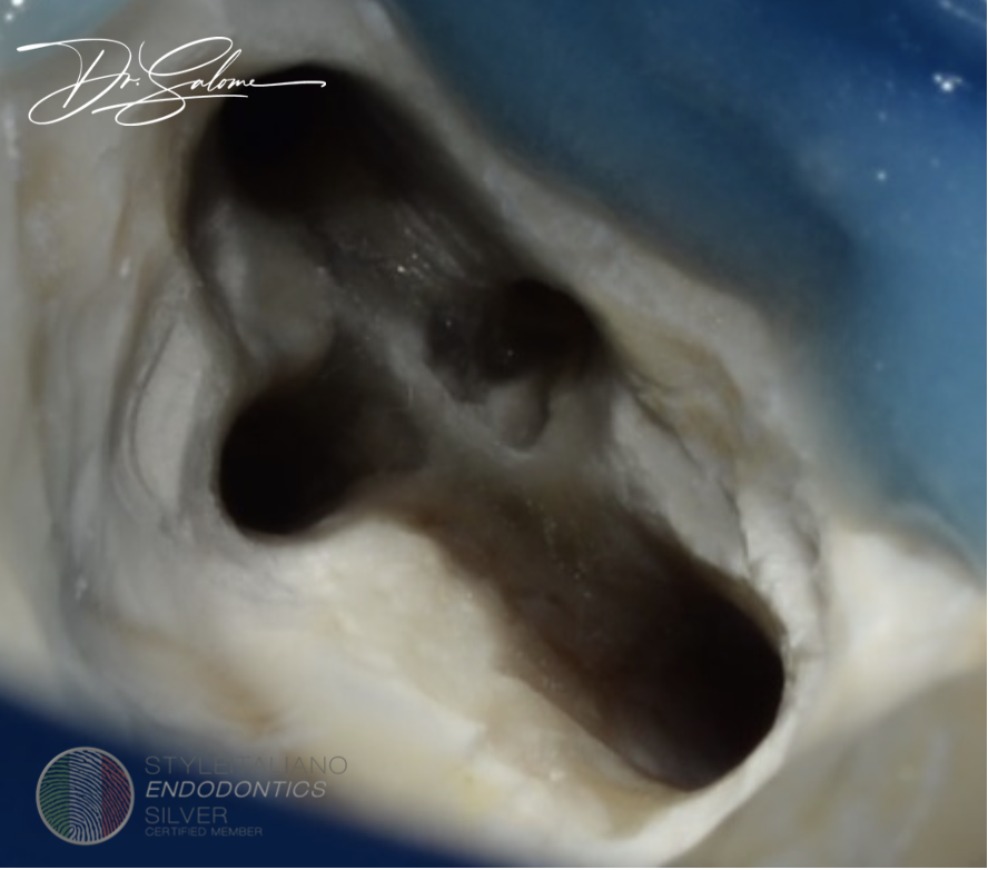
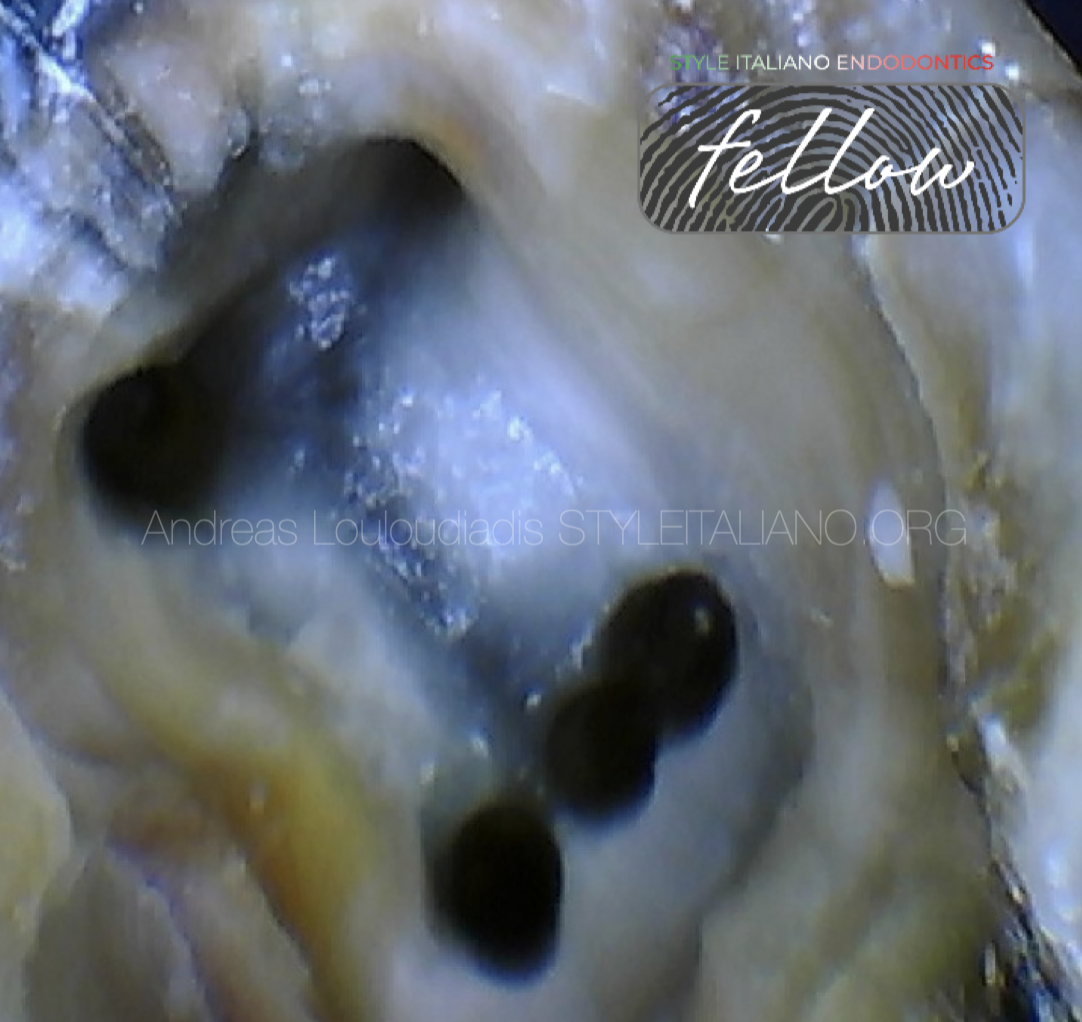
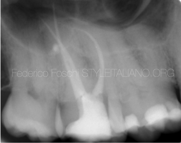
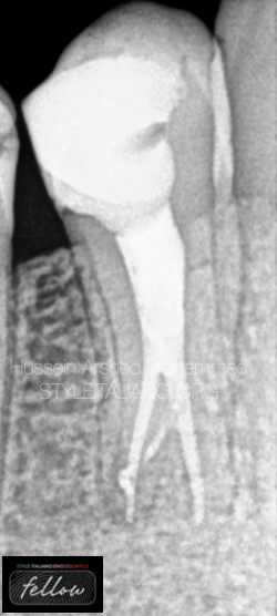
This case report describes a very rare scenario of a mandibular right first molar with five root canals (three in distal root and two in mesial root) and a maxillary left second molar with also five root canals (three in the mesial buccal root, one in the distobuccal root, and one palatal) in the same patient.
 C-SHAPED MAXILLARY SECOND MOLAR
Root canal treatment of maxillary second molar can be very difficult . Beside the limited accesibility of tooth in mouth with limited mouth opening ,with complex root canal anatomy specially […]
 Management of the complex anatomy of lower premolars
Variations in the root canal configurations are a great challenge for the endodontist during endodontic procedures. This necessitates the understanding of canal morphology before initiating the treatment.
 A mandibular first molar with 6 root canals:
One of the most important prerequisites for successful endodontic treatment is the adequate knowledge and understanding of root canal anatomy. The mandibular first molar (MFM) seems to be the tooth […]
 Autotransplantation as an alternative treatment option
Traditional transplantation, surgical extrusion and intentional replantation procedures are important treatment options that clinicians may consider performing in their daily clinical practice.
 Radix entomolaris and paramolaris
Supernumerary root in mandibular molars; prevalence, classification, diagnosis and management.
 Distal canal as a determinant of MB2 location?!
Finding mB2 can be challenging work. Usually, the entrance is covered with thick deposition of secondary dentin, which impede visualization. In clinical practice finding MB2 is of higher importance, while ignoring it can lead to apical periodontitis in vast majority of cases. We want to share more predictable searching method for easy detection of MB2.
 A Mandibular First Molar with Radix Entomolaris and Middle Mesial Canal: a case report
Case report of the endodontic treatment of a mandibular first molar with radix entomolaris and middle mesial canal
 A diagnostic conundrum
Multi-rooted teeth in the upper arch can present diagnostic difficulties. Vitality testing can result in erratic response due to necrobiosis status of the pulp. The aid of CBCT scan to […]
 Bending preservation of gutta-percha in lower first premolar with 3 canals
This article clarifies how to to bend gutta-percha cone and preserve this bending even if the temperature of the body can straighten the master cone in deep apical splitting of the canals.