We all make mistakes - correct protocols help us detect them early
27/11/2023
Fellow
Warning: Undefined variable $post in /home/styleendo/htdocs/styleitaliano-endodontics.org/wp-content/plugins/oxygen/component-framework/components/classes/code-block.class.php(133) : eval()'d code on line 2
Warning: Attempt to read property "ID" on null in /home/styleendo/htdocs/styleitaliano-endodontics.org/wp-content/plugins/oxygen/component-framework/components/classes/code-block.class.php(133) : eval()'d code on line 2
The patient comes to my attention for a fracture of the distal margin due to extensive caries of 1.4.
From the radiographic analysis it is possible to appreciate the subgingival extension of the defect and its close relationship with the vasculonervous plexus of the tooth. It is therefore decided to treat the tooth endodontically and then restore it through a cuspal covering overlay.
Studying the radiographic profiles of the roots reveals a very scholastic anatomy with two roots with a fairly long profile (3).
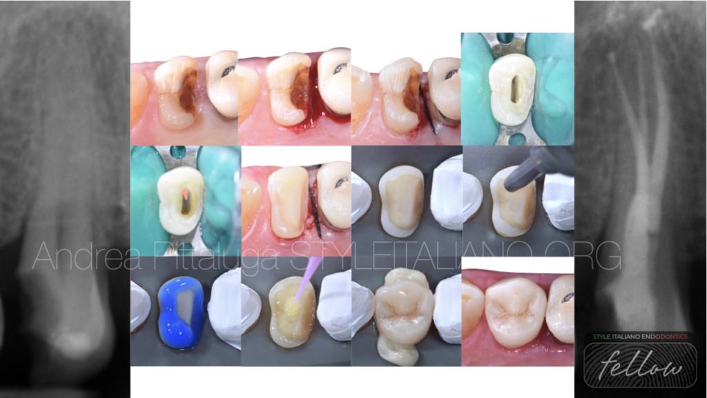
Since the carious lesion develops within the gingival sulcus, it is decided to approach the case with a preliminary gingivectomy to be able to easily isolate the tooth and restore it during the same session.
After proceeding with the cusp reduction, we proceed with the isolation and chamber opening: this last maneuver confirms the radiographic analysis revealing a vestibular and a palatal canal facing each other(4). The palatal canal is shaped mechanically and finished with a 30/06 while the vestibular canal is finished with a 25/06. The canals are therefore sealed with traditional warm vertical condensation and, after relocating the distal margin, it is reconstructed and prepared for the future overlay.
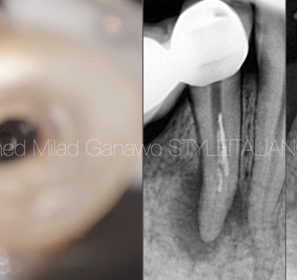
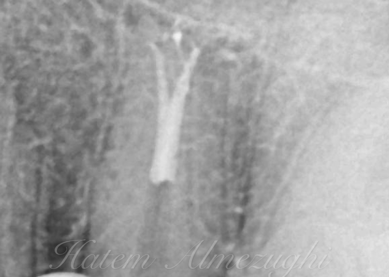
Once the final rx has been completed, the surprise arrives: the hydraulic pressure of the vertical condensation led the gutta-percha and the cement to highlight an "extra" root not found either during the preliminary analysis or during the treatment. In fact, its development is uncommon, starting from the middle third of the vestibular canal.
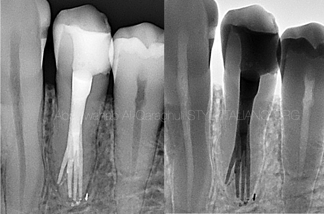
It is therefore decided to remove the adhesive restoration again and to re-approach the vestibular canal: the distovestibular canal is also shaped up to 25/06 and sealed with a warm vertical condensation technique.
It is reconstructed again with an adhesive technique and the tooth is prepared for the definitive restoration.
The following appointment proceeds with the cementation of the lithium disilicate cuspal covering restoration and the case is finally concluded
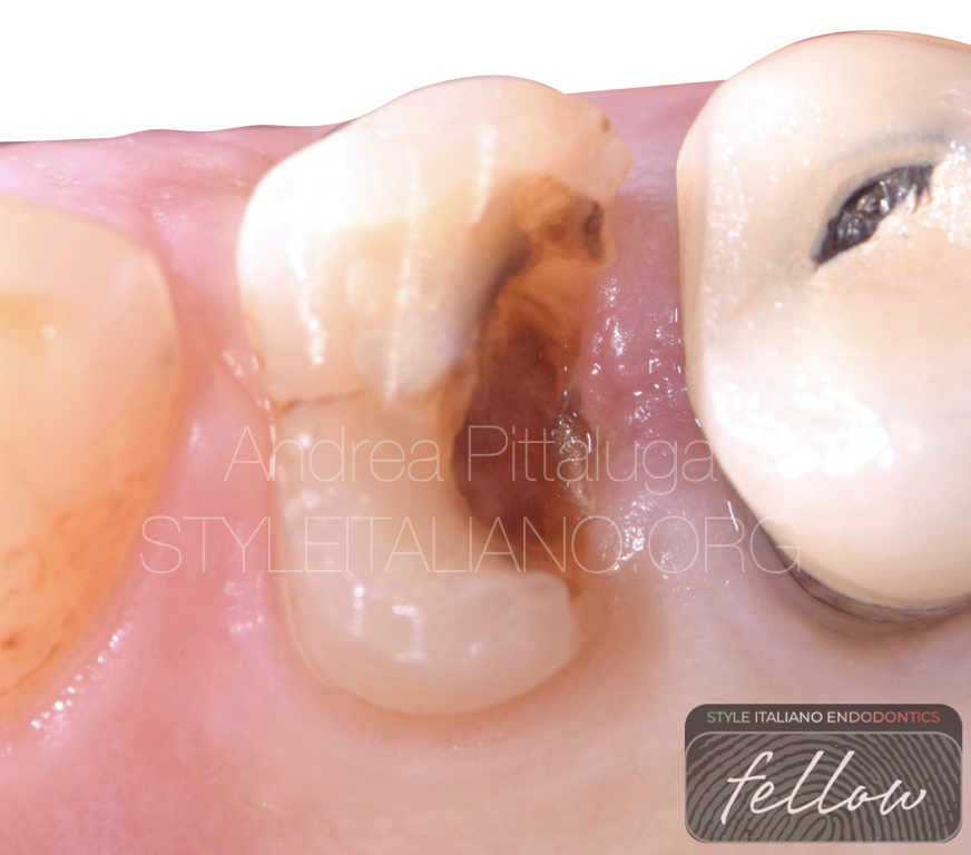
Fig. 1
Initial situation

Fig. 2
Radiographic analysis
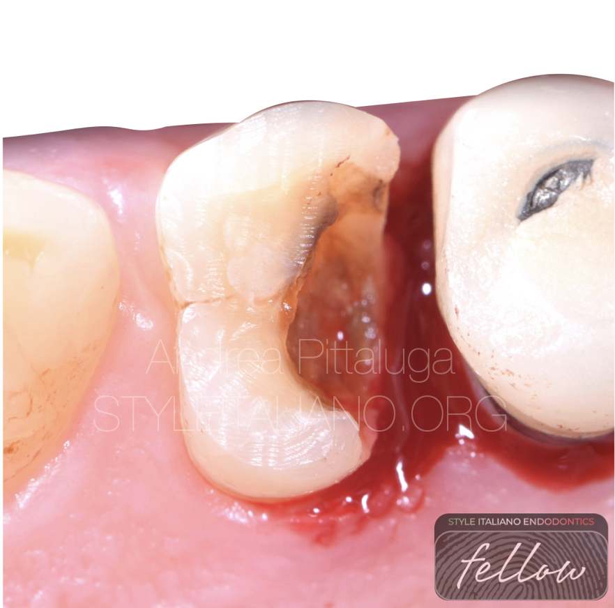
Fig. 3
Gingivectomy
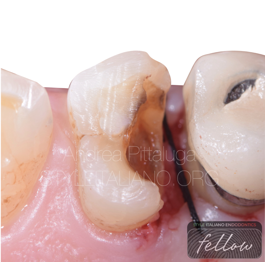
Fig. 4
Suture
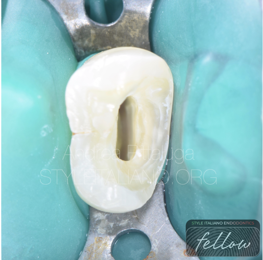
Fig. 5
Isolation and chamber opening
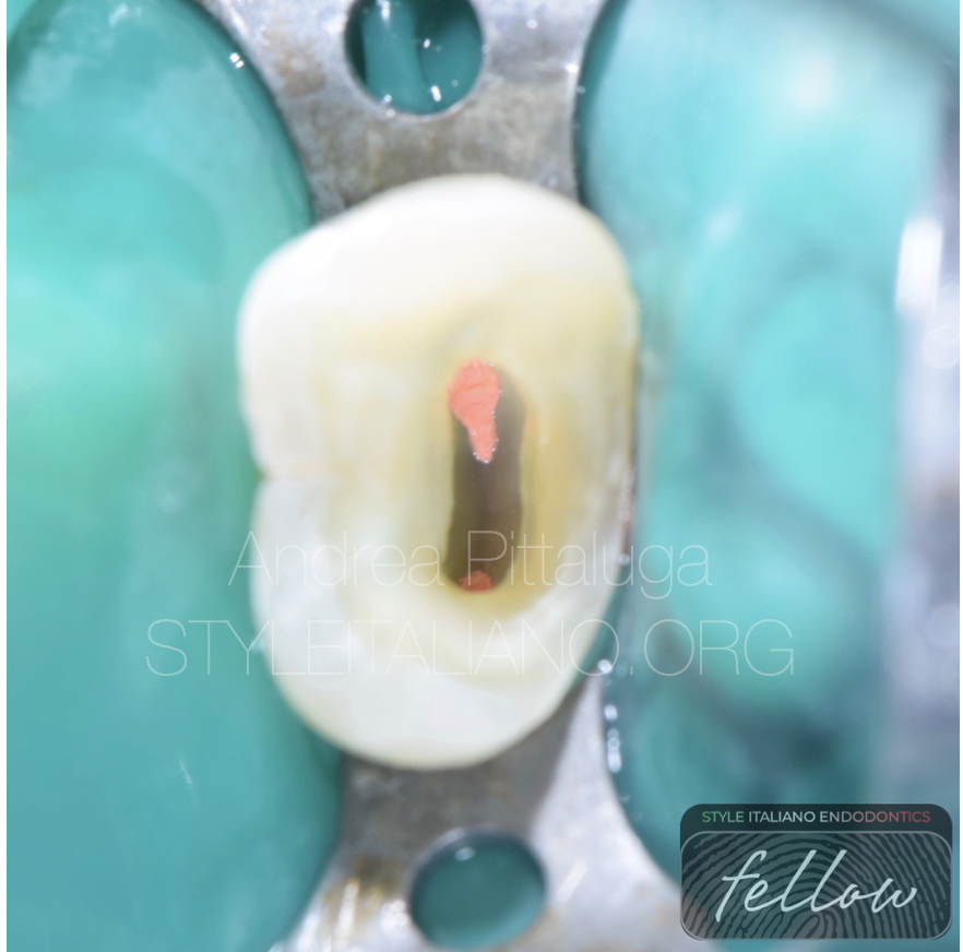
Fig. 6
Obturation
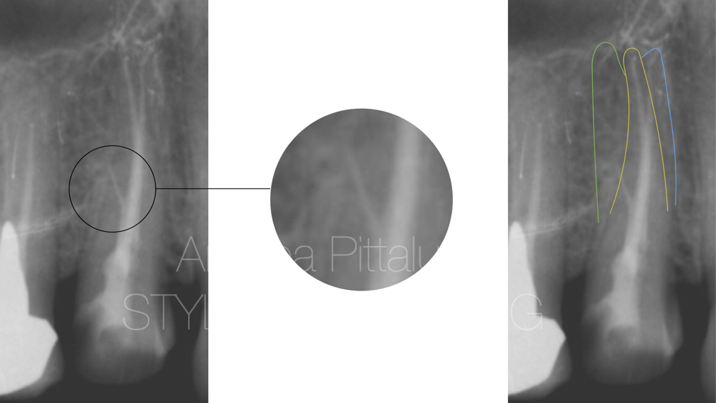
Fig. 7
Post Op X-Ray and missed anatomy detection
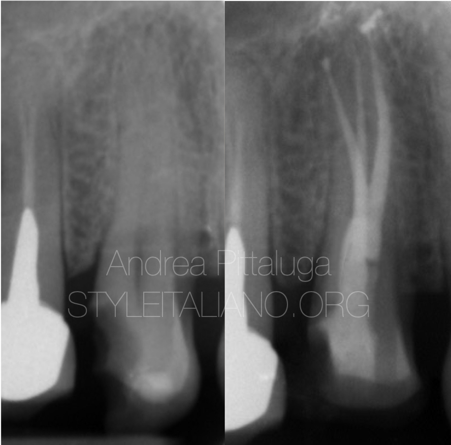
Fig. 8
Correction
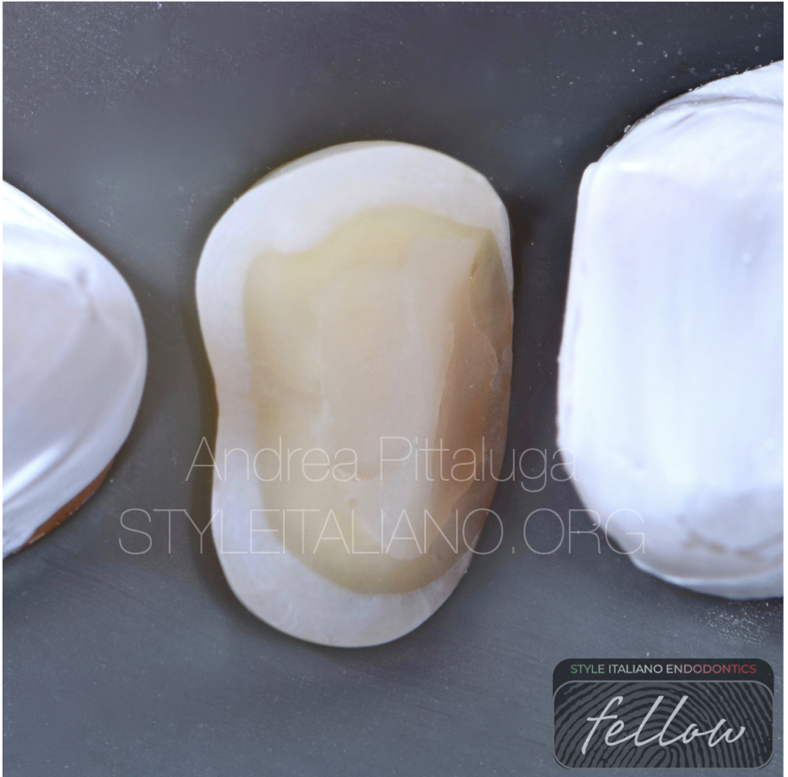
Fig. 9
Build up and Prep
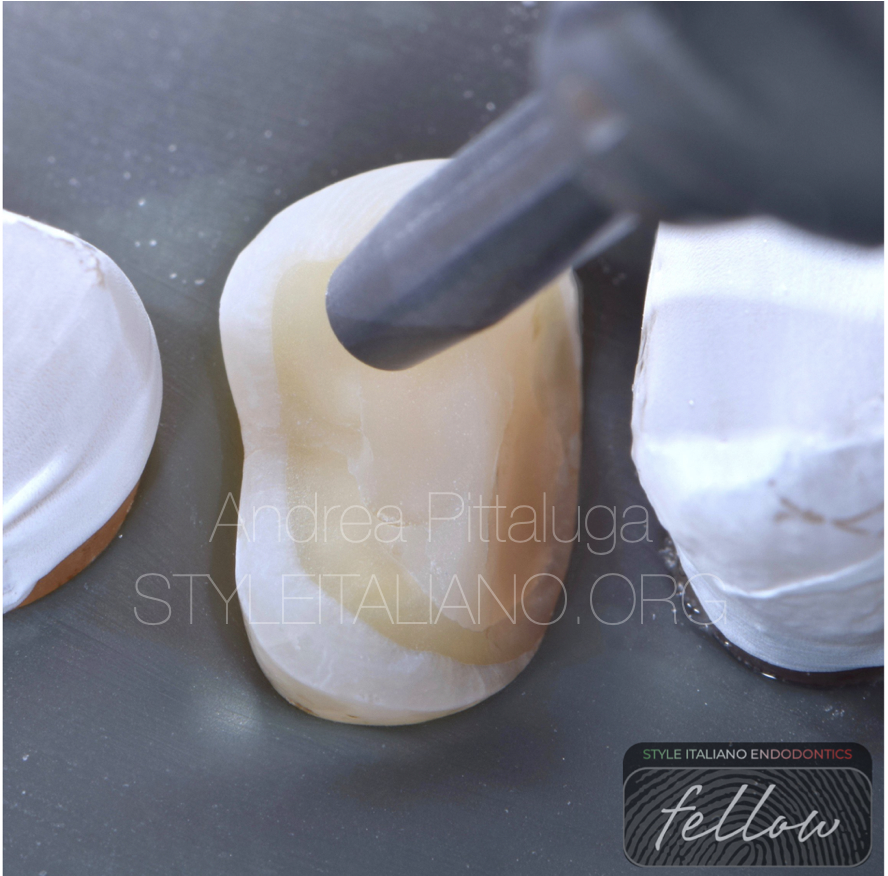
Fig. 10
Sandblasting
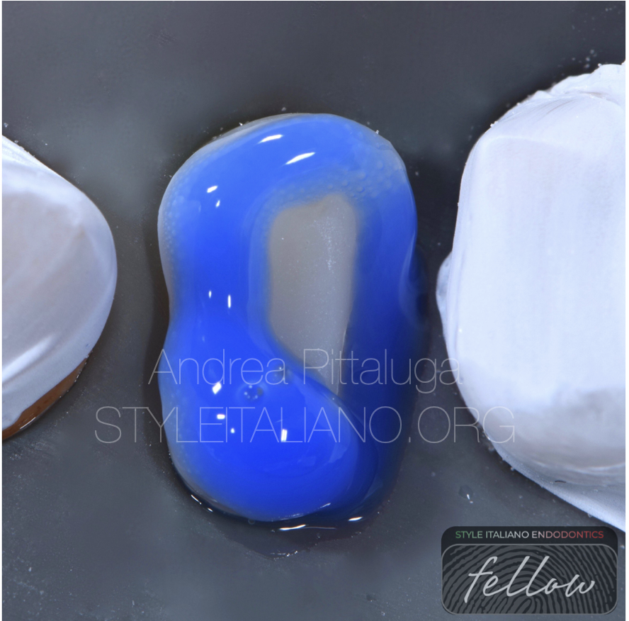
Fig. 11
Etching
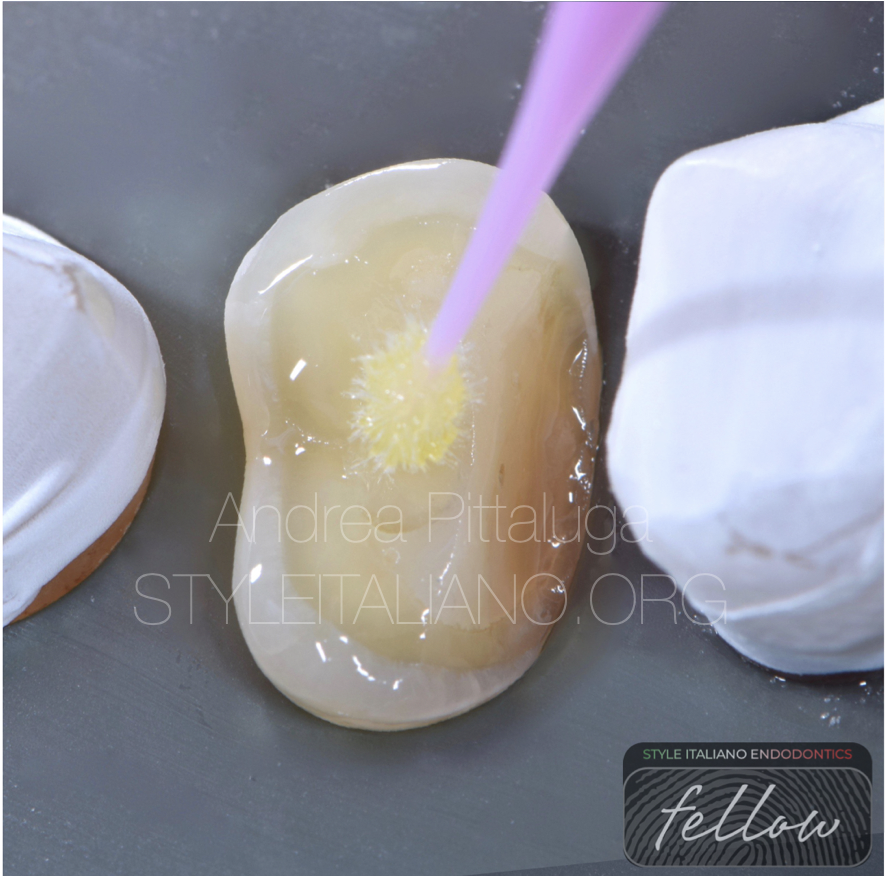
Fig. 12
Adhesive
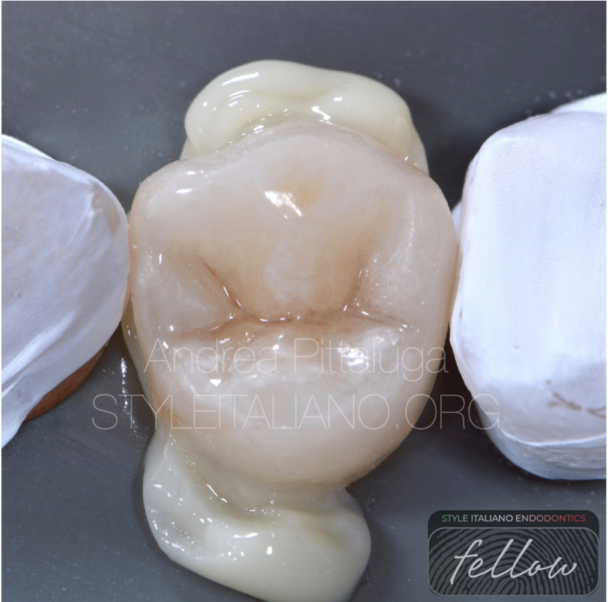
Fig. 13
Bonding
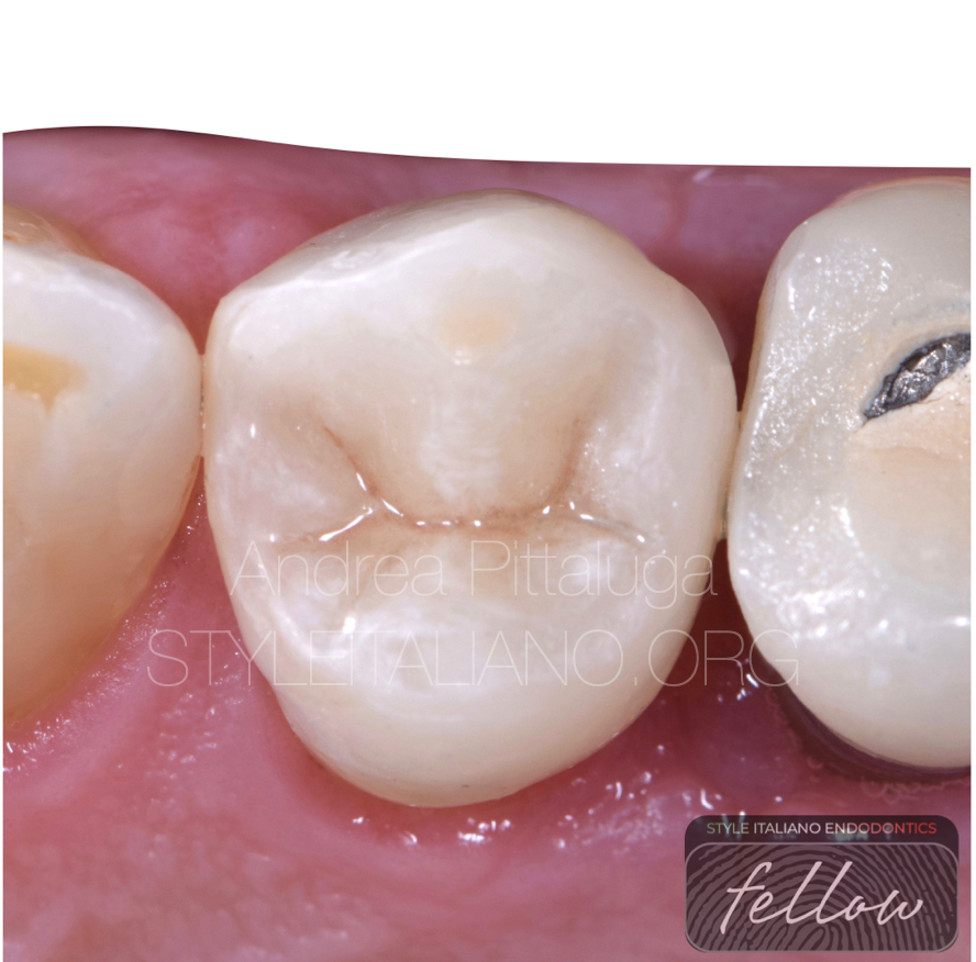
Fig. 14
Final result
Fig. 15
About the author:
Dr Andrea Pittaluga
Conclusions
A correct protocols can highlight anatomies not found during the primary treatment and their early localization can safeguard us from possible failures (5).
Bibliography
1.Mohammed Mashyakhy - Prevalence of Missed Canals and Their Association with Apical Periodontitis in Posterior Endodontically Treated Teeth: A CBCT Study Int J Dent. 2021; 2021: 9962429.
2.Zahed Mohammadi - A Clinical Update on the Different Methods to Decrease the Occurrence of Missed Root Canals Iran Endod J. 2016 Summer; 11(3): 208–213.
3.Vertucci FJ. Root canal anatomy of the human permanent teeth. Oral Surg Oral Med Oral Pathol. 1984;58(5):589–99.
4. RC B. Access openings and tooth morphology. In: Cohen S, RC B, editor. Pathways of the Pulp. 4 ed. St. Louis: The CV Mosby Company; 1987.
5. Slowey R. Root canal anatomy. Road map to successful endodontics. Dent Clin North Am. 1979;23(4):555–73.