Management of deep apical split
12/08/2023
Fellow
Warning: Undefined variable $post in /home/styleendo/htdocs/styleitaliano-endodontics.org/wp-content/plugins/oxygen/component-framework/components/classes/code-block.class.php(133) : eval()'d code on line 2
Warning: Attempt to read property "ID" on null in /home/styleendo/htdocs/styleitaliano-endodontics.org/wp-content/plugins/oxygen/component-framework/components/classes/code-block.class.php(133) : eval()'d code on line 2
Speaking of ANATOMIC VARIATIONS
The clinician should be aware of root canal anatomy and morphology, not to mention the protocol of debridement, disinfection and obturation.
Vertucci5 reported the incidence of one canal at the apex in them at 75%, and two canals at apex at 24%. In the same study, Vertucci5 found maxillary second premolars with three canals at apex to be only 1%. Studies on maxillary premolars show a low incidence of three root canals.
The goal of this study was to enlighten every clinician of anatomical configurations before approaching any maxillary premolars.This figure shows working length where first manual file inserted toward split palatally, then followed by inserting the second manual file toward the split buccally.
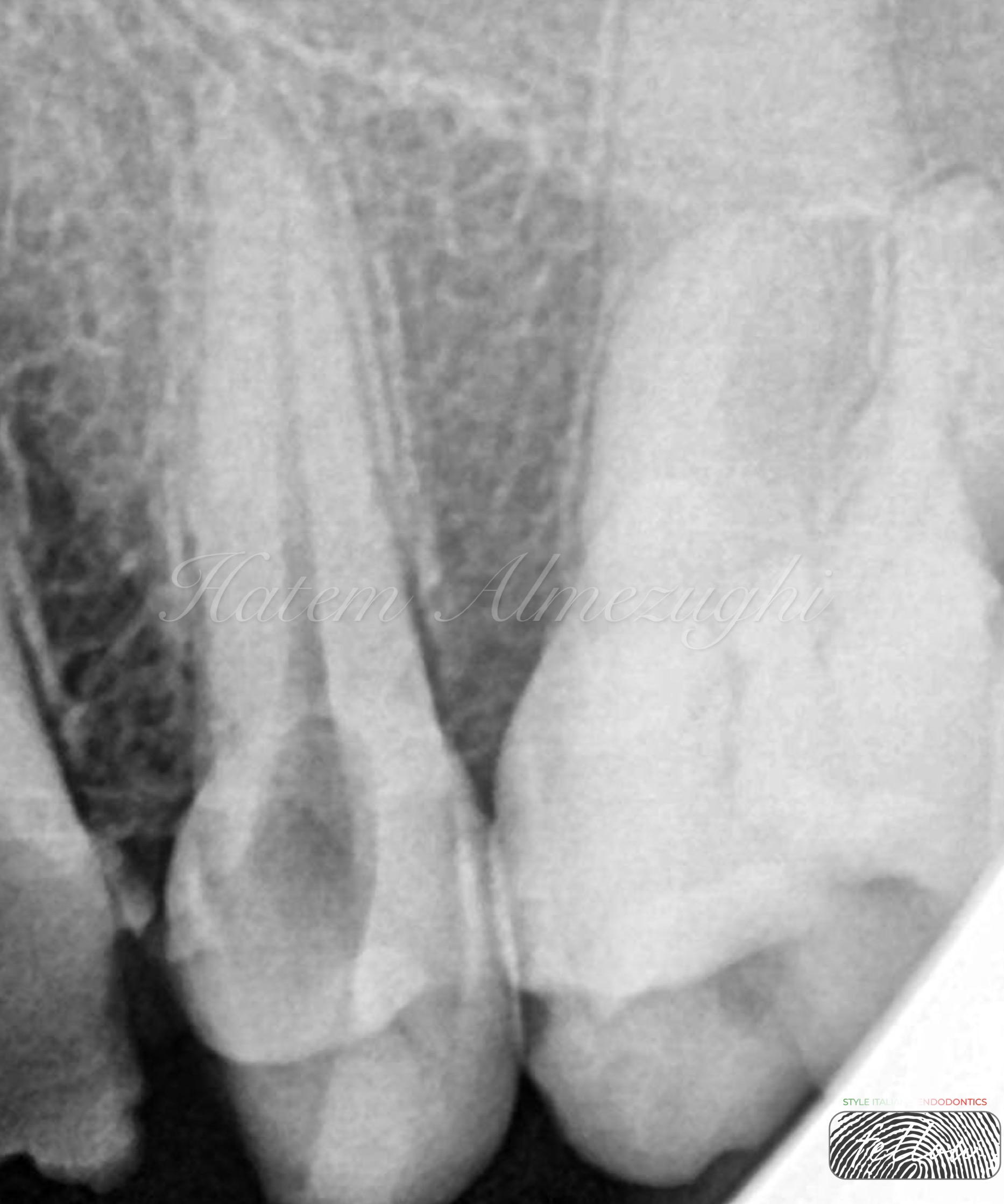
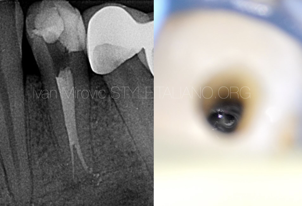
Fig. 1
Pre-operative radiograph shows previously accessed tooth 25 by the referral
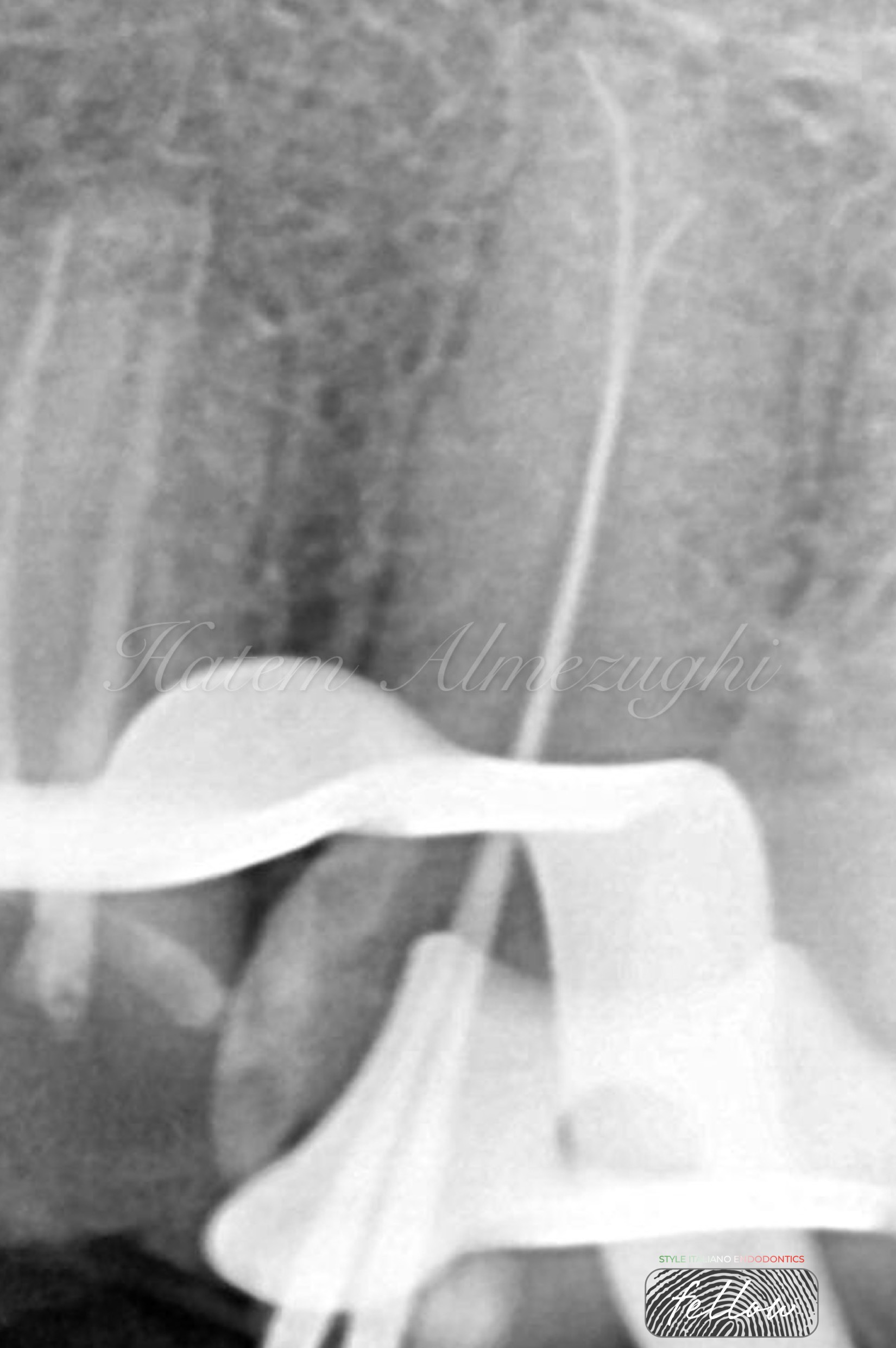
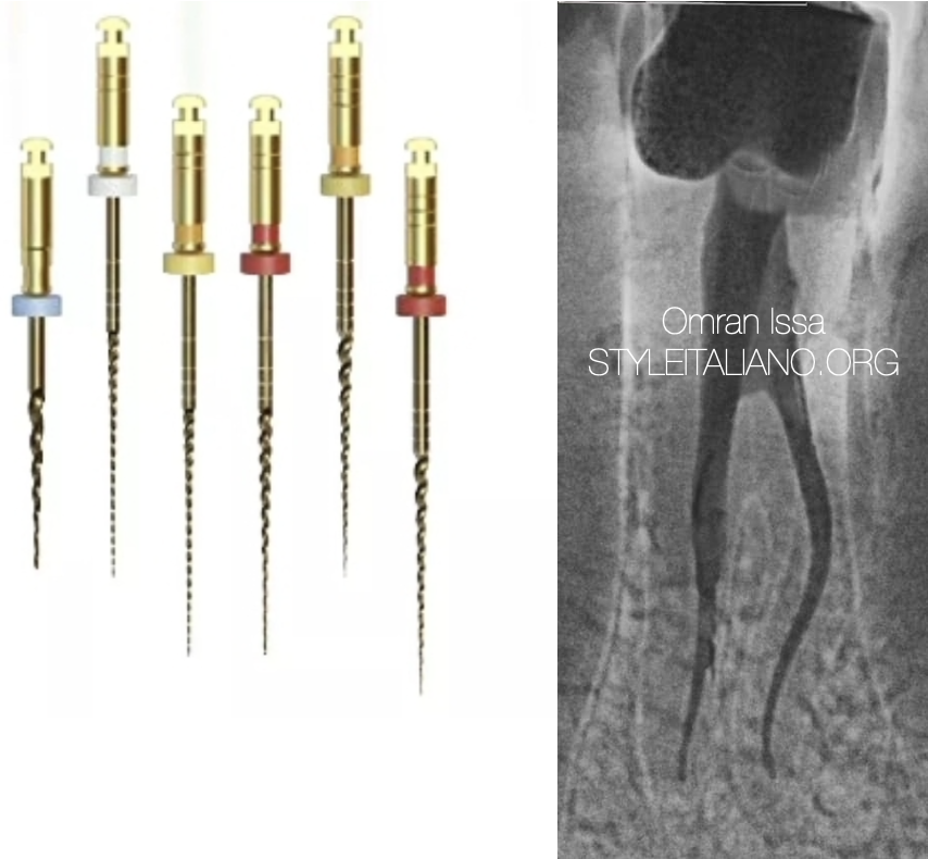
Fig. 2
This figure shows working length where first manual file inserted toward split palatally, then followed by inserting the second manual file toward the split buccally.
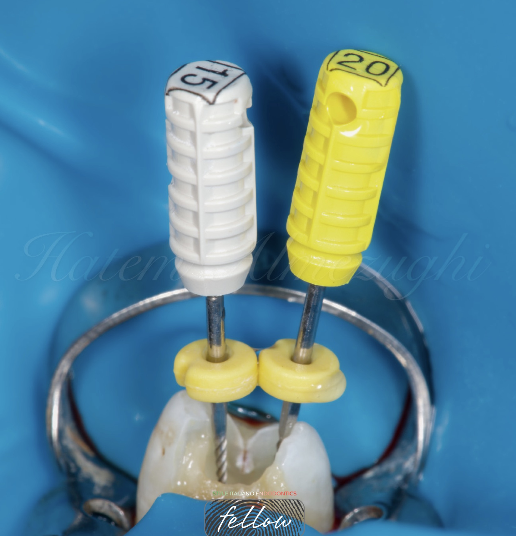
Fig. 3
This figure shows intra oral photo of inserting manual files to measure the working length
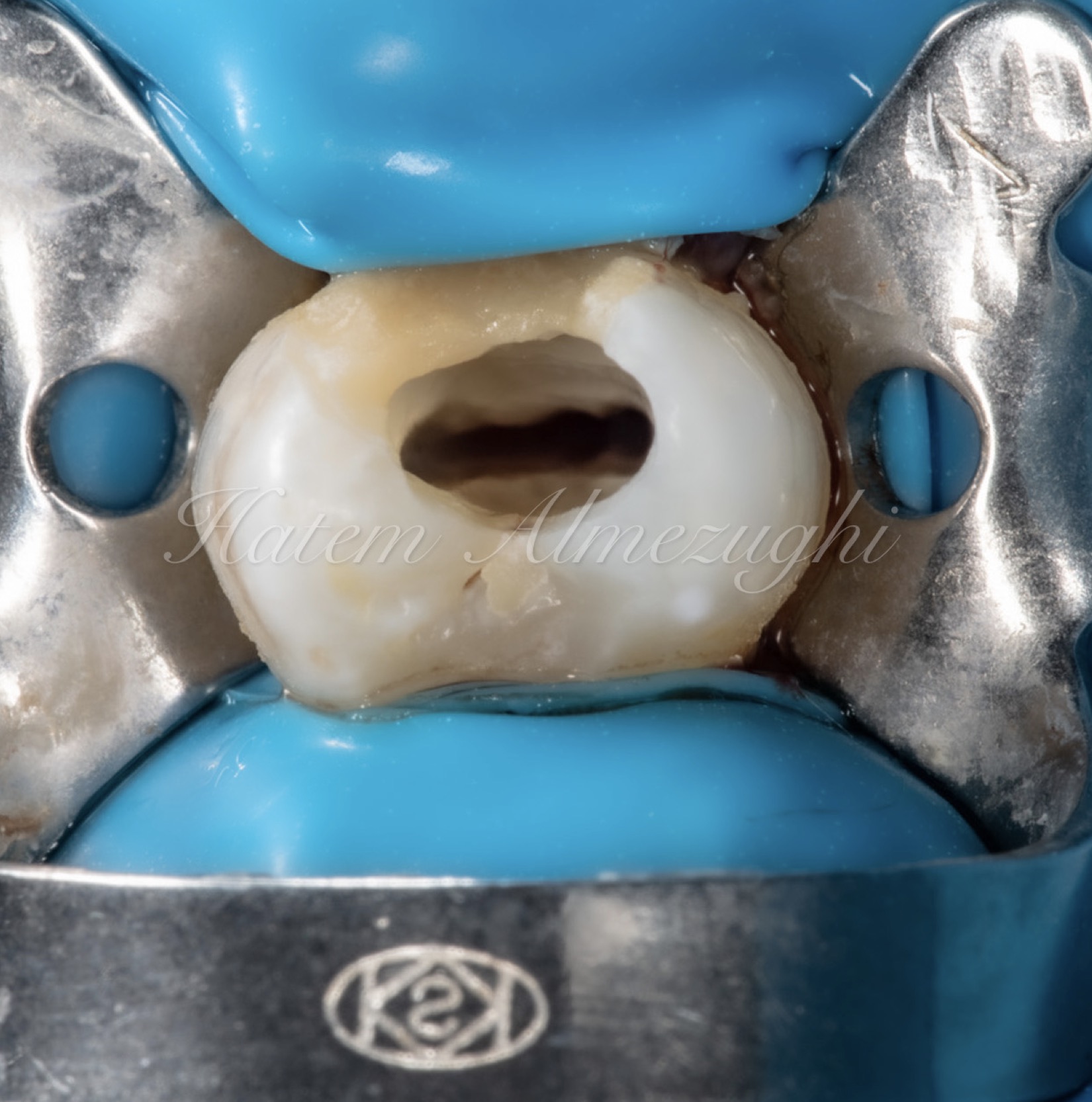
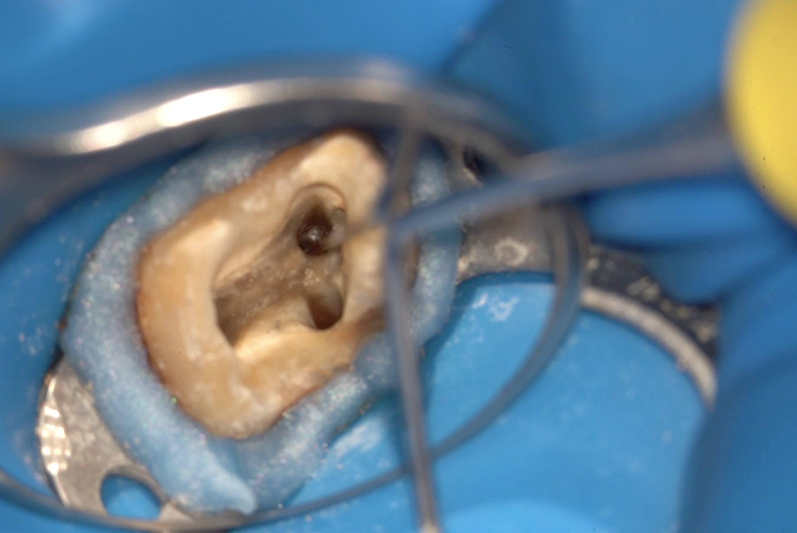
Fig. 4
This figure shows another intra oral photo of tooth where canal is clean ready for cone fit.
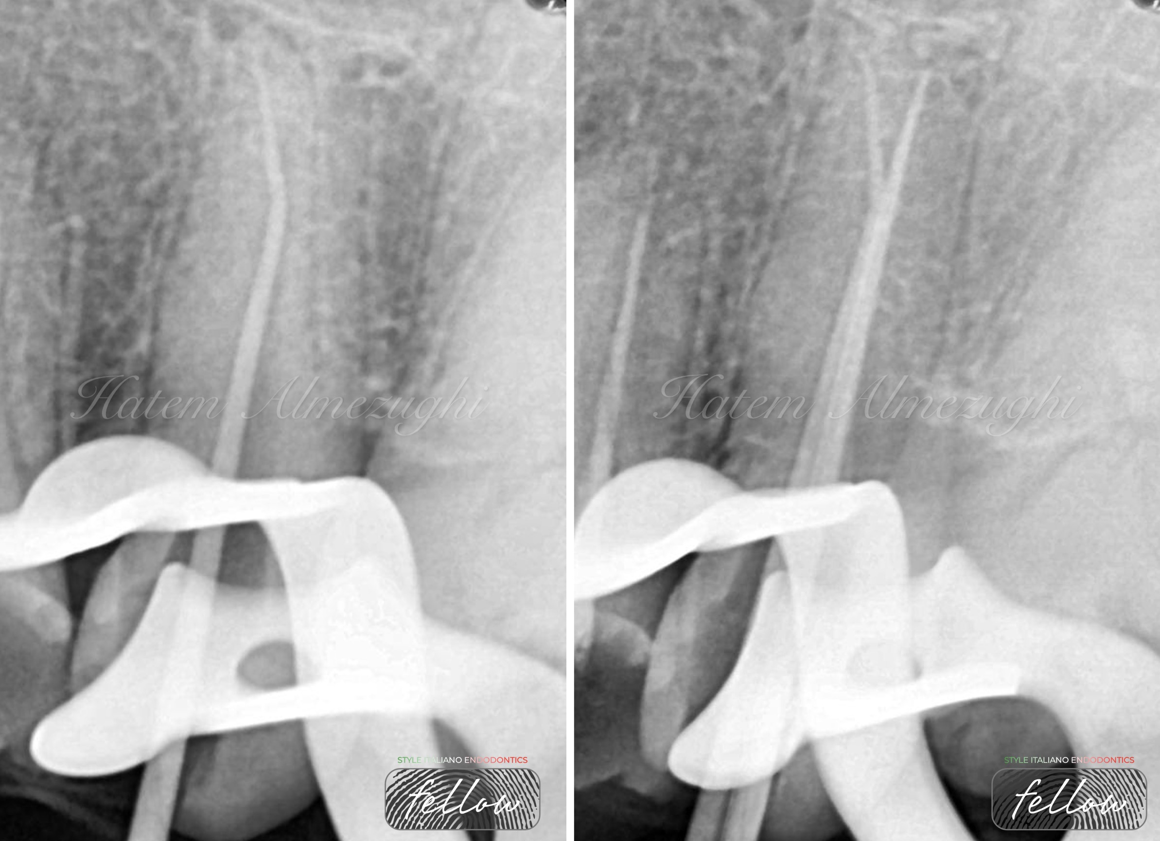
Fig. 5
This figure show cone fitting by inserting gutta percha in apical split then hardly insert the other one in buccal split
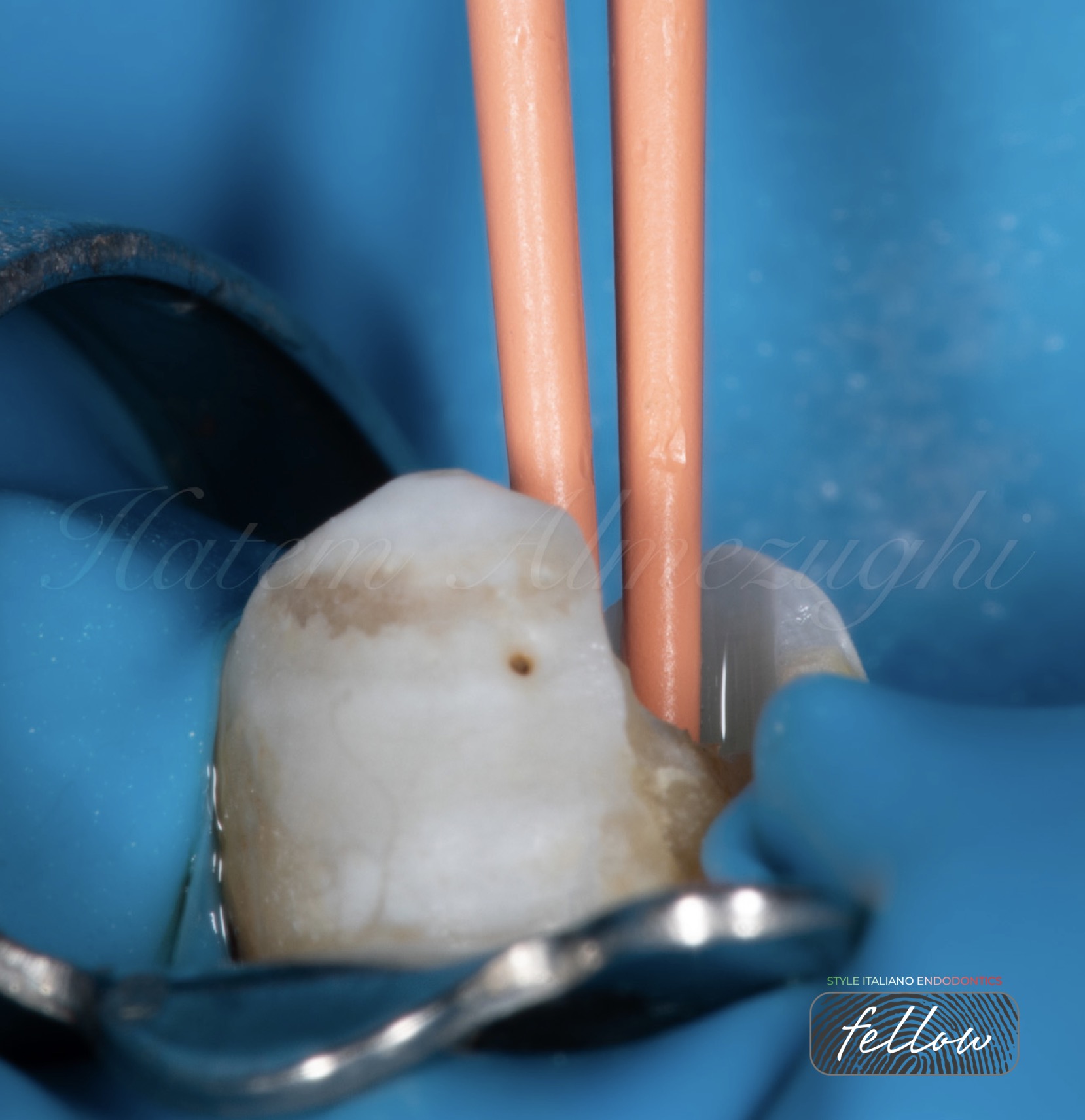
Fig. 6
This figure shows an intra oral photo of cone fit , inserting gutta percha 25/4, after couple of trials I’ve could inserted them both.
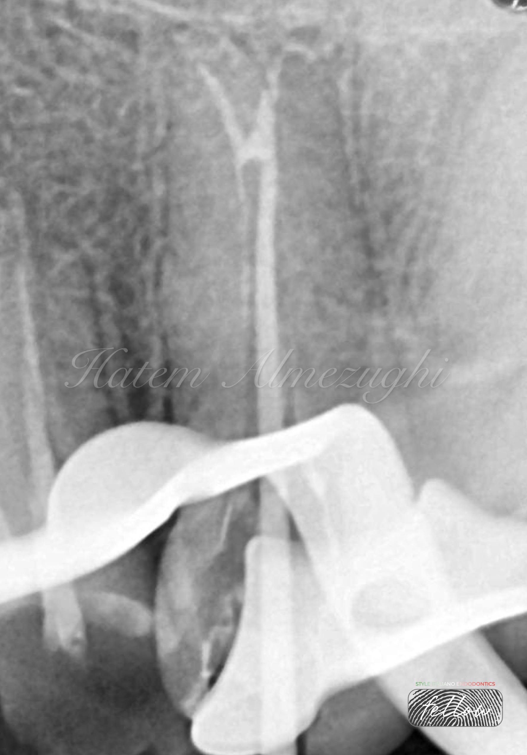
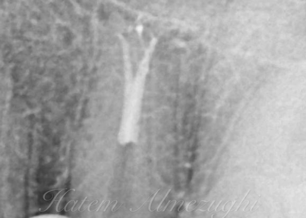
Fig. 7
In this figure another scenario encountered where couldn’t insert GPs together after been coated with sealer, so gp in palatal split inserted and seared off to the split level, to allow inserting the other one , as shown in radiograph
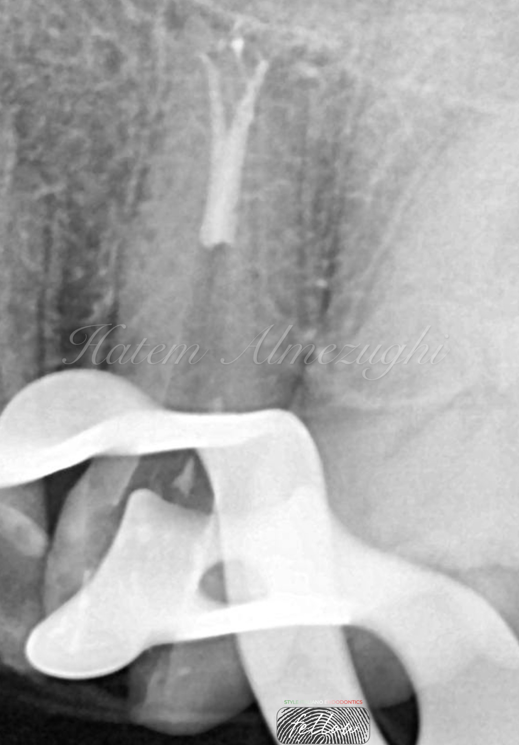
Fig. 8
After the second gp inserted in buccal split and seared off by heat plugger, I started pack fill in phased to insure no voids left between.
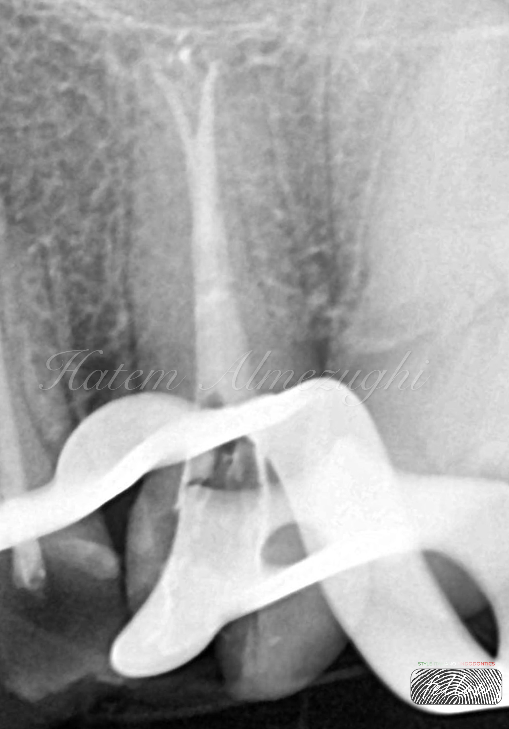
Fig. 9
Pack fill stages nearly done, apart of last stage then gp adaptation by manual plugger.
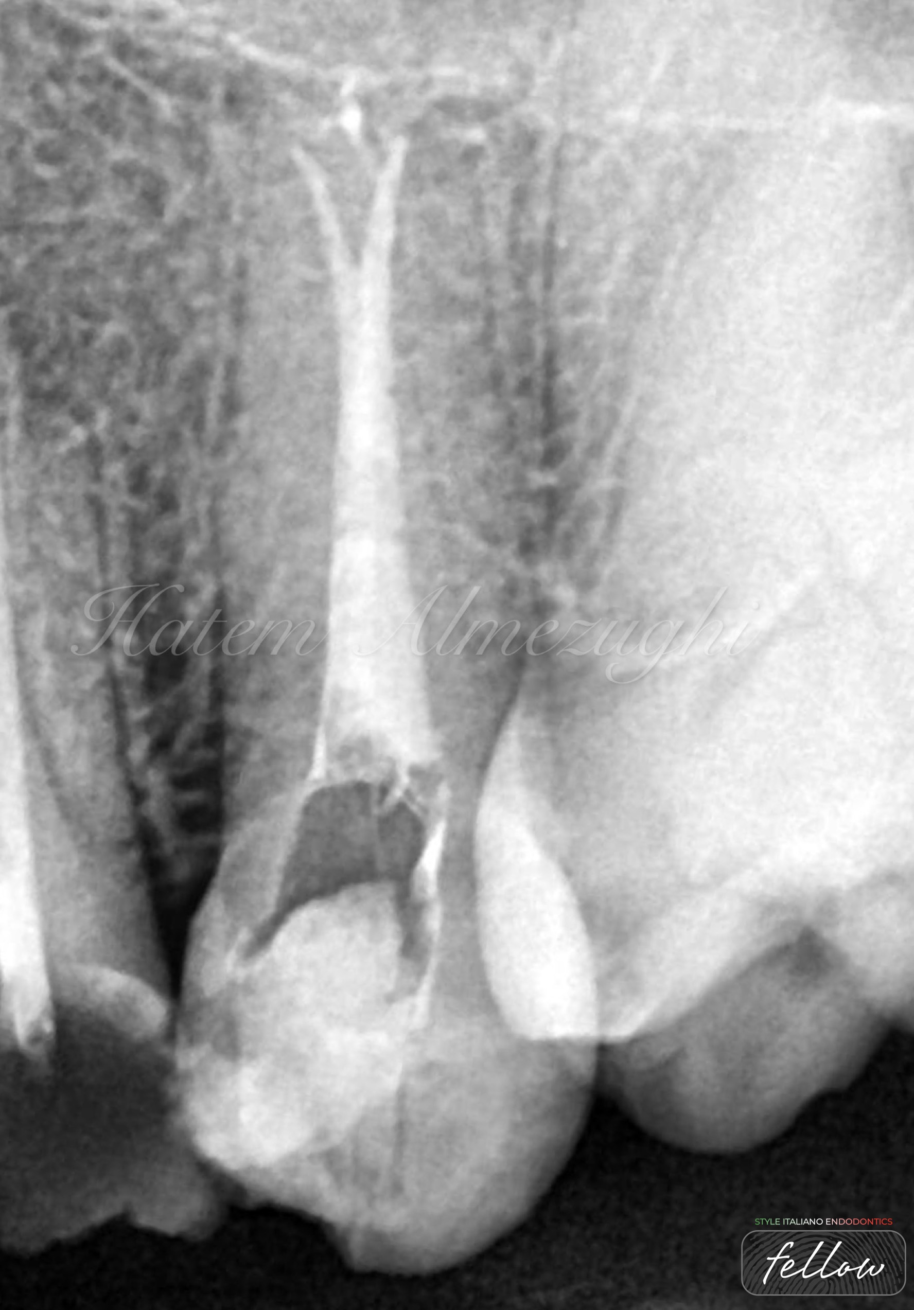
Fig. 10
Last figure shows obturation done .

Fig. 11
Dr. Hatem Almezughi (BDS)
- Was graduated from Tripoli university, Libya.
- Local and international speaker in different conferences and forums .
- instructor in many endodontic courses.
- Dental practice limited to endodontics and restorative .
- Fellow member in style Italiano endodontics.
Conclusions
Being familiar with variations and anatomical morphology make clinicians more aware of unpredictable future unusual clinical cases of all teeth in general and premolars in particular
Bibliography
1. Baratto-Filho F, Fariniuk LF, Ferreira EL, Pecora JD, Cruz-Filho AM, Sousa-Neto MD. Clinical and macroscopic study of maxillary molars with two palatal roots. Int Endod J. 2002.
2. Vertucci FJ. Root canal anatomy of the human permanent te
Maniglia-Ferreira C, Almeida-Gomes F, Sousa BC, Lins CCSA, Santos RA. A case of unusual anatomy in second mandibular molar with four canals. Eur J Dent. 2008.
3. Atieh MA. Root and canal morphology of maxillary first premolars in a Saudi population. J Contemp Dent Pract. 2008.