The tricky lower first premolar with 4 canals
30/10/2023
Abdulwahab Al-Qaraghuli
Warning: Undefined variable $post in /home/styleendo/htdocs/styleitaliano-endodontics.org/wp-content/plugins/oxygen/component-framework/components/classes/code-block.class.php(133) : eval()'d code on line 2
Warning: Attempt to read property "ID" on null in /home/styleendo/htdocs/styleitaliano-endodontics.org/wp-content/plugins/oxygen/component-framework/components/classes/code-block.class.php(133) : eval()'d code on line 2
The mandibular first premolar can be considered one of the most challenging teeth for the endodontist during endodontic procedures; because of the complexity of its root canal morphology and increased incidence of multiple canals. The knowledge of internal anatomy of root canals and their possible variations as well as use of magnification, operating microscope, radiographic examination and illumination, can increase the chances of finding additional canals and contribute to the success of endodontic treatment.
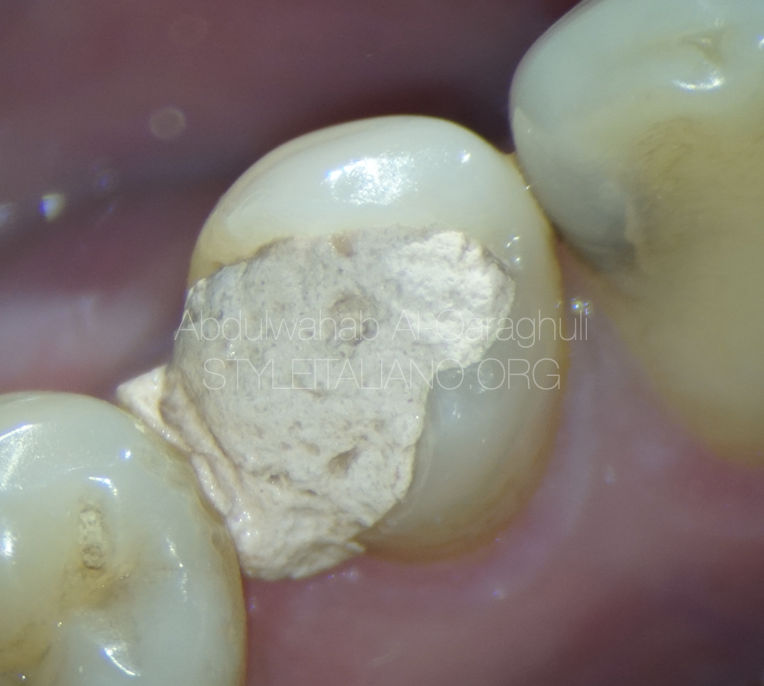
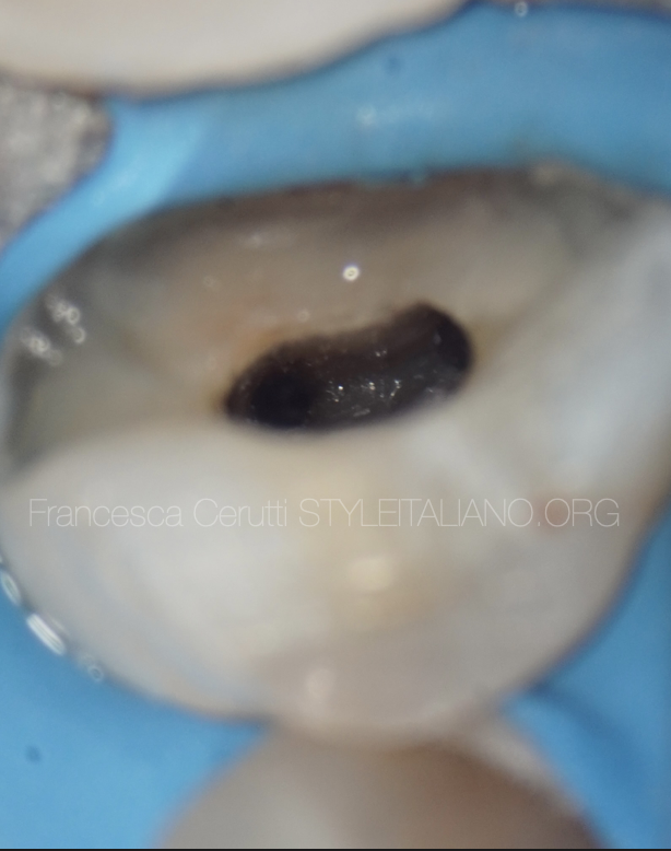
Fig. 1
Initial situation of referred lower first premolar with temporary filling
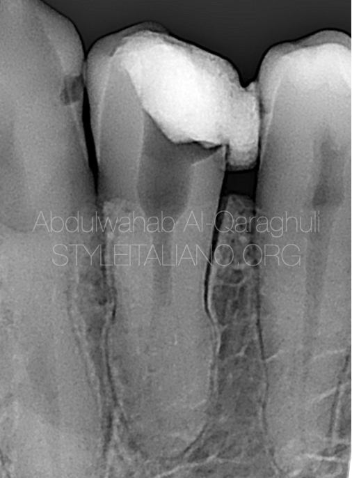
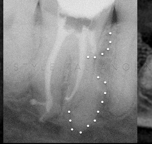
Fig. 2
Pre-op xray showing the abnormal anatomy
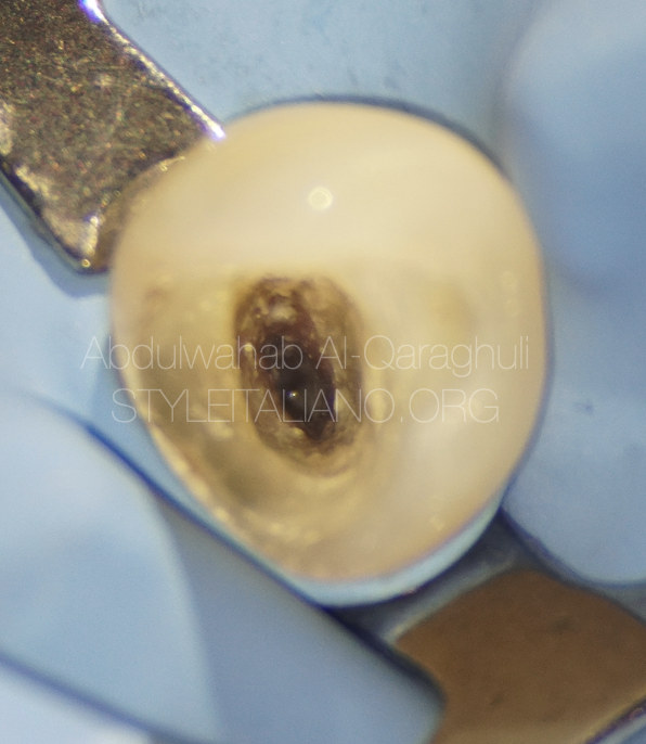
Fig. 3
After temporary filling removal and rubber dam isolation
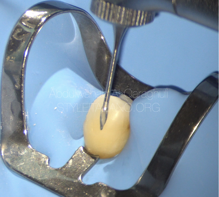
Fig. 4
gates glidden was used to enlarge the coronal third of the canal
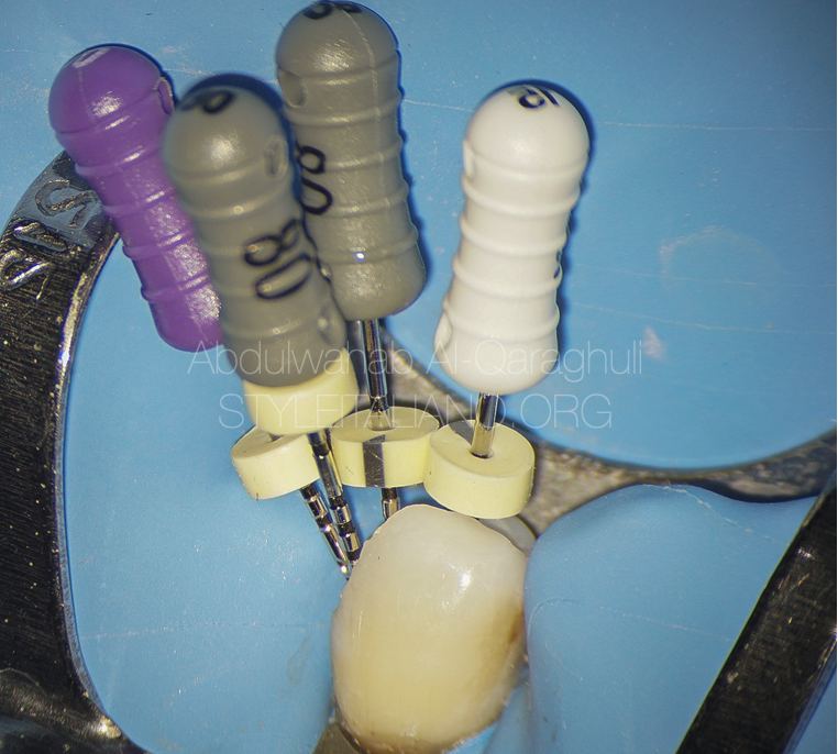
Fig. 5
Files in the canals
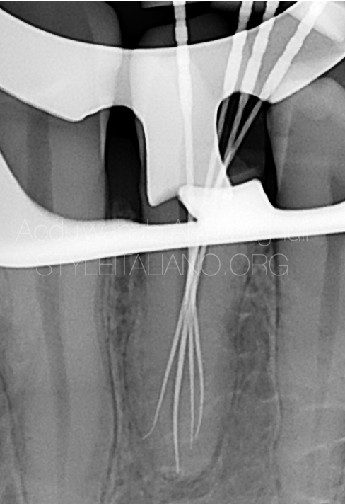
Fig. 6
Working length determination to the four canals
( distal , mesial , buccal , lingual )
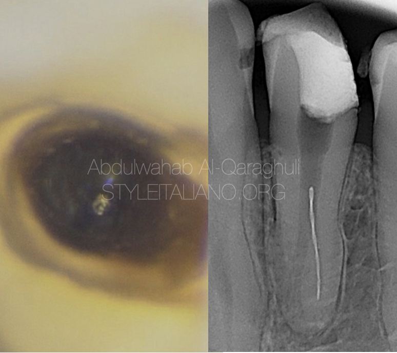
Fig. 7
Rotary file was separated during the instrumentation of distal canal
Separated instrument removal by loop technique
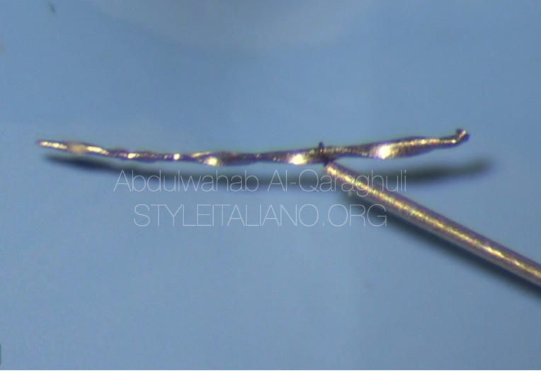
Fig. 8
Separated file was successfully retrieved by BTR pen
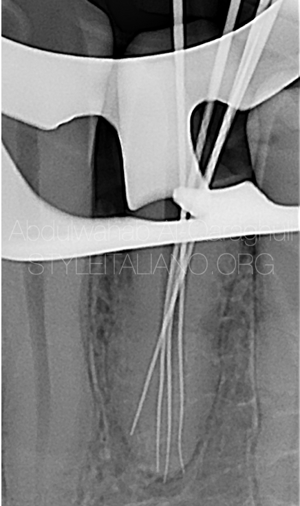
Fig. 9
Files reached the full working length in all canals
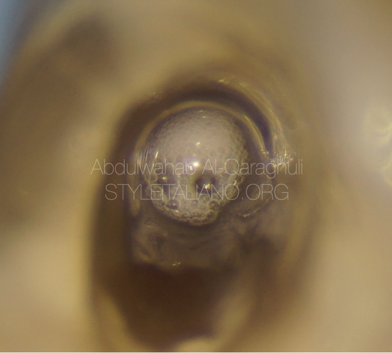
Fig. 10
Sodium hypochlorite in action
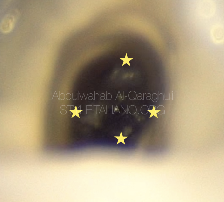
Fig. 11
The stars refer to the canals orifices (Distal , Mesial , Buccal , Lingual)
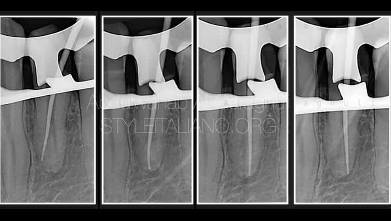
Fig. 12
Cone fit X-rays
Obturation
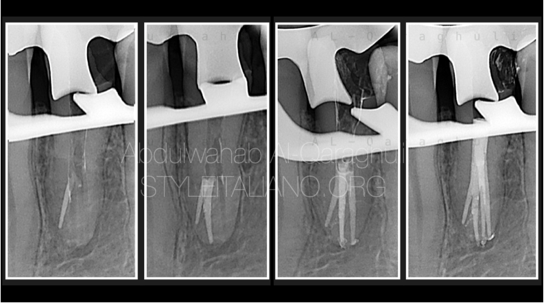
Fig. 13
Obturation in process
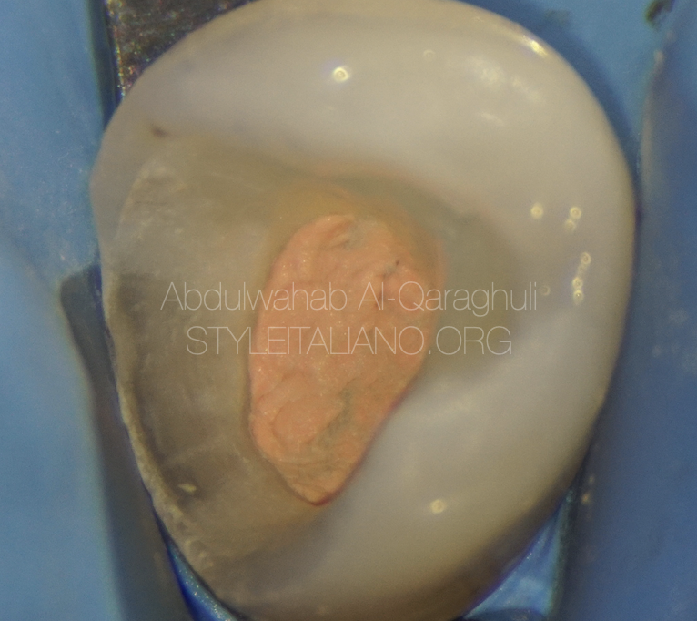
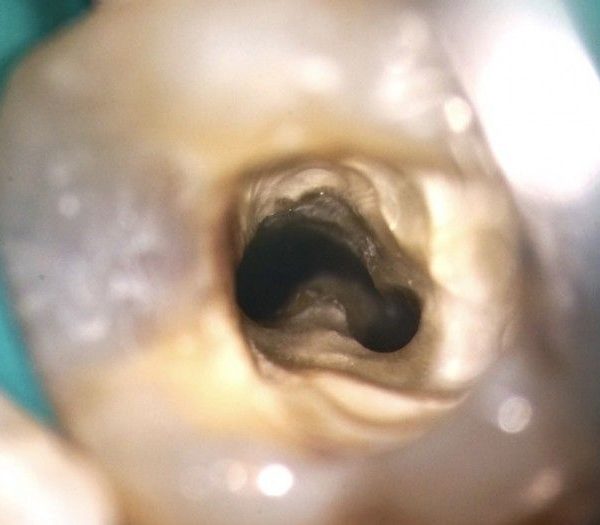
Fig. 14
Clean chamber
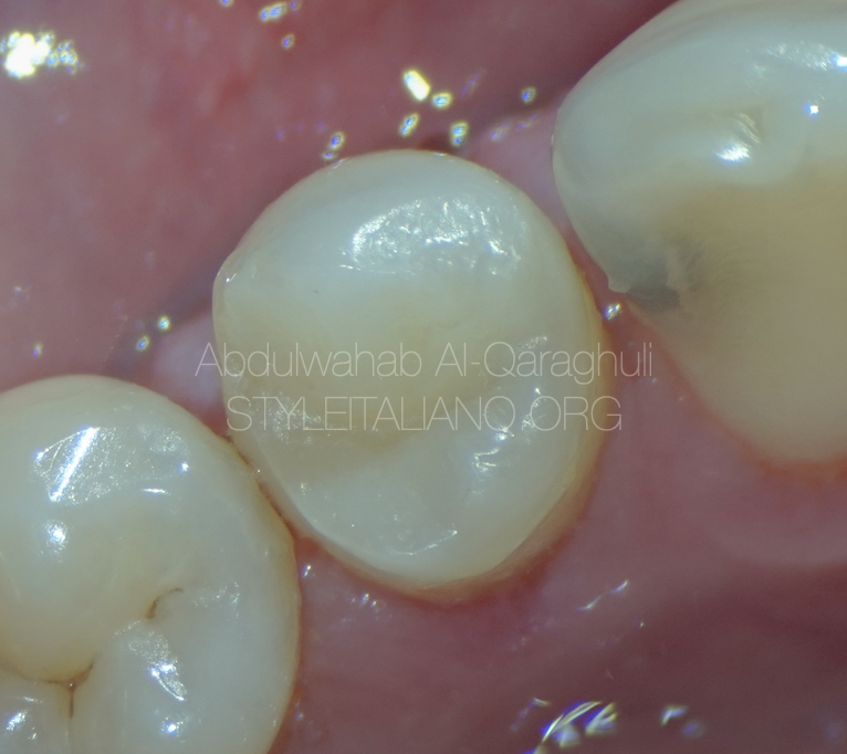
Fig. 15
Final restoration
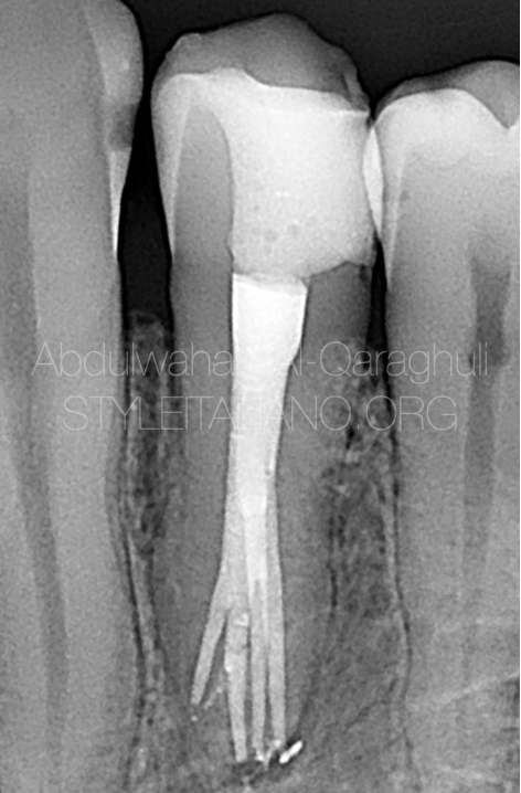
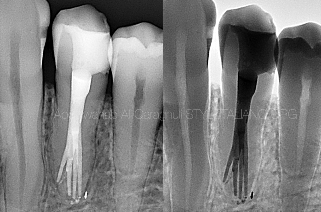
Fig. 16
Post-op X-ray
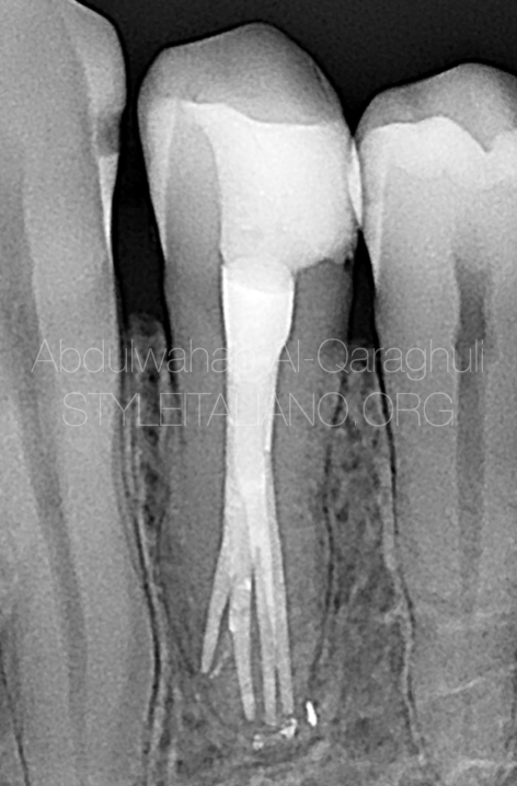
Fig. 17
2 years follow up
Conclusions
This case report presents the successful endodontic treatment of a mandibular first premolar with Four Canals. It is essential to always consider variations in pulp anatomy and morphology before the beginning of root canal treatment. Utilizing magnification tools, e.g. a microscope, to magnify the internal anatomy of a root canal increases the probability of finding additional canals. Furthermore, radiographic examination is imperative when endodontic treatment is to be successful. If canals are identified and negotiated correctly, complex premolars’ anatomy can be treated predictably
Bibliography
Zhang M, Xie J, Wang Y-h, Feng Y. Mandibular first premolar with five root canals: a case report. BMC oral health. 2020;20(1):1–5.
Saati S, Shokri A, Foroozandeh M, Poorolajal J, Mosleh N. Root morphology and number of canals in mandibular central and lateral incisors using cone beam computed tomography. Braz Dent J. 2018;29:239–44.
Yang L, Han J, Wang Q, Wang Z, Yu X, Du Y. Variations of root and canal morphology of mandibular second molars in Chinese individuals: a cone-beam computed tomography study. BMC oral health. 2022;22(1):1–12
Al-Shawwa SS, Al-Khairallah Y, Alenazy MS, Al-Dayel O. Endodontic management of mandibular second premolar with Type IX canal configuration using cone-beam computed tomography. Saudi Endod J. 2019;9(2):144
Bürklein S, Heck R, Schäfer E: Evaluation of the root canal anatomy of maxillary and mandibular premolars in a selected German population using cone-beam computed tomographic data. J Endod. 2017, 43:1448-52.