A complex mandibular 2nd molar
17/03/2020
Fabio Gorni
Warning: Undefined variable $post in /home/styleendo/htdocs/styleitaliano-endodontics.org/wp-content/plugins/oxygen/component-framework/components/classes/code-block.class.php(133) : eval()'d code on line 2
Warning: Attempt to read property "ID" on null in /home/styleendo/htdocs/styleitaliano-endodontics.org/wp-content/plugins/oxygen/component-framework/components/classes/code-block.class.php(133) : eval()'d code on line 2
A particular clinical case .
The second lower molar has been endodontically treated for an invasive caries, but during the therapy the colleague faced some problems in the mesio-buccal canal and for this reason he decided to refer the patient .
From the intra-op x-ray, taken during the previous treatment , it’s evident that the manual file is out of the root, simulating a perforation .
From the pre-op cbct the clinical situation is pretty clear: part of the mesial root is separated from the rest of the tooth. The apical two-thirds have probably been fractured due to a previous trauma, but they are still vital, that’s why it doesn’t represent a problem for the patient .
The coronal one third has been treated as a root with a large foramen.
First, we improved the access cavity removing completely the decayed tissue; after that, we proceeded with the control of the copius bleeding coming from the canal, applying a firm pressure with large paper points.
After a correct cleaning and shaping, a collagen extra radicular barrier was used to improve the apical control of the MTA plug.
The cement has been injected using the MAP-ONE syringe, then condensed and finished with a micro-brush
In a second appointment, the root canal treatment has been completed, despite the difficulties due to the endodontic anatomy.
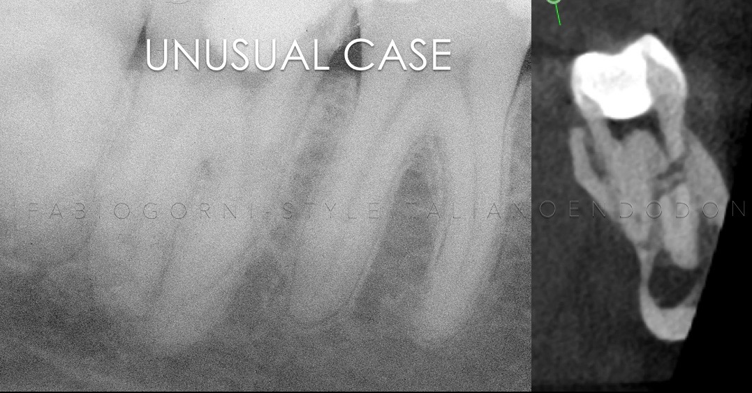
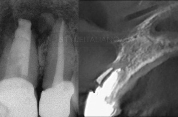
Fig. 1
Pre-op x-ray and CBCT section
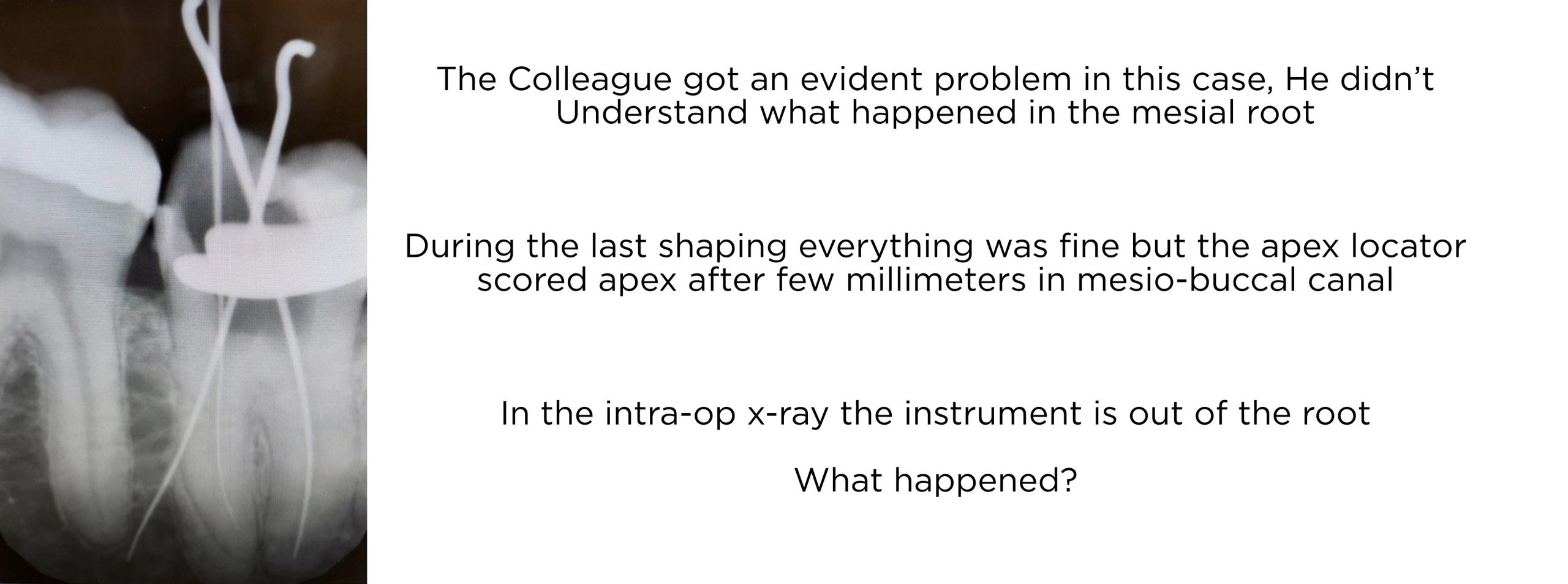
Fig. 2
Intra-op x-ray from the previous treatment
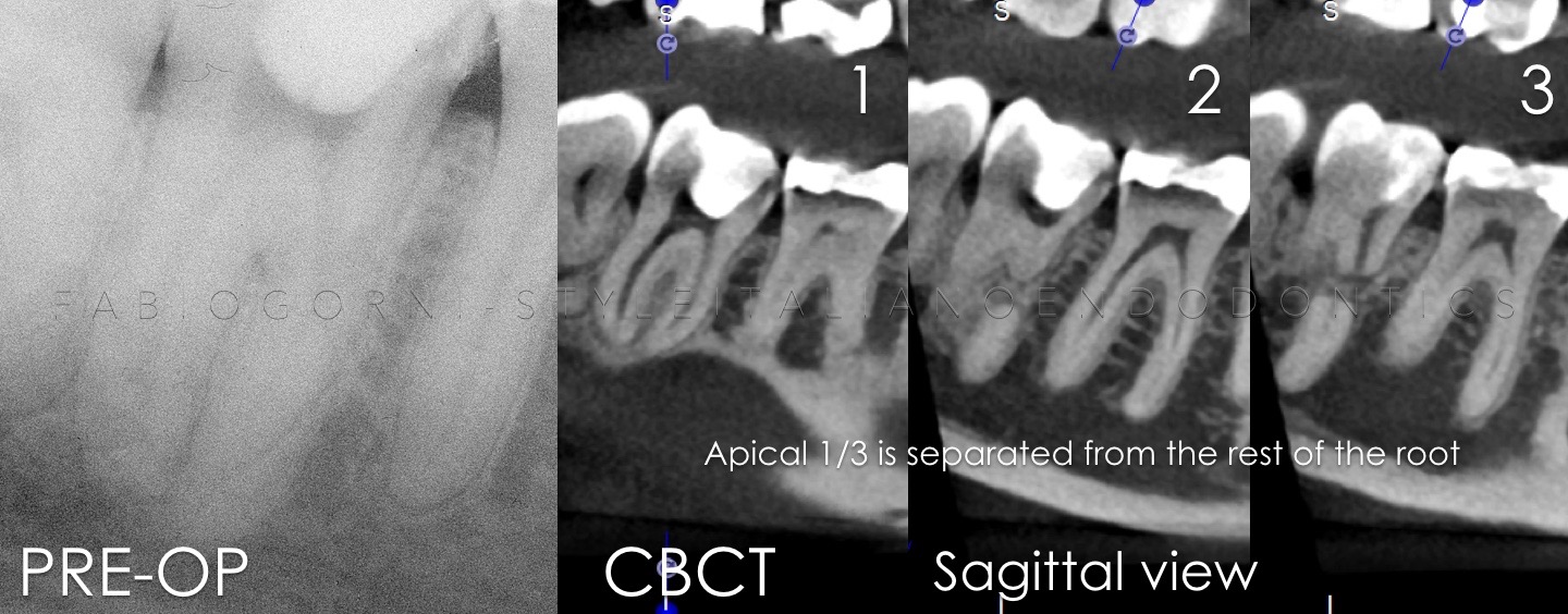
Fig. 3
Pre-op CBCT . In the sagittal views we can clearly see part of the root separate form the coronal part of the root
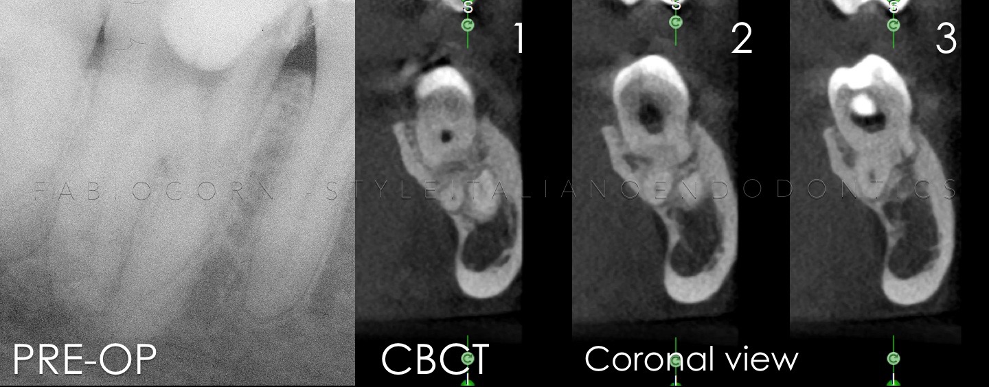
Fig. 4
Pre-op CBCT . In the coronal views it’s appreciable the distance between the apical two-thirds and the coronal one-third of the root
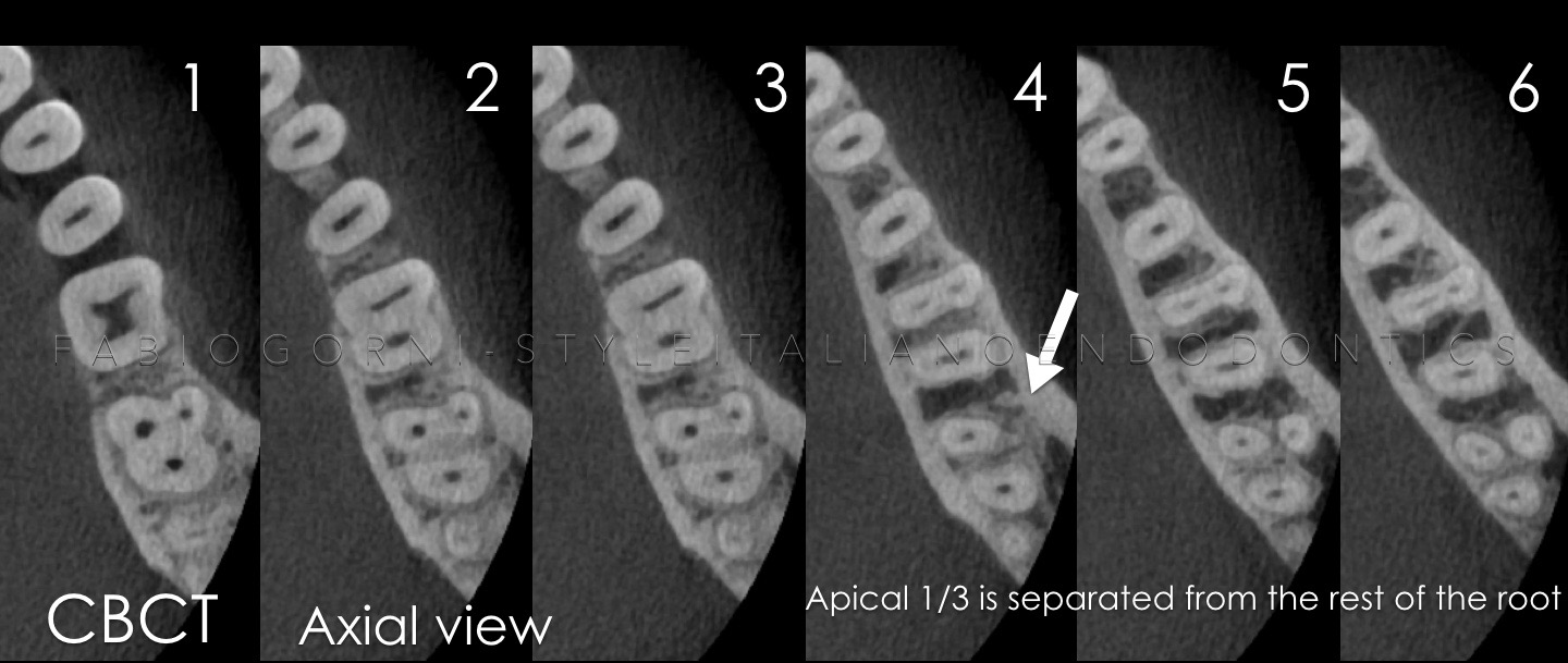
Fig. 5
Pre-op CBCT. Particular of the axial views
Fig. 6
TRIUM CBCT- Acteon
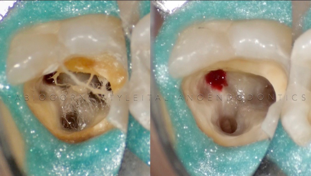
Fig. 7
A correct access cavity improves the pulpal floor visibility
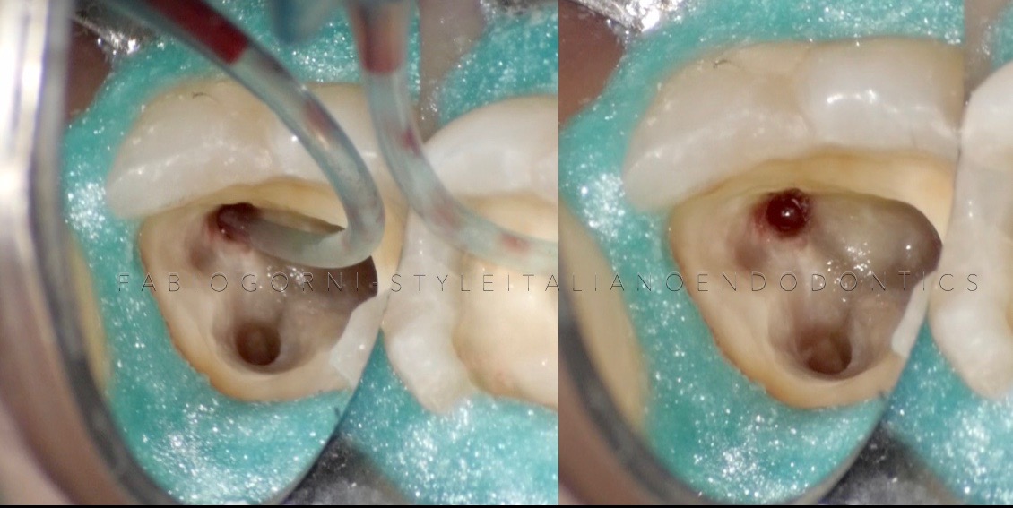
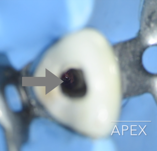
Fig. 8
Copious blood flow out of the mesio-buccal orifice
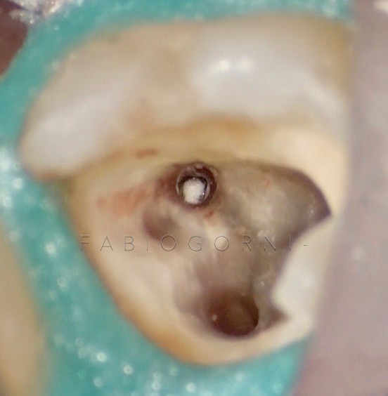
Fig. 9
After cleaning and shaping, we obtained a perfect control of bleeding. The picture shows the resorbable barrier beyond the apex, that will allow us to have a good apical control during the MTA apical plug .
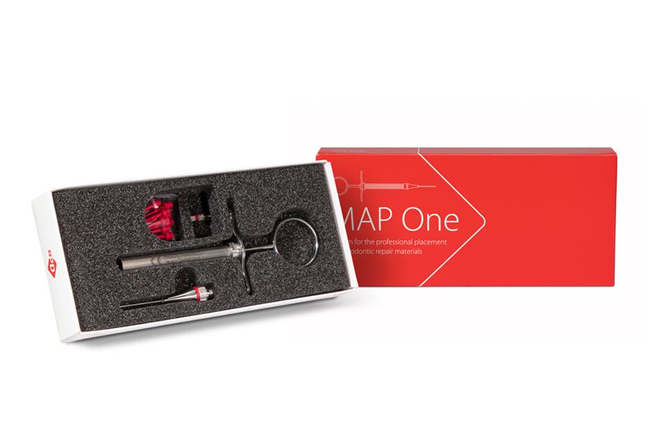
Fig. 10
Map-One - Produits Dentaires SA

Fig. 11
MTA - Produits Dentaires SA
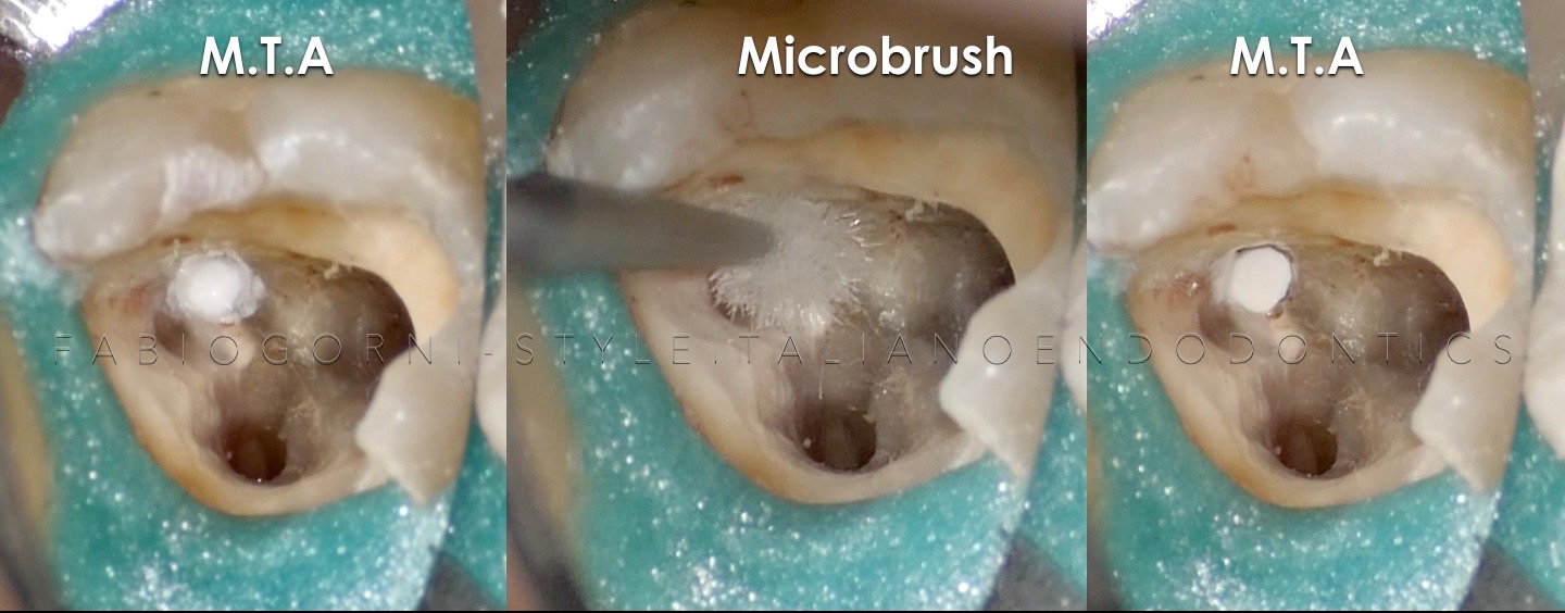
Fig. 12
MTA is extruded in the canal with MAP-ONE and well adapted with a micro brush .
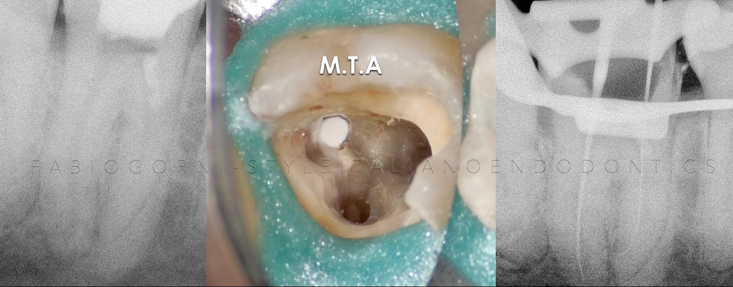
Fig. 13
MTA apical plug and working length
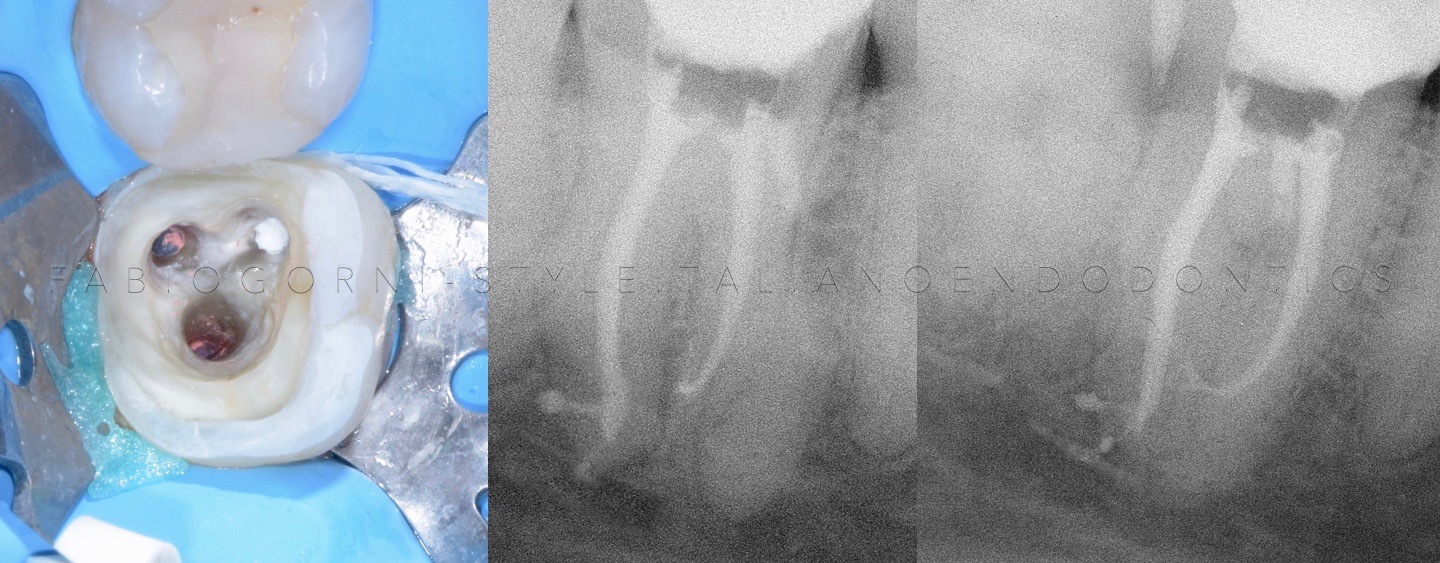
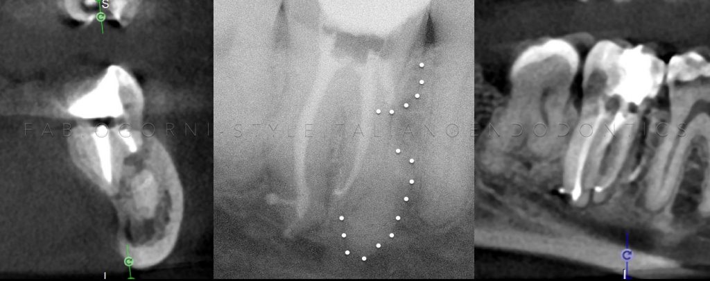
Fig. 14
Post-op x-rays clearly show the apical plug and the complex endodontic anatomy: an strong apical curvature in the mesio -lingual canal and an apical delta in the distal canal
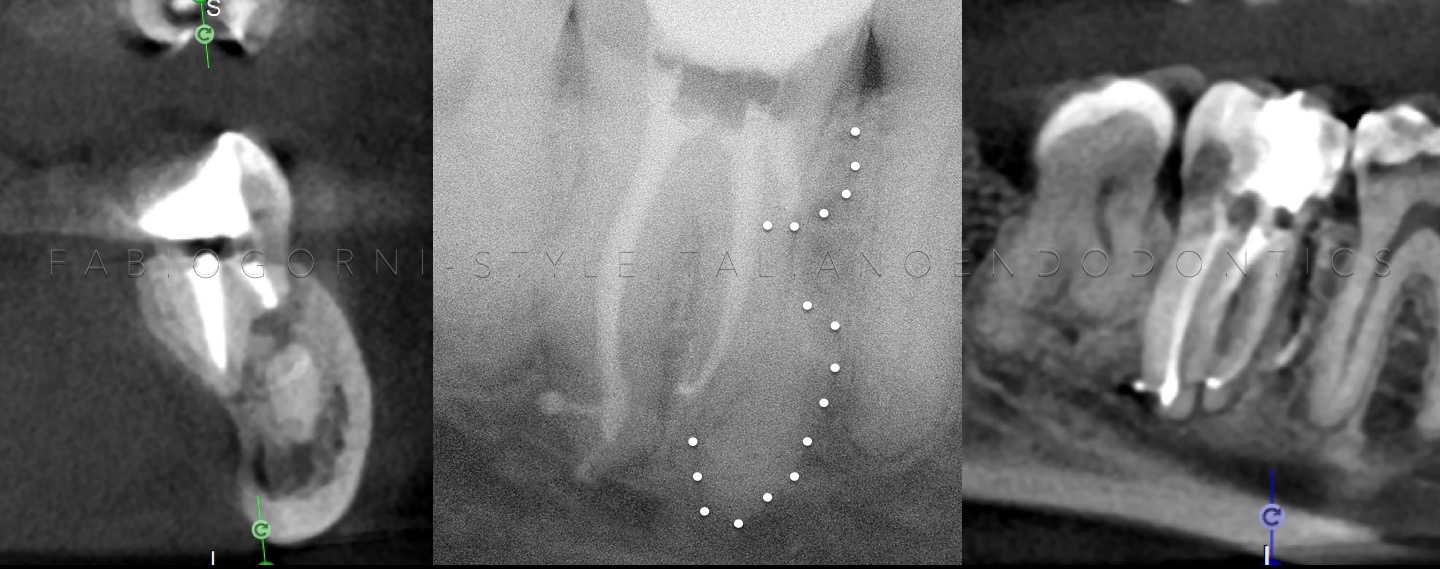
Fig. 15
Post-op CBCT. We decided to execute a CBCT in order to control the overfilling in the distal root , because in the 2D post-op x-rays the cement was close to the mandibular nerve.
The 3D exam confirms that the cement doesn’t invade the mandibular canal and the correct position of the MTA apical plug .
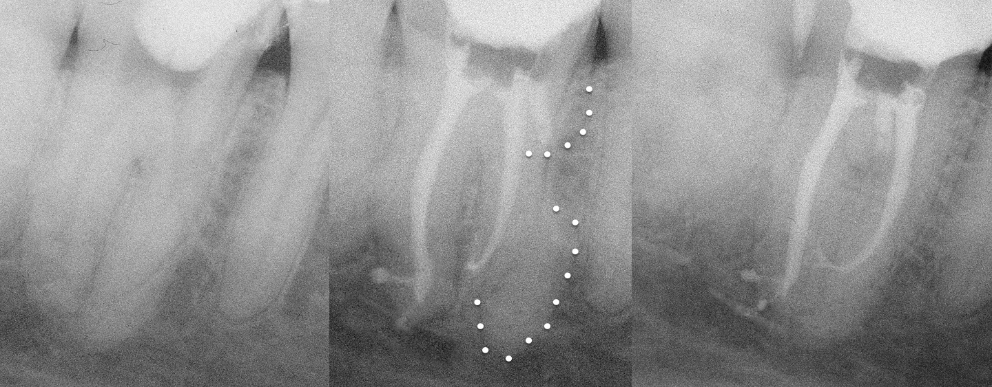
Fig. 16
The final x-rays evidence the endodontic anatomy, the root fragment and the MTA apical plug
Conclusions
An accurate examination of the x-rays and of the CBCT can help the clinician to do a proper diagnosis in complex cases.
After understanding the problem, the sequence to be followed during the therapy is clear. The choice of MAP system has been useful to position MTA in a predictable, precise and correct fashion.
Bibliography
Rodríguez G, Patel S, Durán-Sindreu F, Roig M, Abella F. Influence of Cone-beam Computed Tomography on Endodontic Retreatment Strategies among General Dental Practitioners and Endodontists. J Endod. 2017 Sep;43(9):1433-1437