Dealing with lower premolars
02/02/2023
Francesca Cerutti
Warning: Undefined variable $post in /home/styleendo/htdocs/styleitaliano-endodontics.org/wp-content/plugins/oxygen/component-framework/components/classes/code-block.class.php(133) : eval()'d code on line 2
Warning: Attempt to read property "ID" on null in /home/styleendo/htdocs/styleitaliano-endodontics.org/wp-content/plugins/oxygen/component-framework/components/classes/code-block.class.php(133) : eval()'d code on line 2
The first step to reach the success of root canal therapy is o have a good knowledge of the root and root canal morphology. This helps us to locate all the canals and properly clean, shape, and obturate the canal spaces in all dimensions.
Slowey has suggested that mandibular first premolars, often called as “Endodontist's enigma,” may present the greatest difficulty of all teeth to perform successful endodontic treatment due to their unpredictable anatomy with a wide variety of morphological rarities.
A recently published systematic literature review by Kottoor et al. summarized that lower bicuspids commonly have a single root (97%), with the mandibular first premolars being twice more likely to present with two canals (23.55%) than second premolars (12.64%). Multiplicities of roots (up to 4 roots) and root canals (up to 5 canals) have been documented as case reports in both lower bicuspids. First premolars can present variations such as C-shaped canals, deep mesial radicular invagination (13–27%) and dens invaginatus.
A careful examination of the pre-operative x-ray can help the clinician in identifying pre-operatively such anatomic peculiarities in order to select the strategy and the correct instruments before starting the root canal therapy.
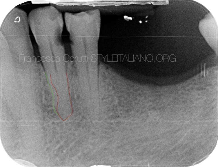
Fig. 1
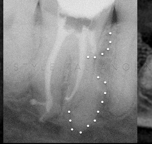
A 57 years old patient came to my attention complaining about pain in the lower left area of the mouth.
The periapical x-ray shows the presence of a proximal decay that involved the first and second lower left premolar.
An accurate analysis of the X-ray showed a fast break in the line representing the root canal of the first premolar, suggesting the presence of a bifurcation.
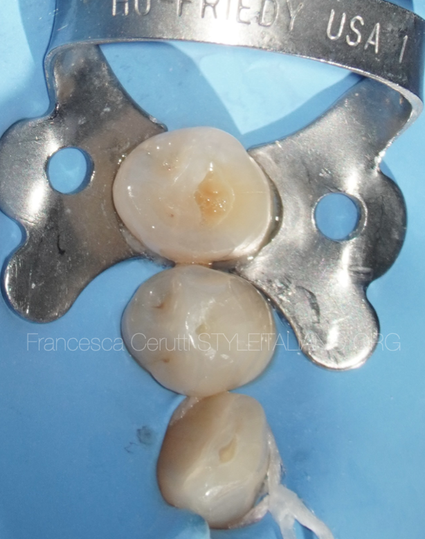
Fig. 2
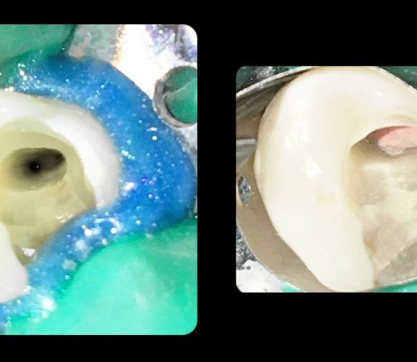
A multiple isolation with rubber dam is done
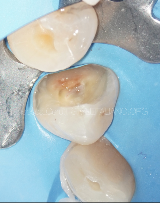
Fig. 3
Once removed the decayed tissue, the pulp of the first premolar is exposed.
Fig. 4
The root canal openings are connected by a line on the pulp chamber floor. This helps in locating the position of the orifices.
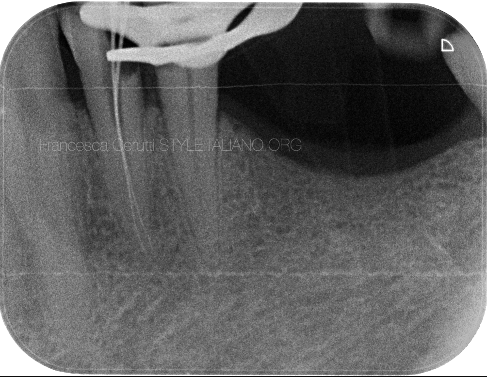
Fig. 5
The intra-operative x-ray shows the recording of the working length, evidencing the presence of two distinct root canals.
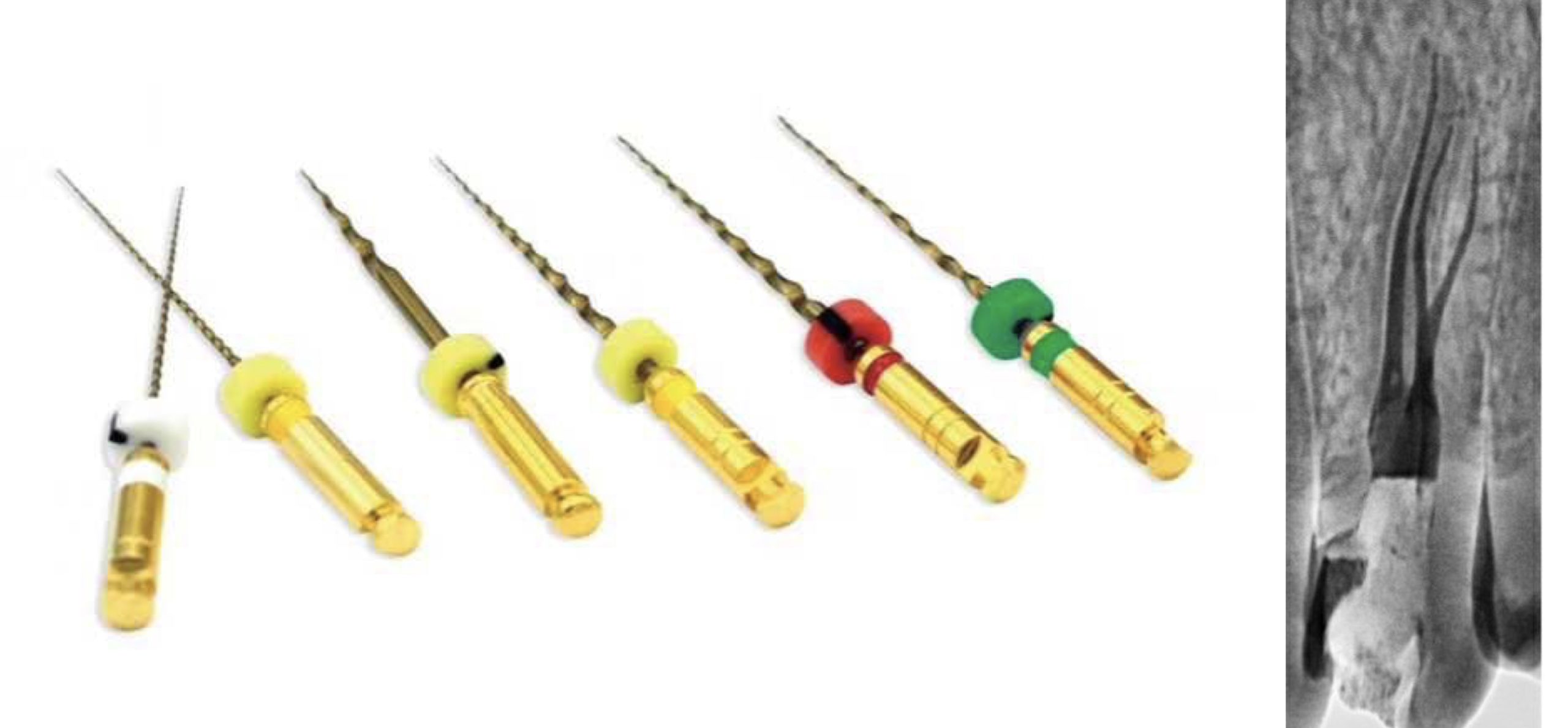
After that the tooth was shaped with rotary files.
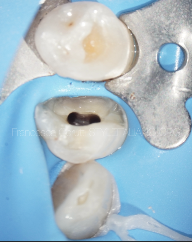
Fig. 6
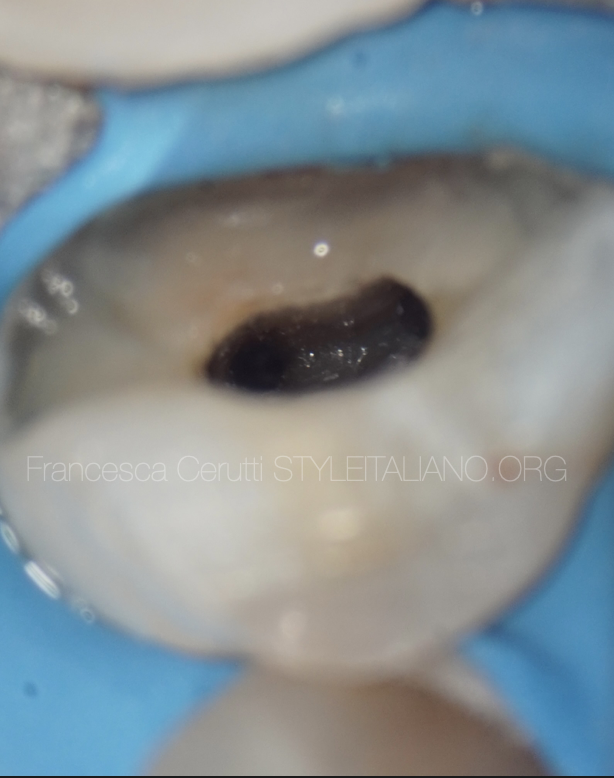
The appearance of the pulp chamber after the phases of shaping and cleaning.
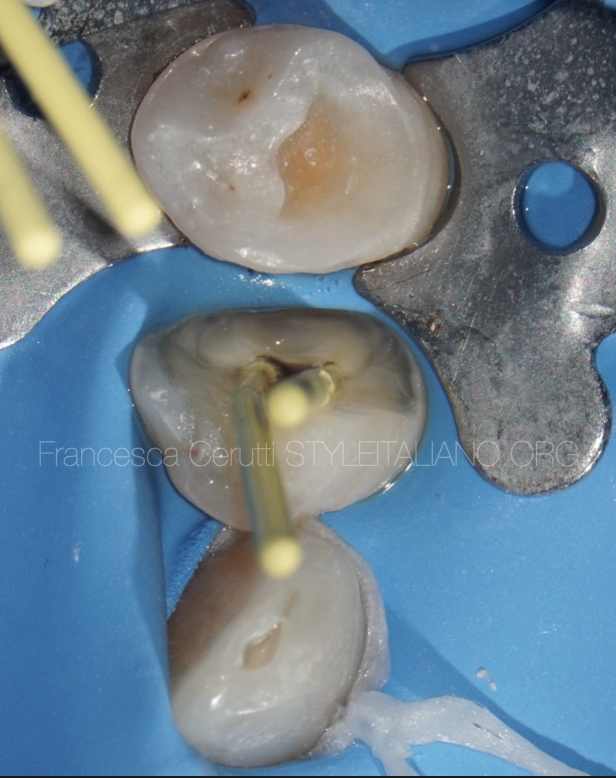
Fig. 7
The gutta percha point fit
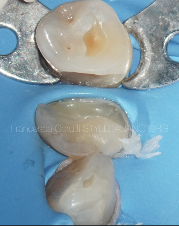
Fig. 8
After the root canal filling, a build up was done on the first premolar, while the decay was removed from the second premolar.
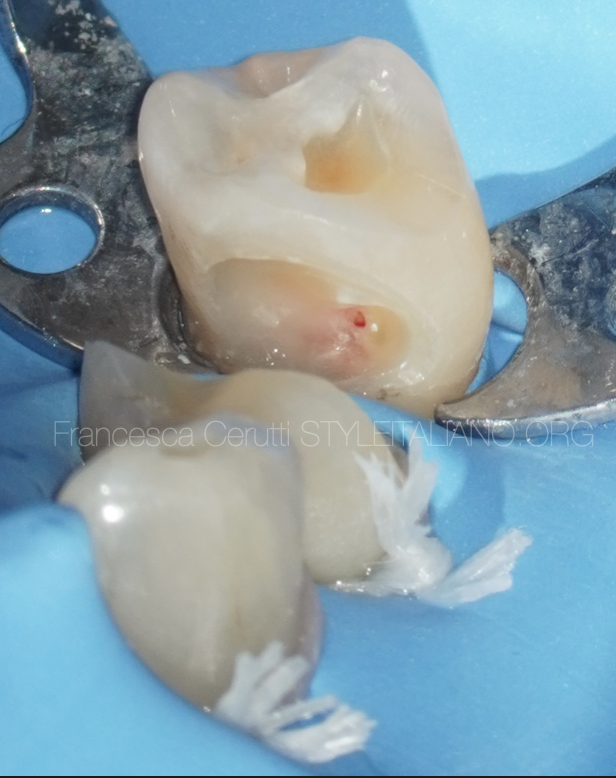
Fig. 9
There was an accidental pulp exposure that was treated with a direct pulp capping with MTA.
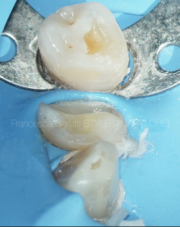
Fig. 10
A direct composite restoration
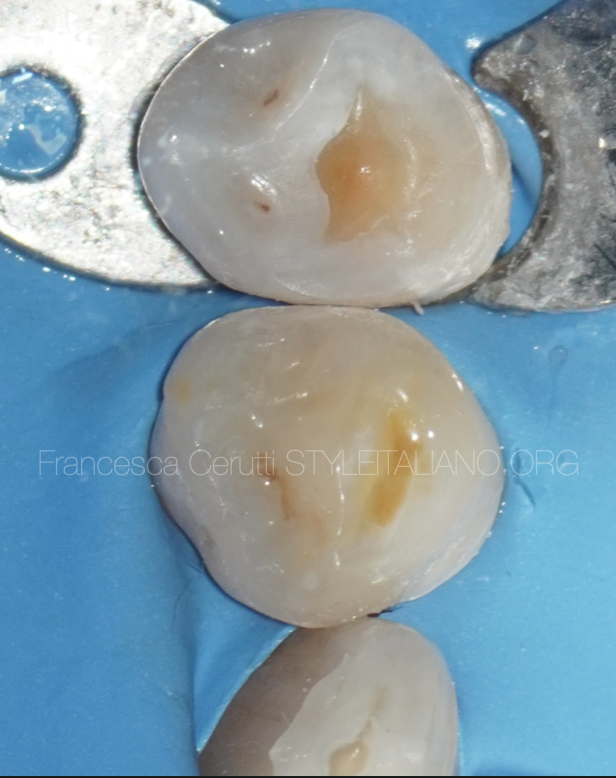
Fig. 11
A composite overlay was luted by using pre-heated composite material
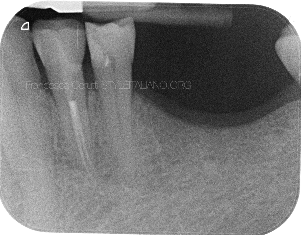
Fig. 12
Post operative x-ray
Conclusions
A careful examination of the pre operative x-ray and a correct endo-restorative workflow can lead to a successful treatment of anatomic variations.
Bibliography
Kararia N, Chaudhary A, Kararia V. Mandibular left first premolar with two roots: A morphological oddity. Contemp Clin Dent. 2012 Apr;3(2):234-6. doi: 10.4103/0976-237X.96840. PMID: 22919233; PMCID: PMC3425116.
Albuquerque D, Kottoor J, Hammo M. Endodontic and clinical considerations in the management of variable anatomy in mandibular premolars: a literature review. Biomed Res Int. 2014;2014:512574. doi: 10.1155/2014/512574. Epub 2014 May 8. PMID: 24895584; PMCID: PMC4034431.
Thanaruengrong P, Kulvitit S, Navachinda M, Charoenlarp P. Prevalence of complex root canal morphology in the mandibular first and second premolars in Thai population: CBCT analysis. BMC Oral Health. 2021 Sep 16;21(1):449. doi: 10.1186/s12903-021-01822-7. PMID: 34530811; PMCID: PMC8444426.
Jang YE, Kim Y, Kim B, Kim SY, Kim HJ. Frequency of non-single canals in mandibular premolars and correlations with other anatomical variants: an in vivo cone beam computed tomography study. BMC Oral Health. 2019 Dec 4;19(1):272. doi: 10.1186/s12903-019-0972-5. PMID: 31801495; PMCID: PMC6894311.