Endodontic management of complex premolar anatomy By MG3 Files
23/08/2021
Warning: Undefined variable $post in /home/styleendo/htdocs/styleitaliano-endodontics.org/wp-content/plugins/oxygen/component-framework/components/classes/code-block.class.php(133) : eval()'d code on line 2
Warning: Attempt to read property "ID" on null in /home/styleendo/htdocs/styleitaliano-endodontics.org/wp-content/plugins/oxygen/component-framework/components/classes/code-block.class.php(133) : eval()'d code on line 2
The success of root canal therapy depends on managing all the existing canals by having the knowledge of the root anatomy, then cleaning, shaping and pack the endodontic space.
Variations in root canal anatomy are common and sometimes can't be noticed while taking 2D radiograph, thus there are cases where CBCT is a must.
This case discusses the endodontic management in maxillary premolar with complex anatomy.
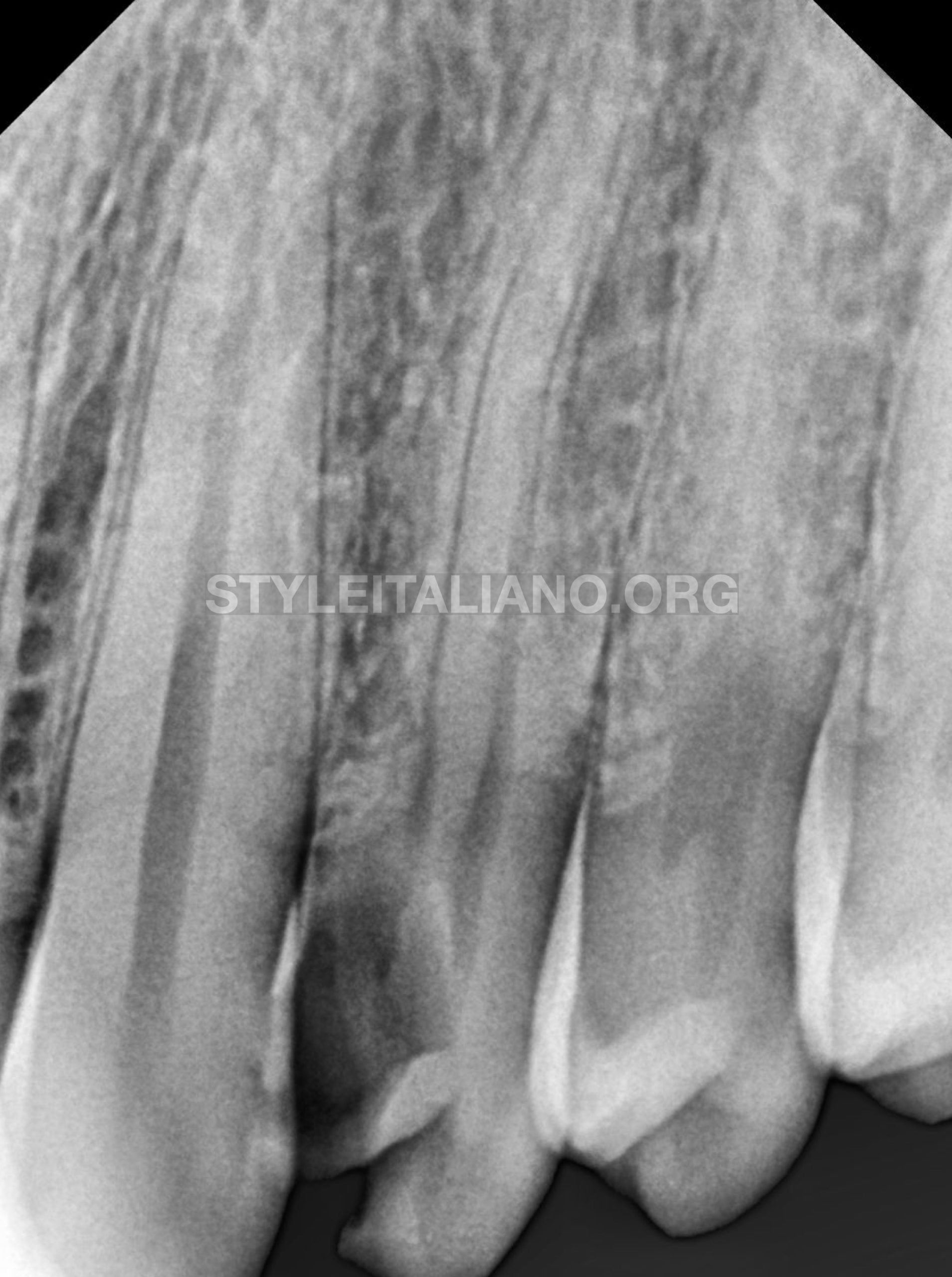
Fig. 1
Initial radiograph shows deep DISTAL CARIES related to tooth 14, as patient was suffering from pain
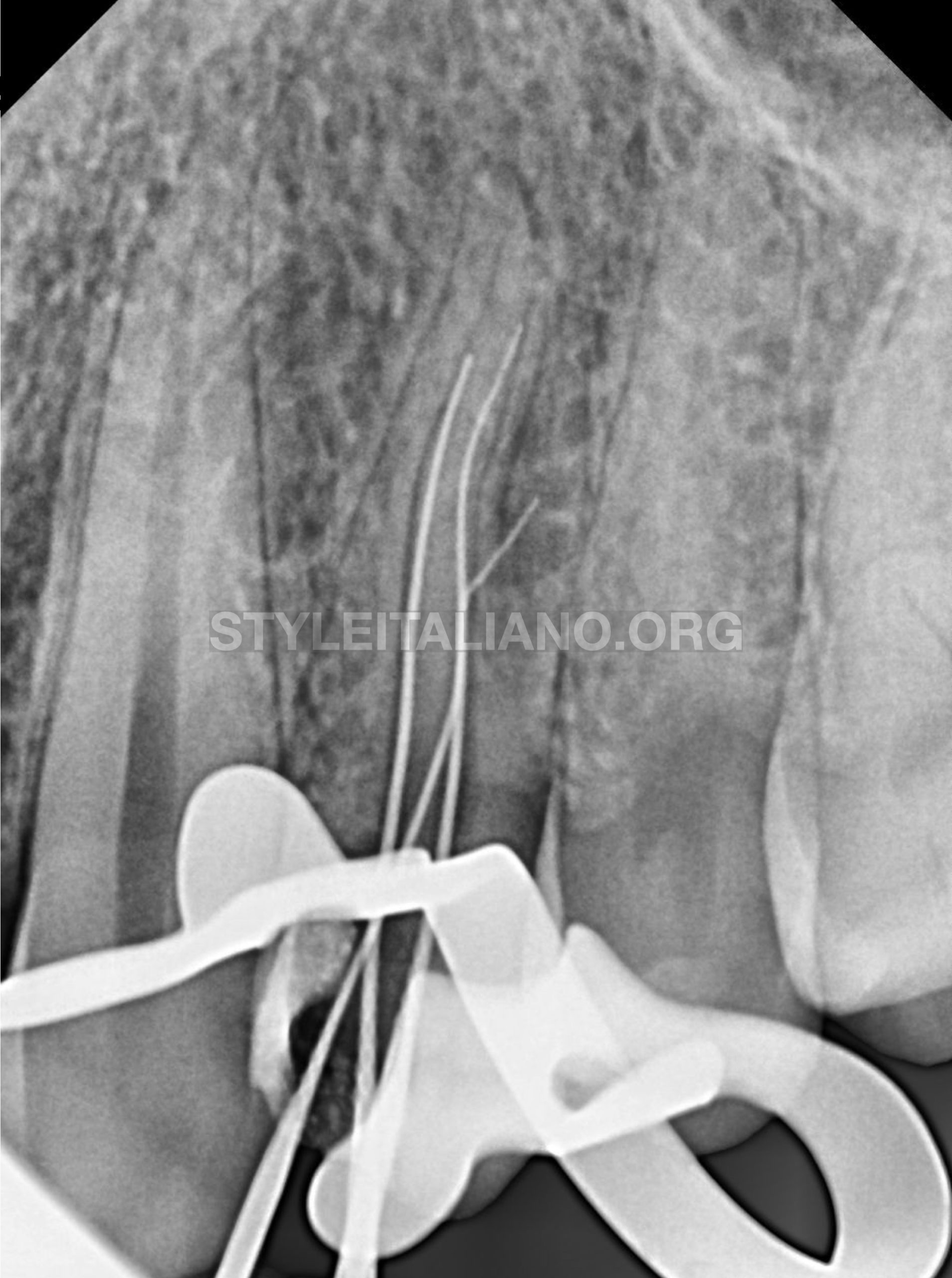
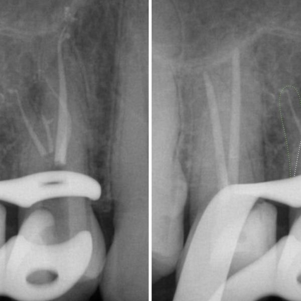
Fig. 2
Canals negotiation to obtain glode path, as you can notice 3rd file was introduced thinking it's LATERAL canal then it turned out it's a split of second canal buccally.
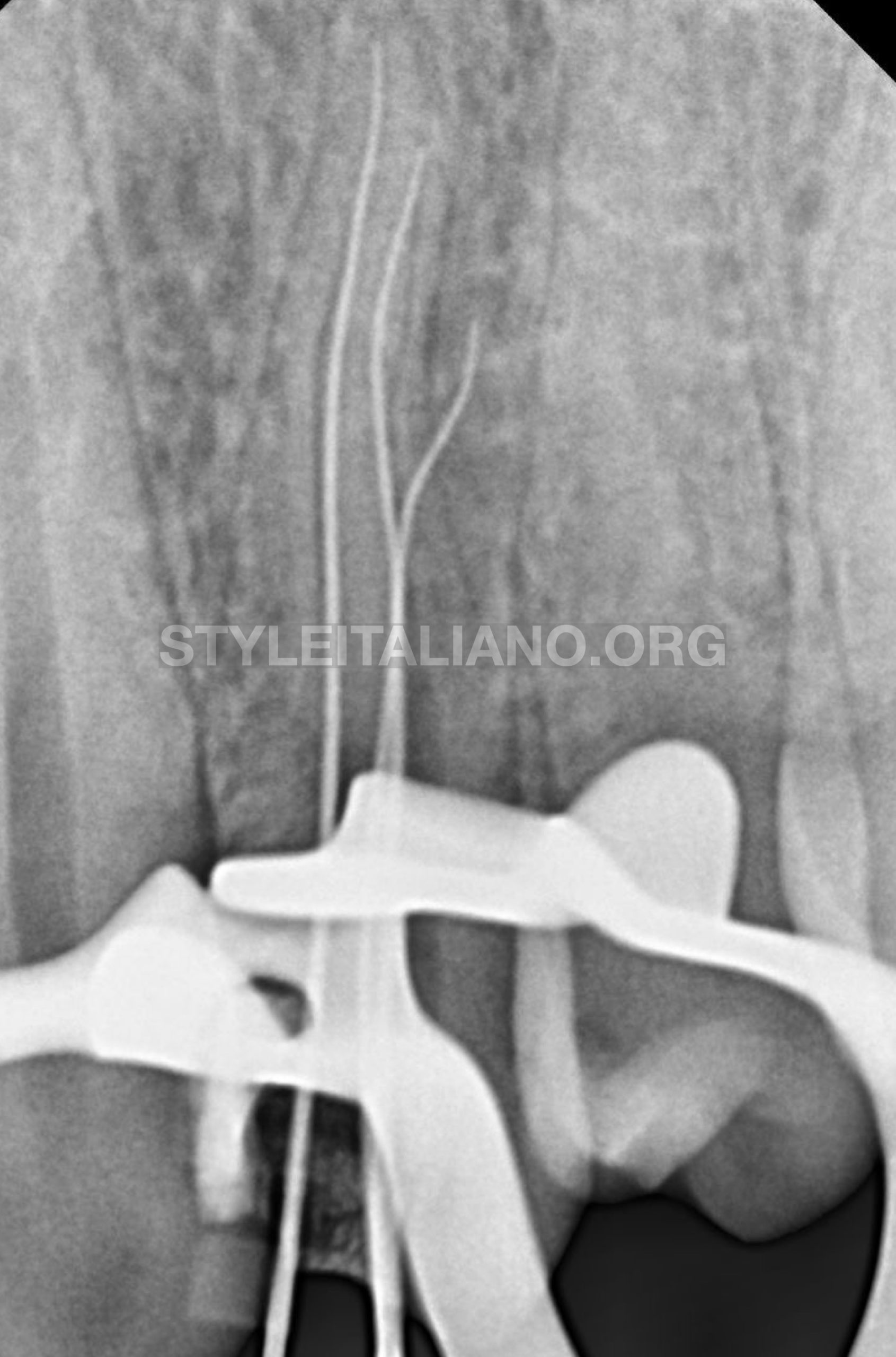
Fig. 3
Different angulation
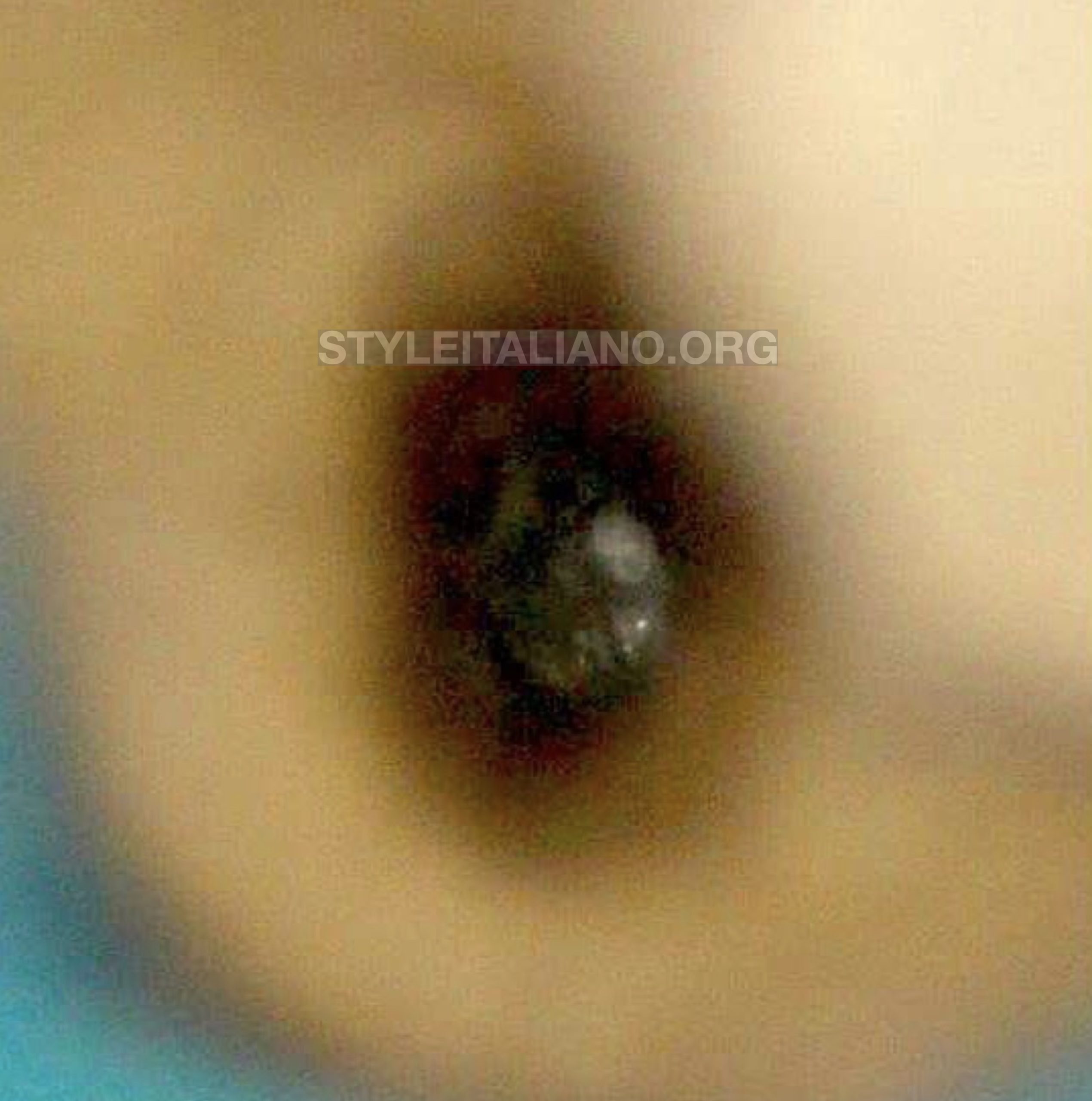
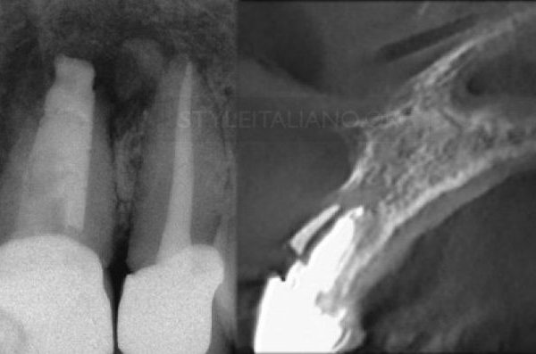
Fig. 4
Intra oral microscopic view shows the split under magnification 25 X
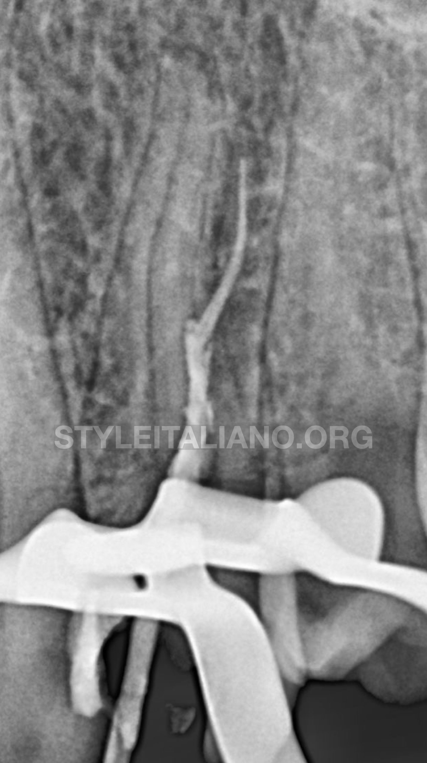
Fig. 5
due to difficulty of inserting 2 guttapercha all at once in split , one has to be introduced first and vertically compacted allowing enough space for the other one to be followed.
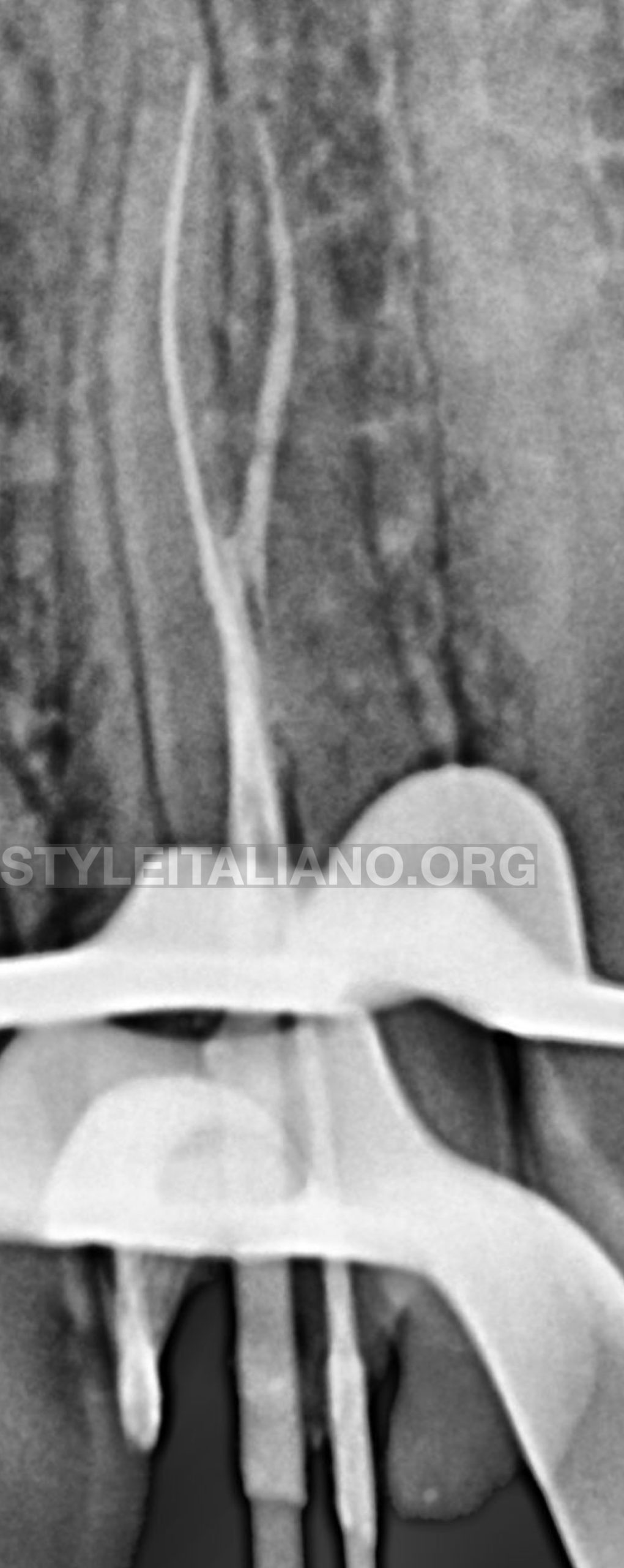
Fig. 6
as you can notice one has been compacted while manual file inserted in the other split to avoid any unnecessary blockage by gp
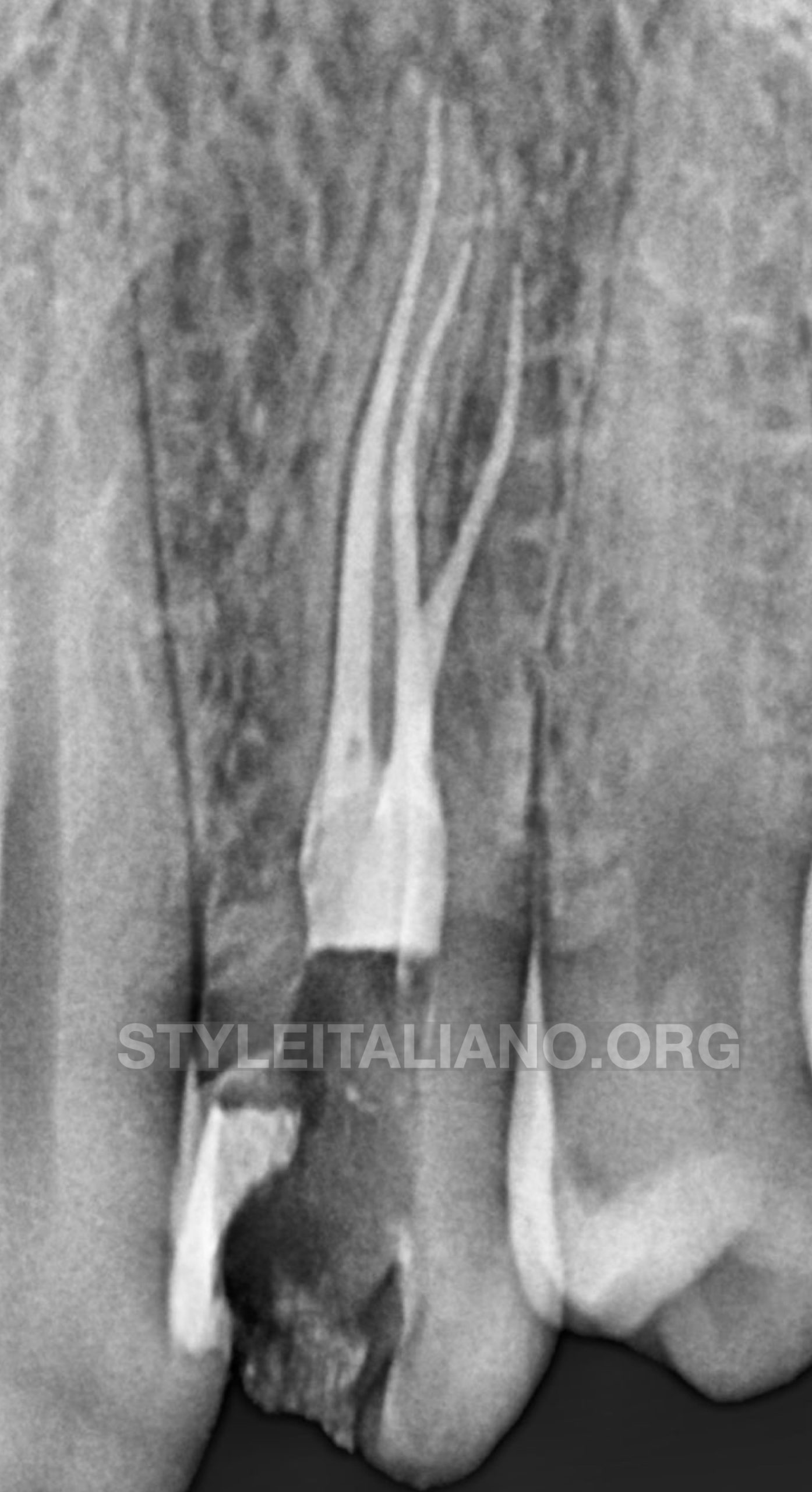
Fig. 7
Final obturation
Conclusions
Root canal morphology of the maxillary first premolar appears with different variations, including root diameter, root canal diameter, canal wall thickness, canal diameter, and cross-sectional root canal shape.
The majority of maxillary first premolars have two canals. Of the 2 roots with 2 canals maxillary first premolars, the furcation groove on the palatal aspect of the buccal root has a great influence on the treatment and prognosis .
Thus , to obtain predictable result we have to dedicate plenty of time for radiograph interpretation not to mention requiring CBCT whenever needed .
Bibliography
Pécora JD, Saquy PC, Sousa Neto MD, Woelfel JB. Root form and canal anatomy of maxillary first premolars. Braz Dent J. 1992;2(2):87-94. Pubmed PMID: 1290917.
Gupta S, Sinha DJ, Gowhar O, Tyagi SP, Singh NN, Gupta S. Root and canal morphology of maxillary first premolar teeth in north Indian population using clearing technique: An in vitro study. J Conserv Dent. 2015 MayJun;18(3):232-6. Pubmed PMID: 26069411.
Hargreaves KM, Cohen S, Berman LH. Cohen's pathways of the pulp.
Mosby Elsevier,; 2011.
[4]. Jayasimha Raj U, Mylswamy S. Root canal morphology of maxillary
premolars in an Indian population. J Conserv Dent. 2010 Jul;13(3):148-51.
Pubmed PMID: 21116391.