4 Canals in a first maxillary premolar
17/05/2020
Calogero Bugea
Warning: Undefined variable $post in /home/styleendo/htdocs/styleitaliano-endodontics.org/wp-content/plugins/oxygen/component-framework/components/classes/code-block.class.php(133) : eval()'d code on line 2
Warning: Attempt to read property "ID" on null in /home/styleendo/htdocs/styleitaliano-endodontics.org/wp-content/plugins/oxygen/component-framework/components/classes/code-block.class.php(133) : eval()'d code on line 2
The comprension of the anatomy of the root canal system is a key factor to achieve the success of the endodontic treatment. For this reason, clinicians must know all the possible anatomic variations and evaluate, at the beginning of each therapy, the canal configuration that the tooth can have. Although maxillary premolars usually have 2 root canals , the presence of 3 distinct root canals has been reported in 1%–6% of the cases. Previous case reports have shown many configurations of 3-canal maxillary premolar: 3 canals in a single root (10), 2 canals in the buccal root and 1 in the palatal root, 3 separate roots and canals (12). Only one report suggests the possibility to have 4 separate canals in a 3-root premolar: 2 canals in the distal root, 1 in the mesial and 1 in the palatal. To my knowledge, no case of a maxillary premolar with 4 distinct roots has ever been reported.
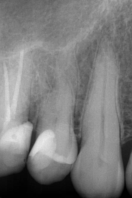
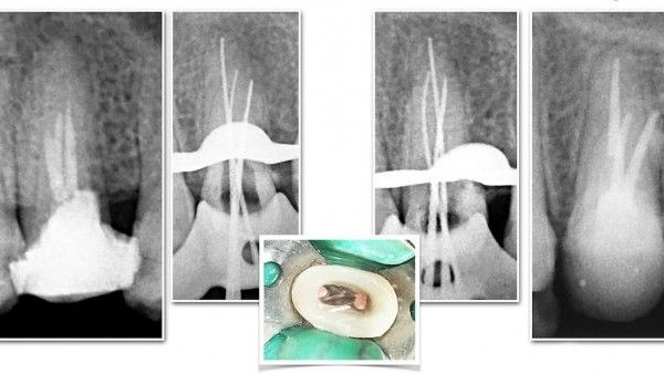
Fig. 1
The preoperative x-ray show a complex anatomy. I thought it was a classical mini-molar with two different vestibular roots.
During the treatment the sensation of the anatomy was a little different from my idea of the the root canal system. the root that I thought was mesial, was exactly at the center of the long axis of the tooth.
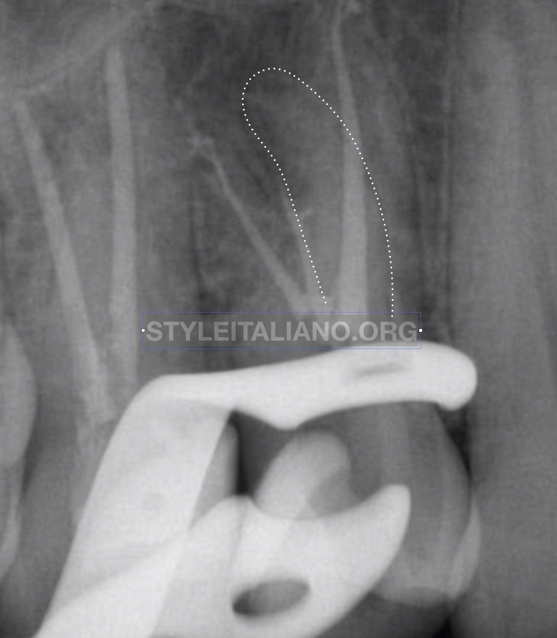
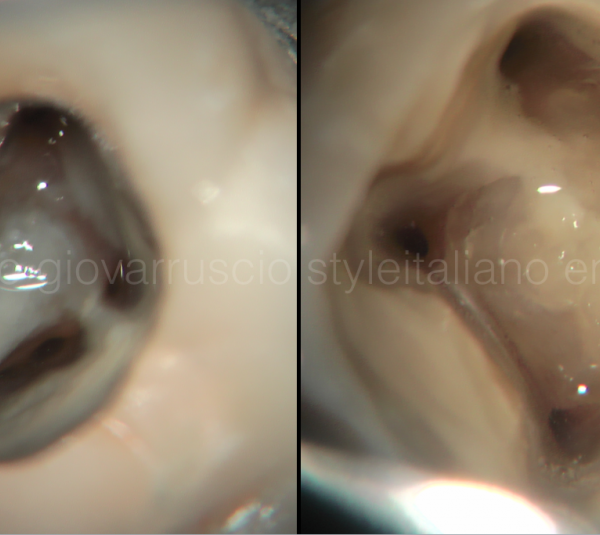
Fig. 2
During the last Xray, the sensation during the shaping was confirmed. From now the anatomy clear. The root canal system was more complex. 4 indipendent canals in 4 indipendent roots.
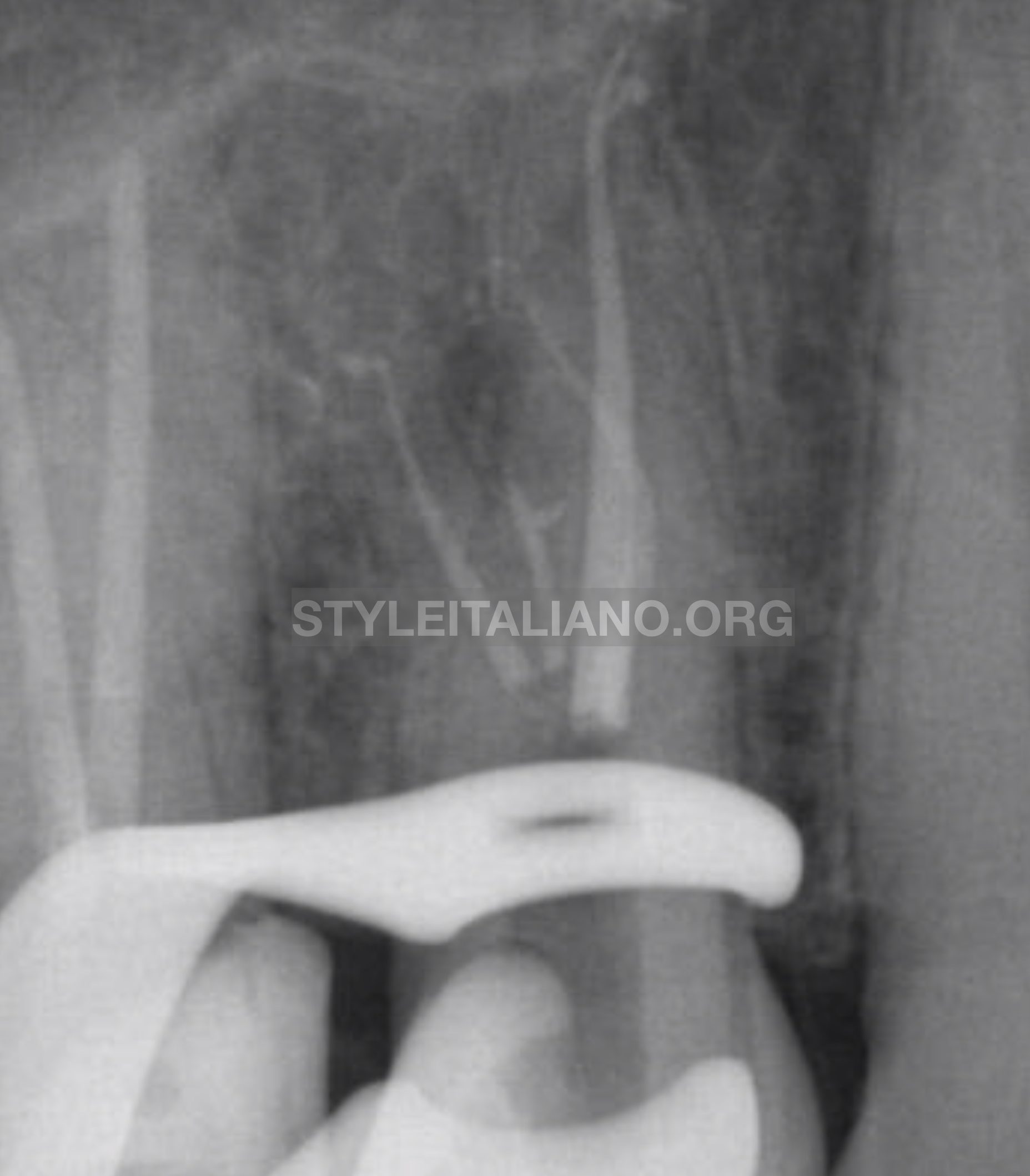
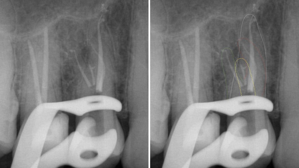
Fig. 3
The difficulties of the treatment was only the identification of the anatomy.Negotiation and shaping of the root canal system were done in a classical manner.
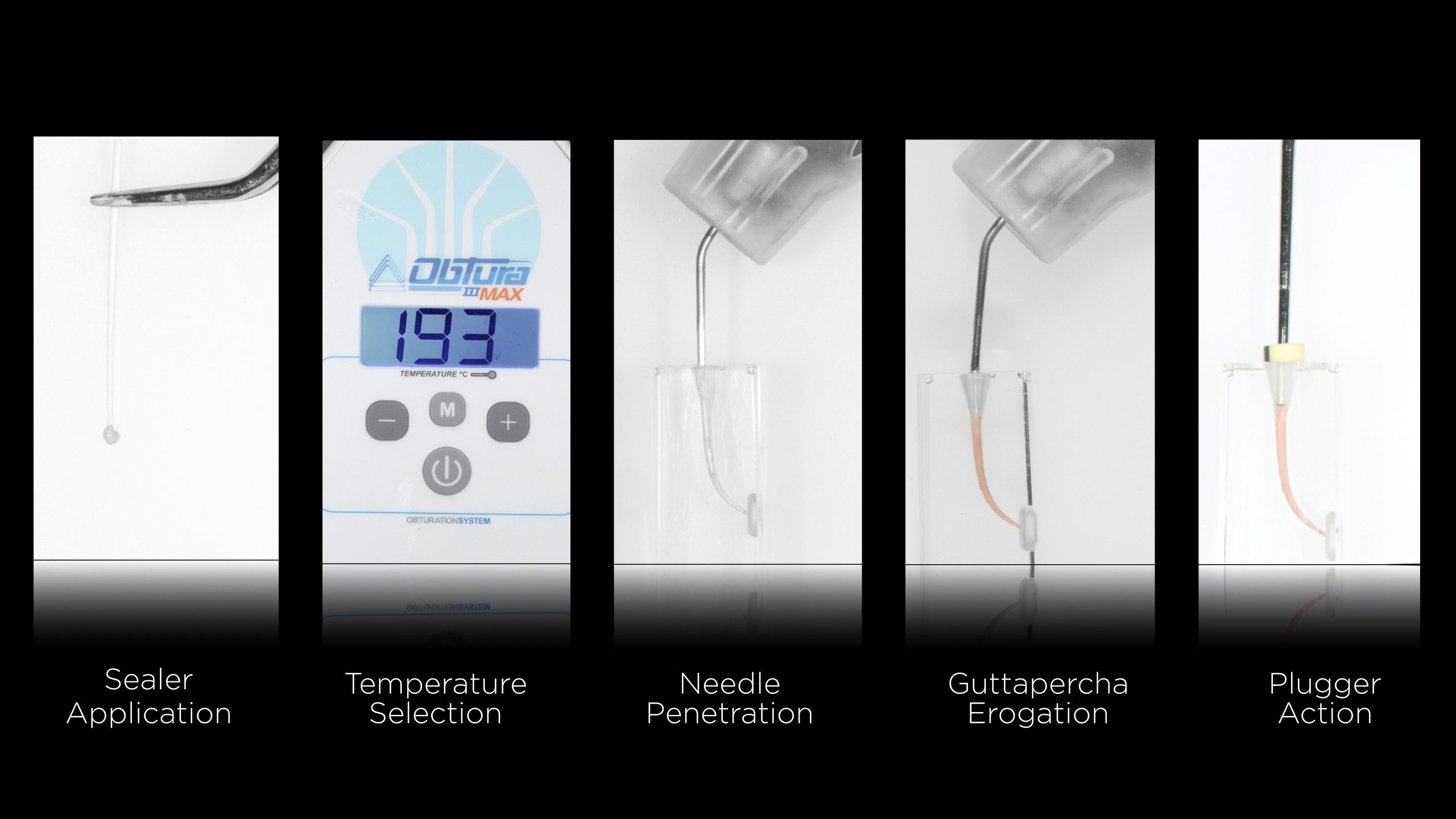
Fig. 4
For the Obturation i decided to do for a modified "thermolasticed guttapercha injection". This protocol called CONE LESS permit to simplify the step of the obturation and fill the endoontic space,
Fig. 5
A magnification of the previous image, underlines the cleaning of the endodontic space.
One of the advantage of this technique is the possibility to perform the technique in a restricted endodontic space, in witch the orofices are very close to the others
Conclusions
The clinical significance of the present case is that this is the first report of a tooth with 4 separate roots in a maxillary premolar. Identification of root canal morphology is a process that begins before starting the treatment and is always in itinere during the therapy.
Bibliography
Ida RD, Gutmann JL. Importance of anatomic variables in endodontic treatment out- comes: case report. Endod Dent Traumatol 1995;11:199–203.
Ahmad IA, Alenezi MA. Root and Root Canal Morphology of Maxillary First Premolars: A Literature Review and Clinical Considerations. J Endod. 2016 Jun;42(6):861-72.
Lea C, Deblinger J, Machado R, Nogueira Leal Silva EJ, Vansan LP. J Endod. 2014 Apr;40(4):591-3.
Yee FS, Marlin J, Krakow AA, Gron P. Three-dimensional obturation of the root canal using injection-molded, thermoplasticized dental gutta-percha. J Endod. 1977 May;3(5):168-74.