Maxillary Premolars - Part 2
31/01/2017
Mohamad Zaafrany
Warning: Undefined variable $post in /home/styleendo/htdocs/styleitaliano-endodontics.org/wp-content/plugins/oxygen/component-framework/components/classes/code-block.class.php(133) : eval()'d code on line 2
Warning: Attempt to read property "ID" on null in /home/styleendo/htdocs/styleitaliano-endodontics.org/wp-content/plugins/oxygen/component-framework/components/classes/code-block.class.php(133) : eval()'d code on line 2
As a continuity to previous article (Mandibular Premolars:Teeth not to underestimate), Today writing about maxillary premolars and challenges which might be faced during locating root canals and possible anatomic variation in such root canal treatment cases.
Hence root canals are the main component of the root canal system so exploration and detection of root canals is so important to ensure maximum debridemt as well as maximum disinfection of the root canal system thus decreasing the risk of treatment failure.
The internal and external morphology of of maxillary premolars were investigated and analyzed showing that Maxillary first premolars Usually have 2 canals in 2 separate roots while the maxillary second premolars shows the anatomy of 1 root with 1 or 2 root canals to be the most, both premolars may exhibit 3 separate roots with 3 root canals.
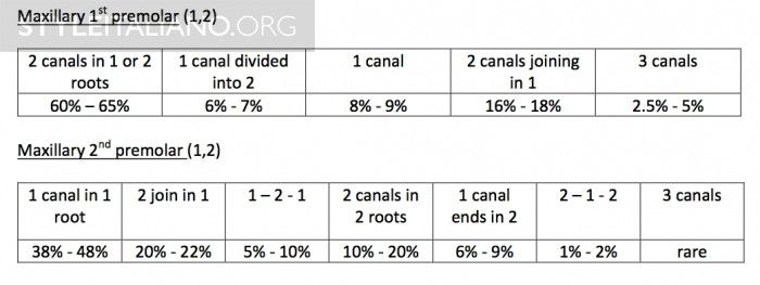
Fig. 1
In April 2014 Carina lea, Jason Deblinger et al. reported a case of 38 years old female with maxillary second premolar having 4 canals in 3 roots, 1 in MB, 1 in P and 2 in DB root.
Canals detected with the aid of Operating microscope. (3)
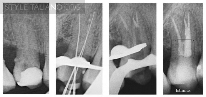
Fig. 2
The external root anatomy on the mesial aspect of maxillary premolars may exhibit a developmental groove on the outer surface.
In case of 1 root or 2 roots with long trunk in maxillary premolars Isthmus connection can be observed between any 2 root canal system occur within a root. (4)
This Isthmus should be considered during canal detection, during cleaning and shaping and during obturation according to its type. Certain external anatomic features which might be evident in some maxillary premolar is the presence of developmental groove on the palatal aspect of the buccal root ( furcal groove or furcation groove) which may divide the buccal root into 2 buccal roots with separate apex or might fade out apically expressing 2 roots joining in one apex or 2 canals in single root which is joining apically.
The incidence of furcal groove is 62% - 100% ( 5, 6 ).
The presence of the furcation groove is of high significance as it lead to inconsequent thickening of the root which should be considered during Biomechanical preparation and during Post placement if it will be placed in the buccal root not to harm the danger zone. (7)
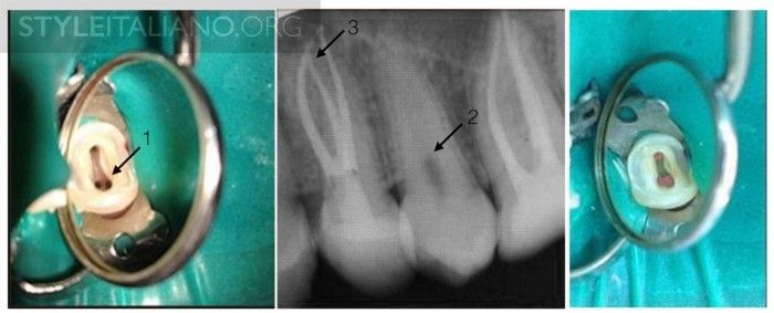
Fig. 3
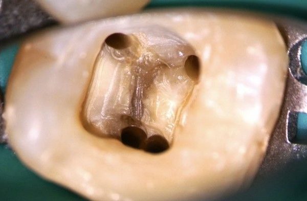
In this case of maxillary first premolar: 2 separate canals present in the buccal root with 2 separate orifices, 3- the 2 canals are joining at the apex sharing the same POE.
Careful widening of the access cavity with Ultrasonics from the buccal aspect mesiodistally would help to locate the extra orifices if present.
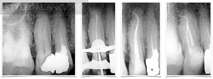
Fig. 4
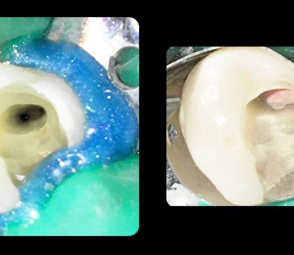
RCT of maxillary 2nd premolar. From preoperative radiographic image more than 2 canals were suspected but after careful negotiation only 2 canals were detected with 2 separate orifice sharing the same POE. Type II Vertucci classification.
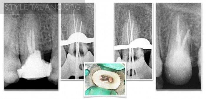
Fig. 5
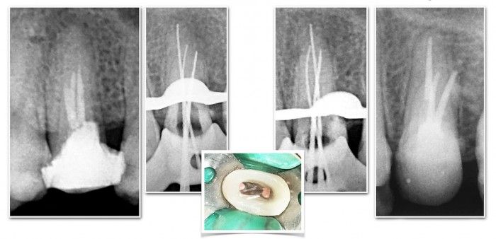
In this case of retreatment of the maxillary first premolar. It was evident the presence of 2 canals or a division in the mid root level in the preoperative radiographic image. Type IV Vertucci classification.
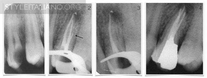
Fig. 6
In this case of RCT for the maxillary 1st premolar seepage of obturation material in different direction was noticed after obturation and before placing the final restoration, retreatment done in the same setting and 2 root canals in the buccal root were successfully located and treated. Type IV Vertucci classification.
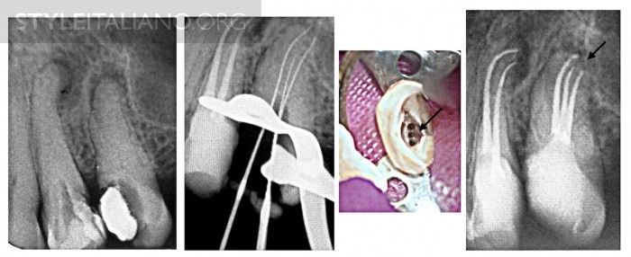
Fig. 7
In this case of RCT of maxillary second premolar 3 orifices were detected in a single root which was calcified, after successful negotiation and obturation, 3 separate POEs were noticed. Type VIII Vertucci classification.
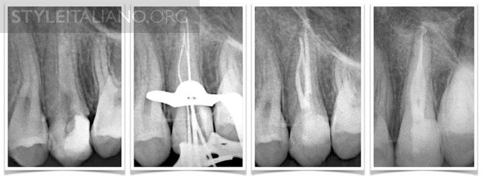
Fig. 8
Analyzing the data from preoperative radiograph was showing a quite broad roots mesiodistally with absence of canal tracing in the radiograph aid in detecting and exploring the 2nd canal in the buccal root.
In this case the canals share the same orifice, by precurving the negotiating files exploring the internal root surface 2 canal were detected sharing the same Apical foramen.
Type III Vertucci classification.
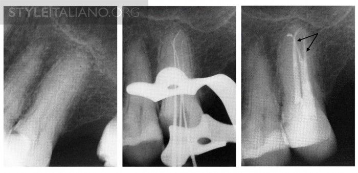
Fig. 9
Maxillary second premolar which was suspected to have more than 2 canals because of the broad mesiodistal diameter of the roots. Type IV root canal system in the buccal root was explored and treated.
Conclusions
Knowledge of the variations in endodontic anatomy of maxillary premolars will assist in diagnosing and managing these cases during root canal treatment.
Bibliography
Refrences.
1.Vertucci FJ. Root canal anatomy of the human permanent teeth. Oral Surg Oral Med Oral Pathol Oral Radiol Endod 1984: 58: 589599.
2.Sert S, Bayirli GS. Evaluation of the root canal configurations of the mandibular and maxillary permanent teeth by gender in the Turkish population. J. Endod 2005: 30: 391399.
3. Carina lea, Jason Deblinger et al. Maxillary Premolar with 4 Separate Canals . JOE Volume 40, Number 4, April 2014
4. Balkan journal of stomatology , Apical Disposition of Isthmus in Maxillary Premolars: A Surgical Endodontic Problem, ISSN 1107 - 1141
5. Joseph I, Varma B, Bhat KM. Clinical significance of furcation anatomy of the maxillary first premolar: a biometric study on extracted teeth. J Periodontol 1996;67: 3869.
6. Awawdeh L, Abdullah H, Al-Qudah A. Root form and canal morphology of Jordanian maxillary first premolars. J Endod 2008;34:95661.
7. Tamse A, Katz A, Pilo R. Furcation groove of buccal root of maxillary first premolars:
a morphometric study. J Endod 2000;26:35963.