Dealing with MM canal in a lower first molar
22/11/2023
Fellow
Warning: Undefined variable $post in /home/styleendo/htdocs/styleitaliano-endodontics.org/wp-content/plugins/oxygen/component-framework/components/classes/code-block.class.php(133) : eval()'d code on line 2
Warning: Attempt to read property "ID" on null in /home/styleendo/htdocs/styleitaliano-endodontics.org/wp-content/plugins/oxygen/component-framework/components/classes/code-block.class.php(133) : eval()'d code on line 2
Several studies published in the Literature described the morphology of mandibular first molars, reporting the presence of three or four canals. In 1974, Vertucci and William described the presence of an independent middle mesial canal.
Since then, there have been multiple case reports of aberrant canal morphology of the mandibular first molar, describing aberrant canals in the mesial or even in the distal root.
The middle mesial canal can be independent, mesiolingual confluent or mesiobuccal confluent: the first step to treat it is to determine the location of the mesiolingua canal and the mesiobuccal canal. Then, clean the isthmus area using ultrasonic and heavy irrigation to get a clearer view that makes it easier to pick up the middle canal. In some cases, CBCT can help locating the MM canal.
The clinician should be aware of this varied anatomy in order to manage correctly the case.
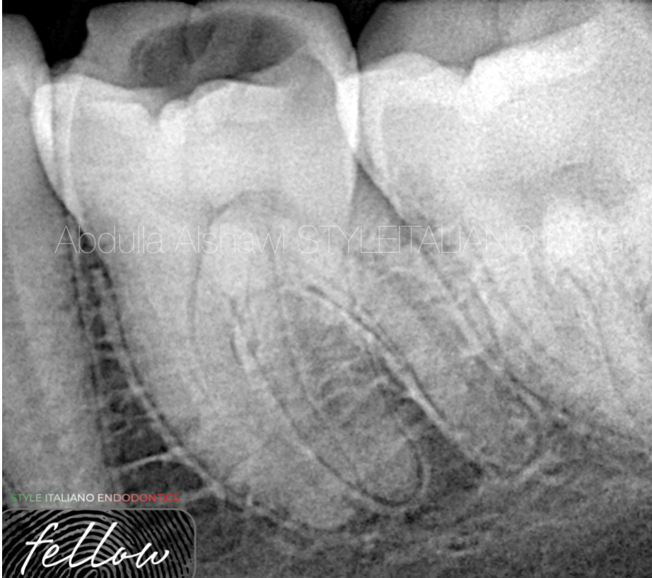
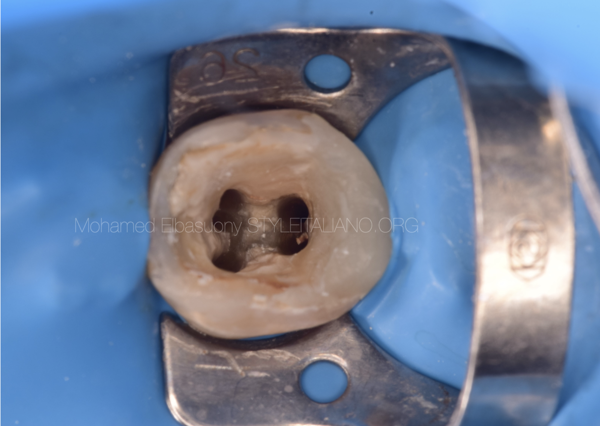
Fig. 1
A 22-year-old female patient came to the clinic with pain from cold stimulation and percussion.
Pre opertive x-ray showed the abnormal anatomy of the first lower left molar
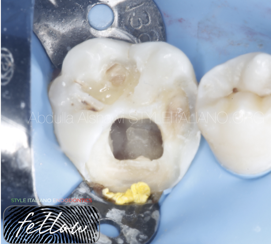
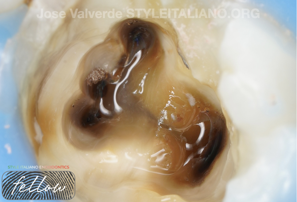
Fig. 2
After caries removal and cleaning pulp chamber.
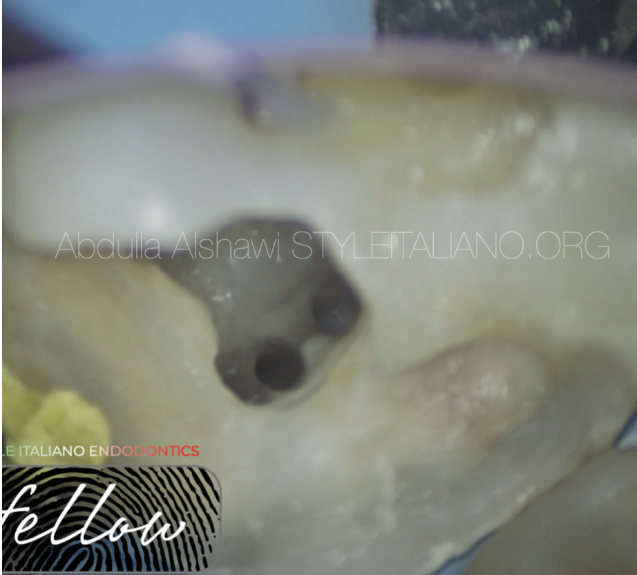
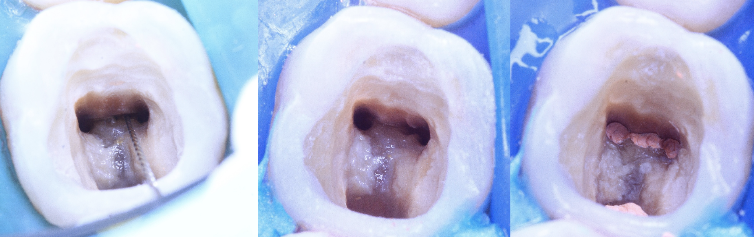
Fig. 3
Cleaning and shaping of middle mesial canal
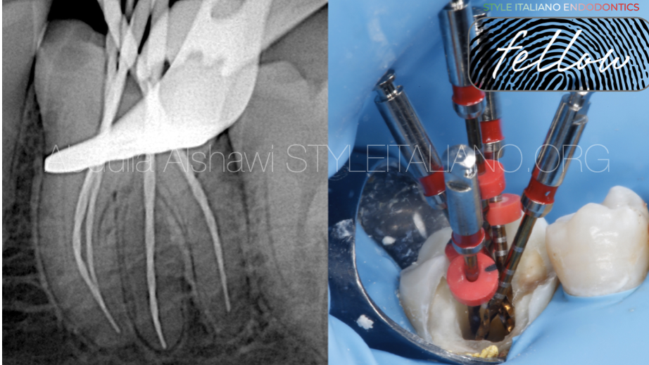
Fig. 4
Checking of W.L and apical control to achieve apical sealing and to avoid over obturation

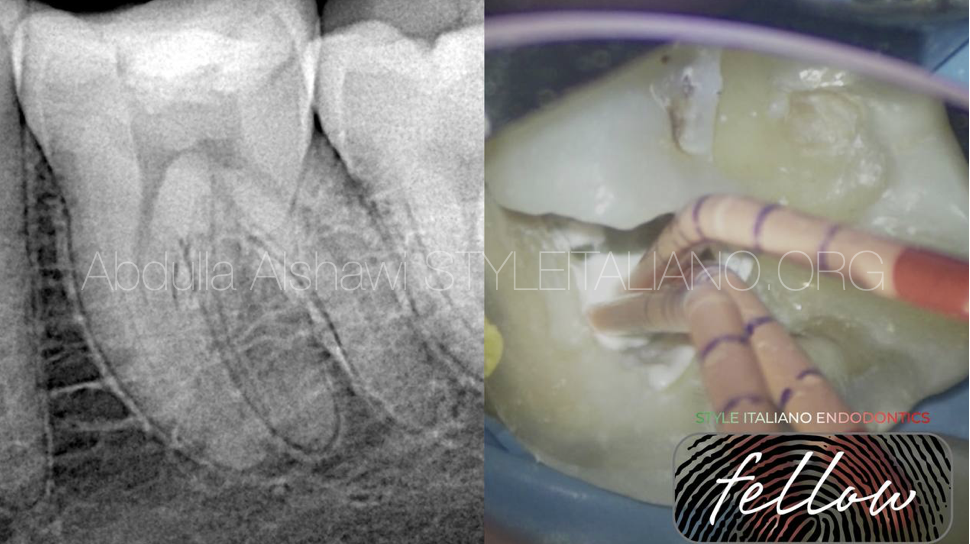
Fig. 5
When checking master Cone, we notice that MMC joins ML and MB canals
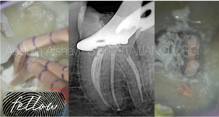
Fig. 6
Obturation done with BC sealer and Modified Warm Vertical compaction
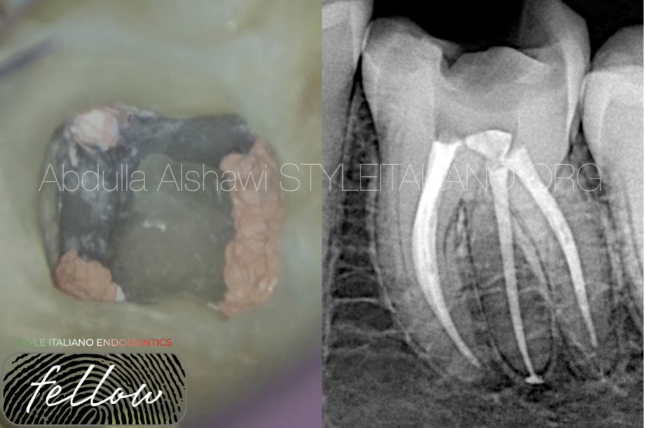
Fig. 7
Final X-ray

Fig. 8
About the author : Dr Abdulla Alshawi
B.D.S
Graduated from Uruk dental collage-Iraq 2018-2019
Conclusions
Understanding the anatomy of the teeth is the most important point in the treatment of the roots of the teeth, and the things that increase the success rate of endodontic treatments are that you have sufficient knowledge and the required equipment, these factors make endodontic treatment more accessible.
Bibliography
Akbarzadeh N., Aminoshariae A., Khalighinejad N., Palomo J.M., Syed A., Kulild J.C., Ghazal Sadeghi G., Mickel A. The association between the anatomic landmarks of the pulp chamber floor and the prevalence of middle mesial canals in mandibular first molars: an in vivo analysis. J. Endod. 2017;43:1797–1801.
Barker B.C., Parsons K.C., Mills P.R. Williams GL. anatomy of root canals. III. Permanent mandibular molars. Aust Dent J. 1974;19:408–413.
Baugh D., Wallace J. Middle mesial canal of the mandibular first molar: a case report and literature review. J. Endod. 2004;30:185–186.
Karunakaran JV, Shobana R, Kumar M, Kumar S, Mankar S. Management of middle mesial canal in mandibular second molar. J Pharm Bioallied Sci. 2012 Aug;4(Suppl 2):S161-4. doi: 10.4103/0975-7406.100259. PMID: 23066241; PMCID: PMC3467889.
Gulabivala K, Opasanon A, Ng YL, Alavi A. Root and canal morphology of Thai mandibular molars. Int Endod J. 2002;35:56–62.