Endodontic retreatment of a mandibular first molar with missing Mid-Mesial canal
14/08/2023
Fellow
Warning: Undefined variable $post in /home/styleendo/htdocs/styleitaliano-endodontics.org/wp-content/plugins/oxygen/component-framework/components/classes/code-block.class.php(133) : eval()'d code on line 2
Warning: Attempt to read property "ID" on null in /home/styleendo/htdocs/styleitaliano-endodontics.org/wp-content/plugins/oxygen/component-framework/components/classes/code-block.class.php(133) : eval()'d code on line 2
The presence or absence of periradicular disease is determined according to clinical and radiographic findings.
This article will discuss the non surgical endodontic retreatment of a mandibular first molar with mid-mesial canal.
Endodontic retreatment is a procedure performed on a tooth that have a previous attempt of definitive treatment resulting in a condition that requires further endodontic intervention to achieve a successful outcome.
Non surgical retreatment holds unique considerations that distinguish it from initial root canal treatment:
- An extensive restoration may have to be removed.
- Technical challenge from previous treatment may be present, such as morphologic alteration in the form of perforation or ledge or canal obstructions.
- Root filling must be removed from the canals.
- Healing rate is generally slower than initial treatment because of the greater difficulty in eliminating resistant infection.
Bearing in mind, radiographs are the eyes of the dentist. They are essential for diagnosis, determining anatomy, managing treatments and assessing outcome.
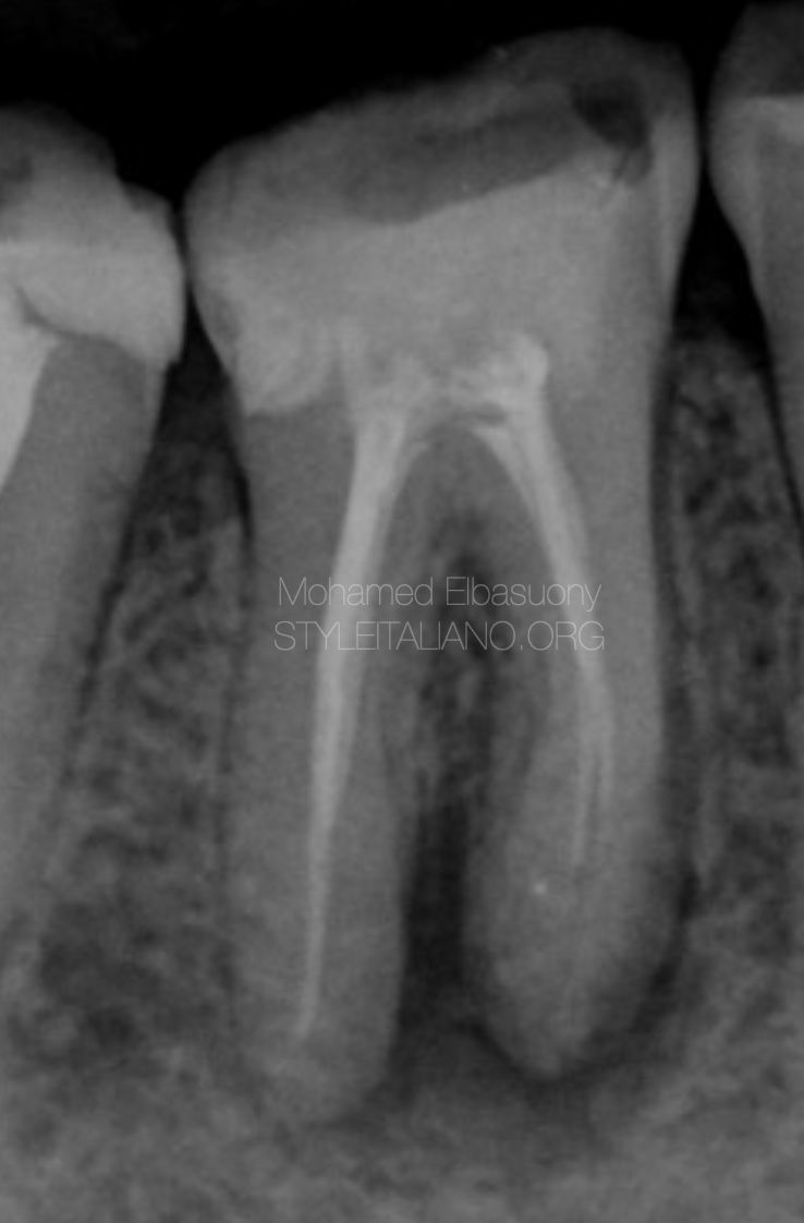
Fig. 1
A 39 years old female came to my attention complaining about pain on the first lower right molar.
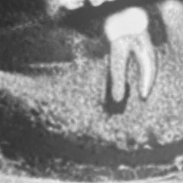
The pre-operative x-ray showed an periapical radiolucency, and suggested the presence of apical periodontitis.
On clinical examination, The tooth was tender to percussion.
I decided to retreat the tooth to relief the symptoms referred by the patient.
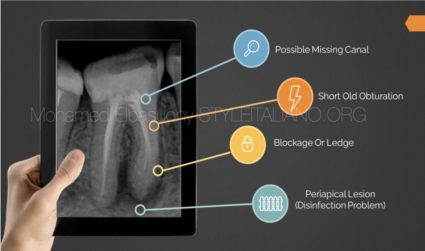
Fig. 2
Possible problems in this case may be
- Possible missing canal
- Short old obturation
- Blockage or ledge
- Periapical lesion

Fig. 3
Local anesthesia was administered.
The tooth was isolated with rubber dam and removal of coronal restoration was done.
Before starting the retreatment procedures, the length of the old root filling was measured on a precise primary radiograph.
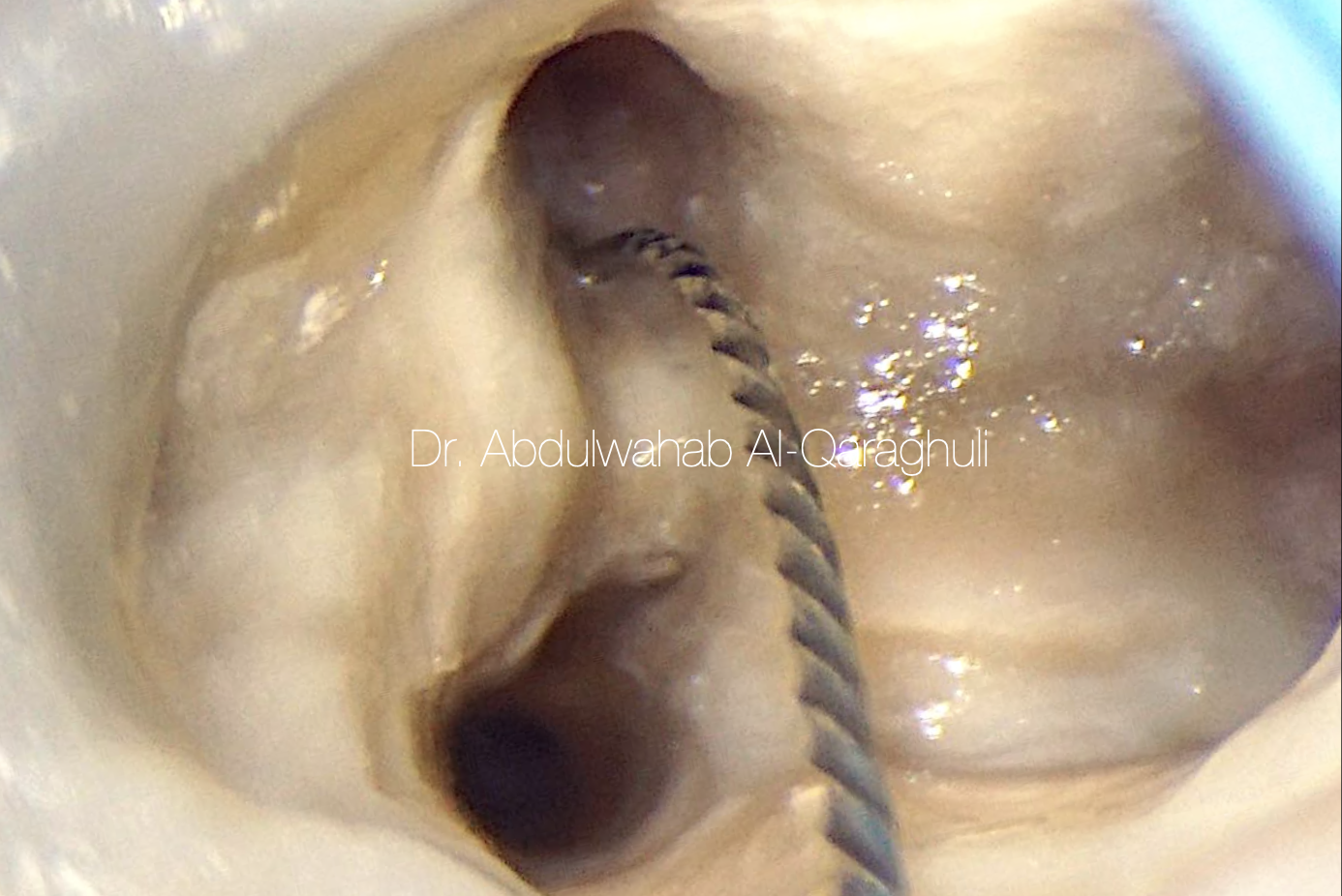
I removed the existing gutta-percha with a rotary Ni-Ti file designed for retreatments 1 mm short of the measured length to remove filling material quickly.
I don’t like to use solvent because the solvent will make the gutta percha fluid so make our penetration easier but also our instrument will push this flowable gutta percha on the walls to unreachable anatomy so reduce the quality of removal of filling material.
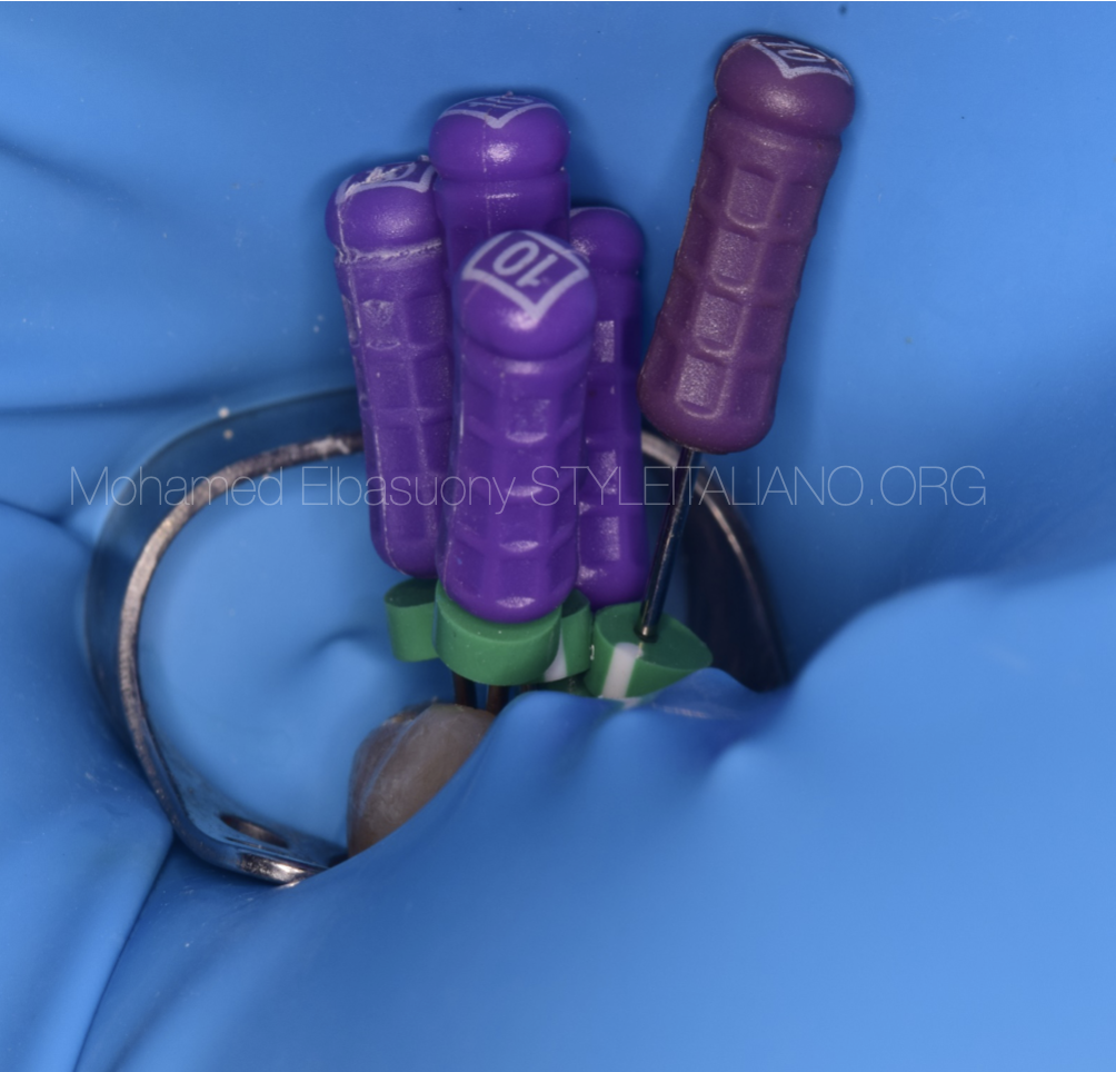
Fig. 4
Manual files .02 SS (size 10 to 15) was used to penetrate last mm of the material to the length measured on X-ray and then mechanically.
Manual & Mechanical technique because :
- If the old filling ends near the apex, it will reduce risk of extrusion of debris.
- If the old filling ends away from the apex, it will permit manual scouting of last mm of the material to understand why it stopped there (presence of ledge or blockage)
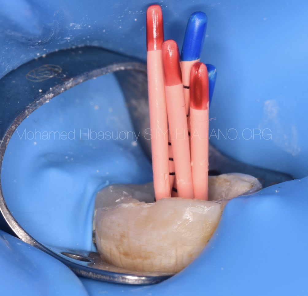
Fig. 5
The mesial canals were shaped up to 25.04 and distal canals were shaped up to 30.04
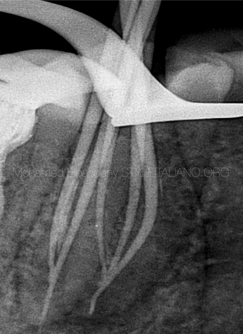
Fig. 6
Master cone radiograph
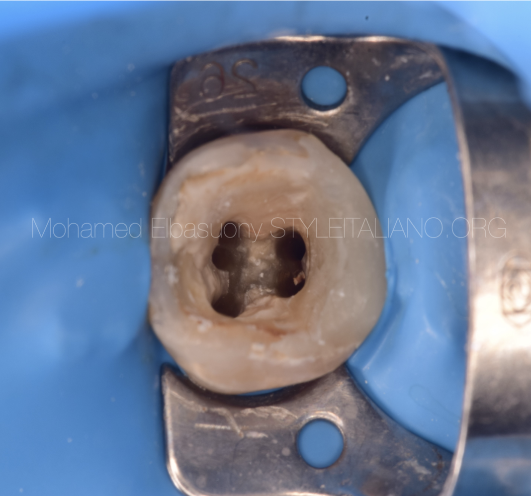
Fig. 7
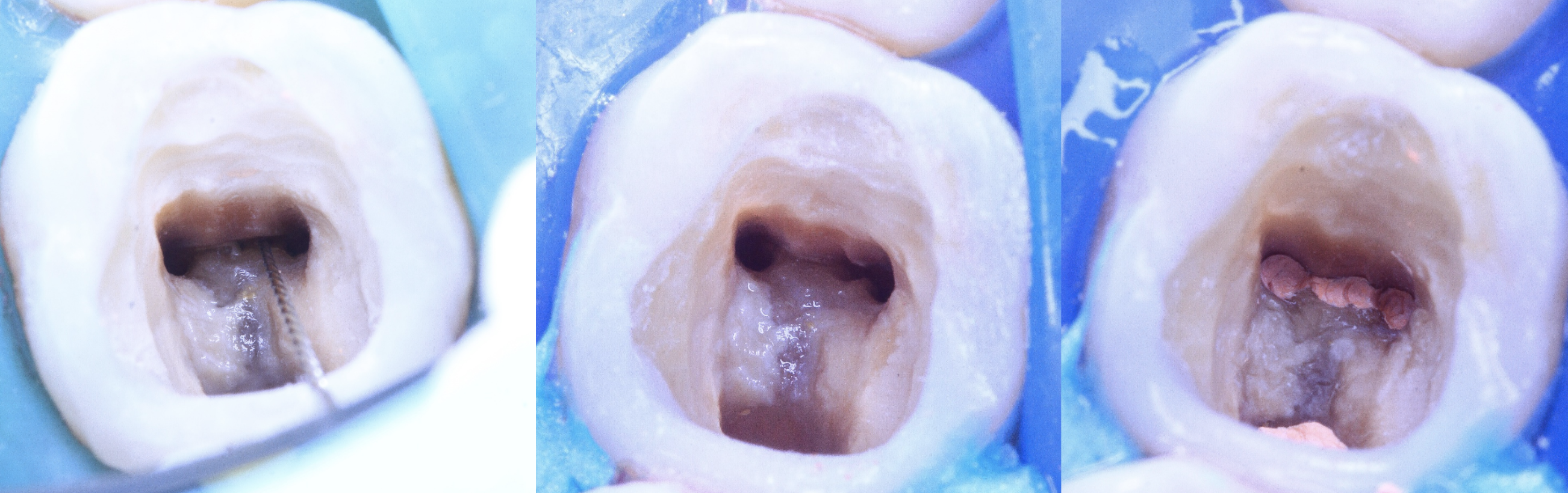
Ultrasonic tip ET 25 was used for isthmus cleaning.
The literature reported that the frequency of post-treatment apical periodontitis in teeth with at least one untreated canal was 98%: missed canal have a significant impact on treatment prognosis.
Proper irrigation dynamics is crucial for effective cleaning and disinfection of the root canal system, as it helps to ensure that the irrigant reaches all areas of the canal and effectively removes debris, bacteria, and necrotic tissue.
Sonic activation was used.
Supplementary use of XP-Endo Finisher R file increases the removal of root canal filling materials from root canal system during retreatment procedures.
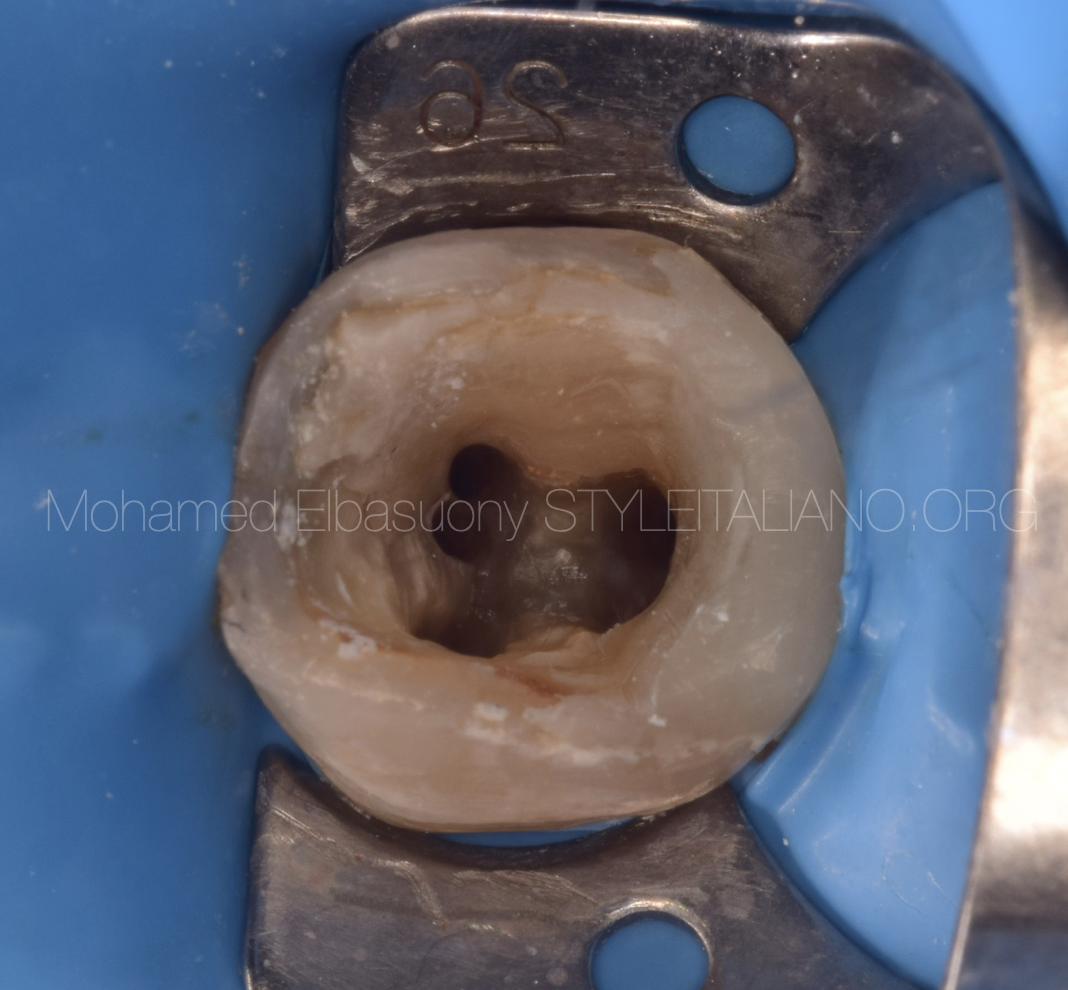
Fig. 8
Canals were dried using paper points and ready to be obturated
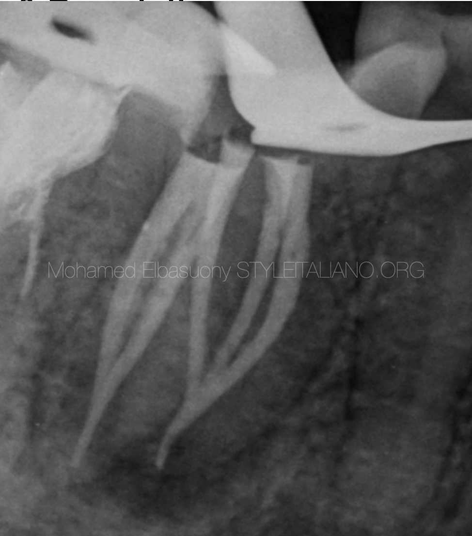
Fig. 9
Obturation radiograph
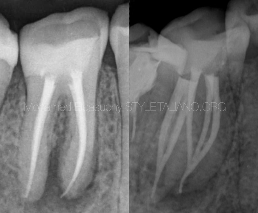
Fig. 10
Final restoration radiograph
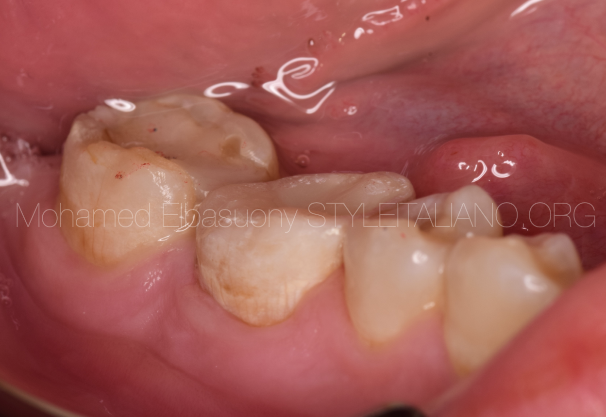
Fig. 11
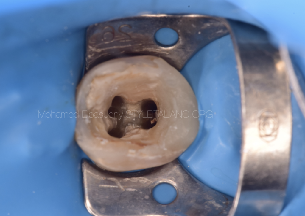
Final restoration
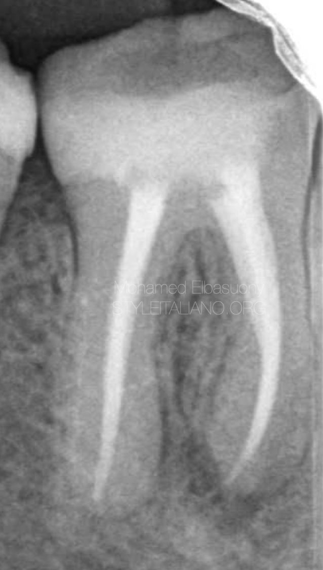
Fig. 12
One year follow up
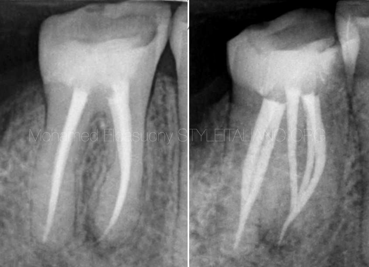
Fig. 13
Two years follow up

Fig. 14
About the author:
Diploma in Endodontics at faculty of dentistry, Mansoura university in 2018
Enrolled in the MSc in Endodontics at faculty of dentistry, Mansoura university in 2020
StyleItaliano Endodontics Fellow
Endodontic Specialist
Conclusions
Knowledge of the anatomy and of the retreatment techniques is mandatory to solve such cases
Bibliography
- 1.Cameron JA. The use of ultrasound for the removal of the smear layer. The effect of sodium hypochlorite concentration: SEM study. Aust Dent J 1988;33:193-200.
- 2.Lumley PJ, Walmsley AD, Walton RE, Rippin JW. Effect of precurving endosonic files on the amount of debris and smear layer remaining in curved root canals. J Endod 1992;18:616-9.
- 3.Al-Dahman, Y., & Al-Omari, M. (2021). Retreatability of bioceramic and GuttaFlow bioseal root canal sealers using ProTaper universal system retreatment files: An Ex vivo study. Saudi Endodontic Journal, 11(1), 42-48.
- 4.Pedullà, E., Abiad, R. S., Conte, G., Khan, K., Lazaridis, K., Rapisarda, E., & Neelakantan, P. (2019). Retreatability of two hydraulic calcium silicate‐based root canal sealers using rotary instrumentation with supplementary irrigant agitation protocols: a laboratory‐based micro‐computed tomographic analysis. International Endodontic Journal, 52(9), 1377-1387.