Dens invaginatus
02/11/2023
Fellow
Warning: Undefined variable $post in /home/styleendo/htdocs/styleitaliano-endodontics.org/wp-content/plugins/oxygen/component-framework/components/classes/code-block.class.php(133) : eval()'d code on line 2
Warning: Attempt to read property "ID" on null in /home/styleendo/htdocs/styleitaliano-endodontics.org/wp-content/plugins/oxygen/component-framework/components/classes/code-block.class.php(133) : eval()'d code on line 2
Dens invaginatus in tooth was first described by dentist ‘socrates’ 1856
Dens invaginatus is a developmental anomaly resulting from the invaginations of the enamel organ into the dental papilla during the soft tissue stage of development.As the hard tissues are formed, the invaginated enamel organ produces a small tooth within the future pulp chamber.
The Etiology of dens invaginatus remains unclear. Genetic factor has been proposed to be the cause.
Classification ;The first documented attempt to classify dens invaginatus was by Hallet (1953).
Oehlers (1957a)describe a system appears to be the most widely used, possibly because of its simple nomenclature and ease of application. This system categorizes invaginations into 3 classes as determined by how far they extend radiographically from the crown into the root .
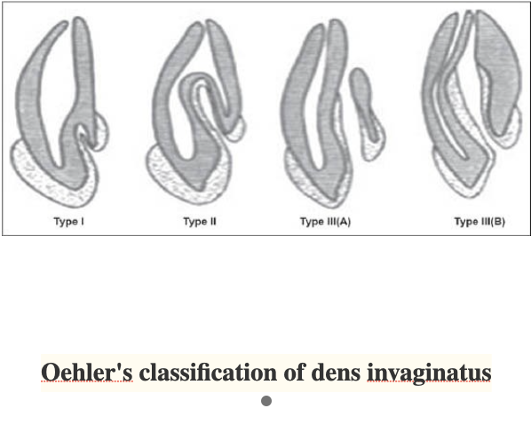
Fig. 1
Type I: The invagination is minimal and enamel-lined; it is confined within the crown of the tooth and does not extend beyond the level of the external amelo-cemental junction.
Type II: The invagination is enamel-lined and extends into the pulp chamber, but remains within the root canal with no communication with the periodontal ligament.
Type III A: The invagination extends through the root and communicates laterally with the periodontal ligament space through a pseudo-foramen. There is usually no communication with the pulp, which lies compressed within the root.
Type III B: The invagination extends through the root and communicates with the periodontal ligament at the apical foramen. There is usually no communication with the pulp.
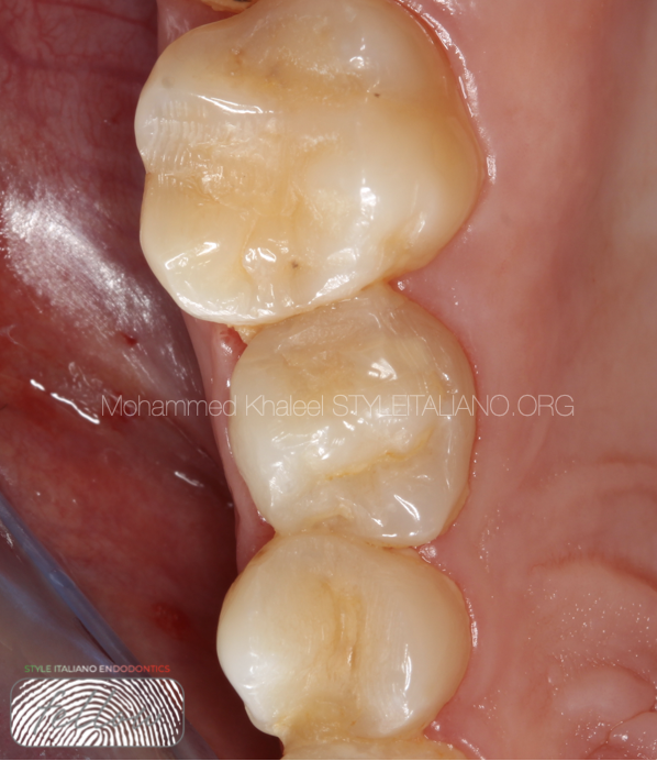
Fig. 2
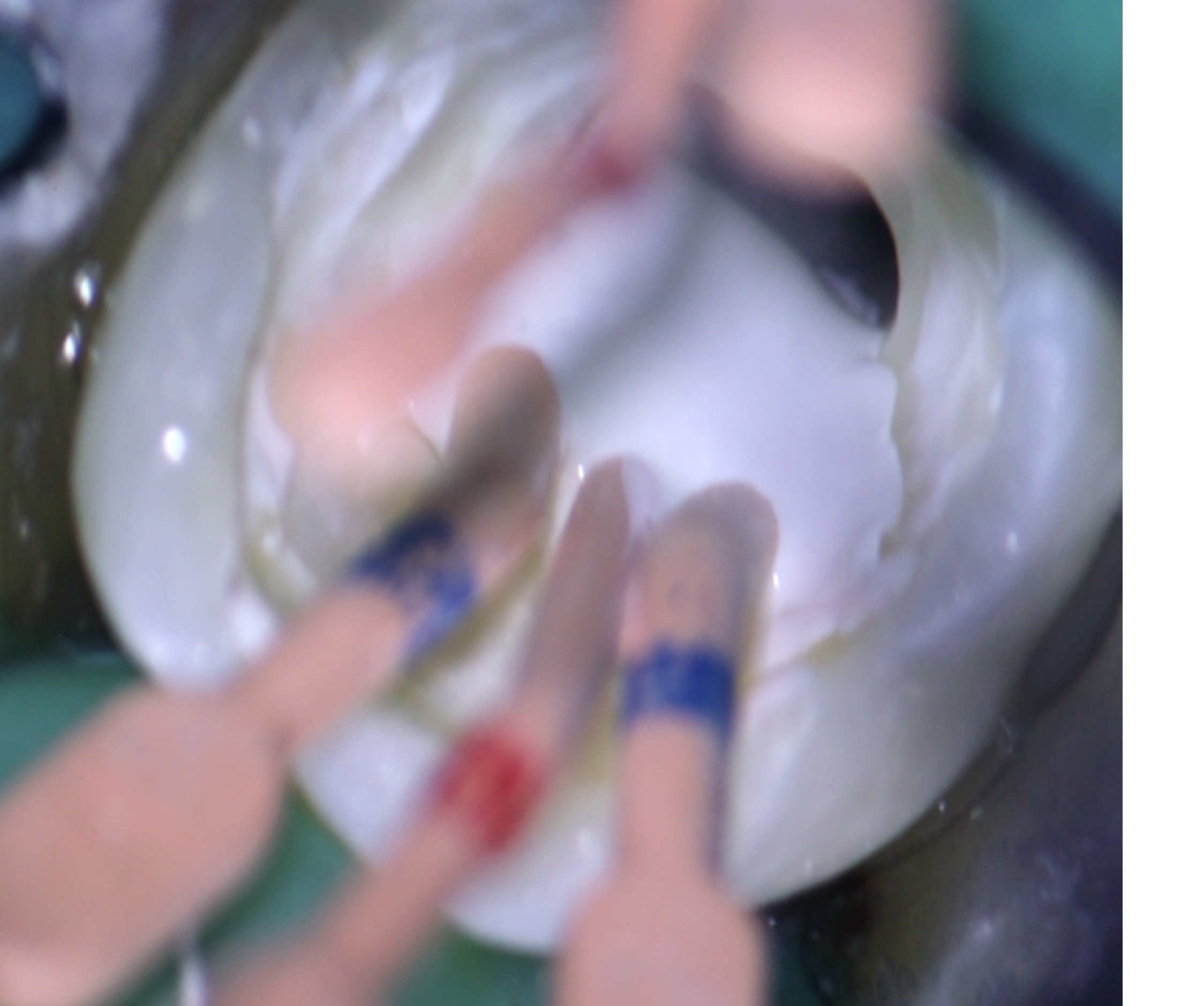
Pre operative clinical photo
A 32-year female with a chief complaint of pain in upper 2nd premolar , the patient was in good general health. Intra-oral examination revealed a wider maxillary 2nd premolar .the tooth was tender to precussion and did not respond to thermal stimuli.
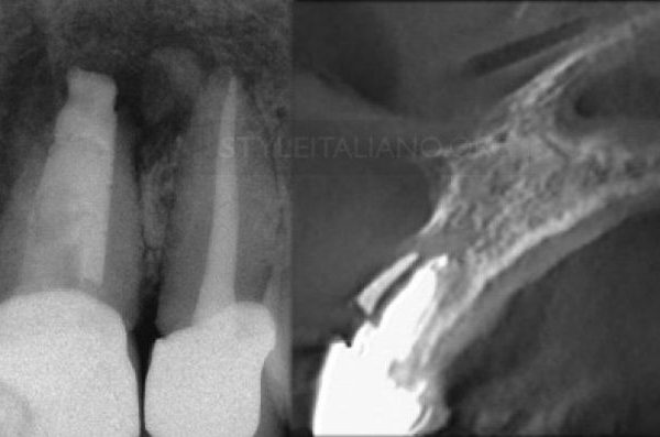
Peri apical radiograph revealed type II DI with periapical pathology related to maxillary right 2nd premolar. Endodontics treatment was started immediately .
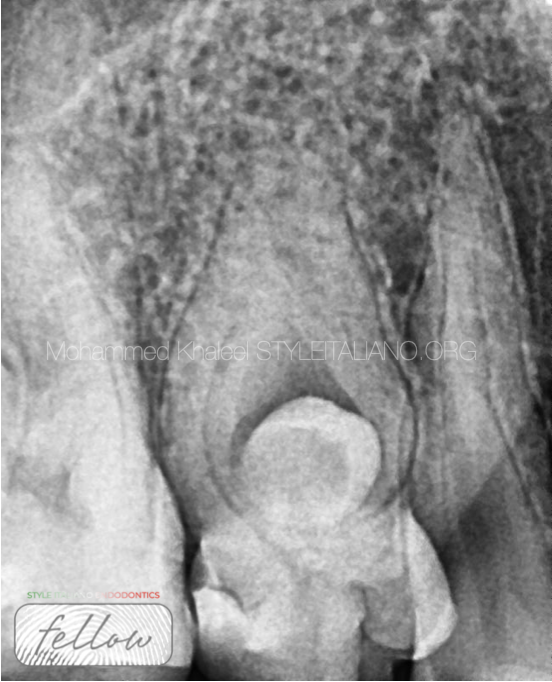
Fig. 3
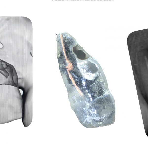
Pre operative X-ray shows tooth within a tooth and abnormal pulp morphology, so I decided take CBCT.
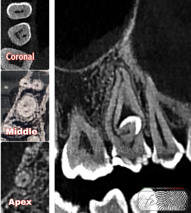
Fig. 4
CBCT shows dens in dente
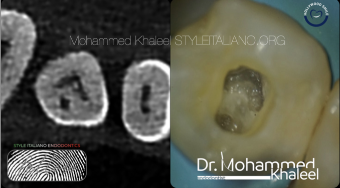
Fig. 5
Access opening by the aid of CBCT
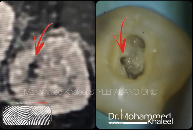
Fig. 6
Finding the main canal
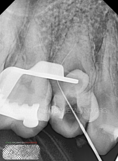
Fig. 7
Canal negotiation by small k file
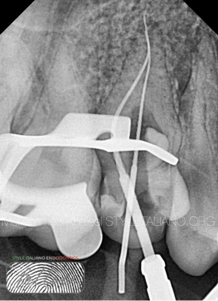
Fig. 8
Instrumentation by manual k file #6,8,10,15 then rotary 20 and 25 taper 4

Fig. 9
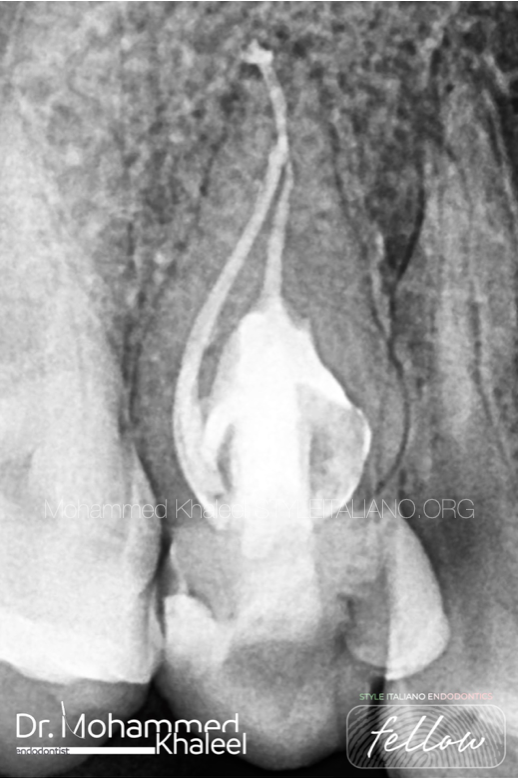
Obturation : main canal (distal one ) by single cone with BC sealer and other by hot technique with BC sealer.

Fig. 10
About the author:
Mohammed khaleel
B.D.S Hawler medical university /college of dentistry 2011
Key opinion leader for Denco
Conclusions
The knowledge of classification and anatomical variations of teeth with dens invaginatus is important for early detection and early management for the practitioners. Also CBCT made the process easy .
Bibliography
1. Shulze C. Developmental abnormalities of the teeth and the jaws. In: Gorlin O, Goldman H, editors. Thoma's Oral Pathology. St.Louis: Mosby; 1970. pp. 96–183.
2. Kronfeld R. Dens in dente. J Dent Res. 1934;14:49–66.
3. Rushton MA. A collection of dilated composite odontomas. Br Dent J. 1937;63:65–85.
4. Oehlers FA. Dens invaginatus I. Variations of the invagination process and associated anterior crown forms. Oral Surg Oral Med Oral Pathol. 1957;10:1204–18.
5. Oehlers FA. Dens invaginatus II. Associated posterior crown forms and pathogenesis. Oral Surg Oral Med Oral Pathol. 1957;10:1302–16.