Print & Try
28/03/2020
Riccardo Tonini
Warning: Undefined variable $post in /home/styleendo/htdocs/styleitaliano-endodontics.org/wp-content/plugins/oxygen/component-framework/components/classes/code-block.class.php(133) : eval()'d code on line 2
Warning: Attempt to read property "ID" on null in /home/styleendo/htdocs/styleitaliano-endodontics.org/wp-content/plugins/oxygen/component-framework/components/classes/code-block.class.php(133) : eval()'d code on line 2
How much technology can help us? Print&Try technique represents a really useful method in order to simulate the clinical treatment that will be done next. From CBCT it’s obtained a file that is transformed in an STL and printed after with an high definition 3d printer. Clinician can try and find the best approach in order to be more precise during the real treatment. In this article will be described the management of a dens invaginatus with Print&Try technique.
Dens invaginatus is a developmental malformation that occurs during tooth development prior to calcification due to an infolding of the enamel organ into the dental papilla. The affected teeth show a deep invagination of the enamel and dentine starting from the crown and sometimes extending deep into the root. The prevalence of dens invaginatus can be variable, ranging from 0.3% to 10%, most commonly affecting the permanent maxillary lateral incisors, followed by the maxillary central incisors. (Diseases and Conditions in Dentistry: An Evidence‐Based Reference)
The aim of this report is to present a case of Type III dens invaginatus in a maxillary lateral incisor with a periapical lesion and its successful treatment using a combination of surgical and non-surgical therapy.
The complete removal of the diseased pulp tissue can be a challenge for clinicians due to the complexity of internal canal anatomy and may require either non-surgical endodontic therapy alone or that therapy may have to be combined with surgical endodontics to achieve an adequate sealing of the root canal system.
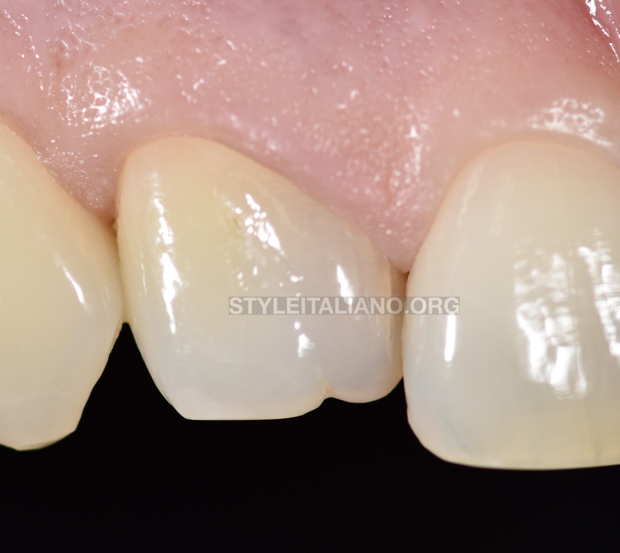
Fig. 1
35 years old patient with 1.2 with a typical aspect of dens invaginatus with an anatomical malformation in the crown.
The tooth responded negatively to vitality tests and it was also discromic.
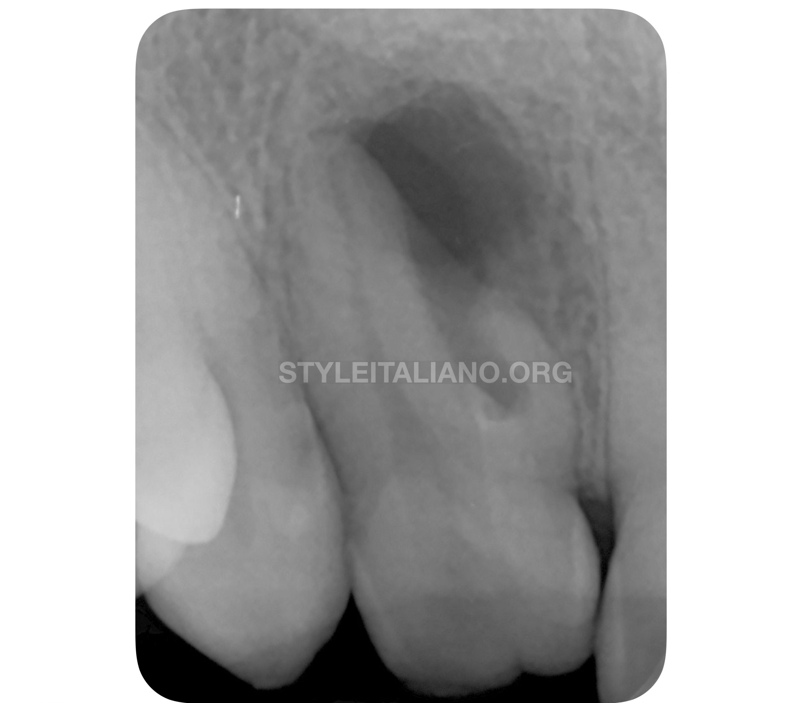
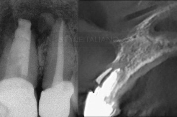
Fig. 2
Radiograph evaluation confirmed the presence of dens invaginatus and a periapical radiolucent lesion.
Radiography has an important role in the diagnosis and assessment of the irregular morphology of the root canal system, but conventional planar radiography only provides a two-dimensional representation of the complex anatomy
Cone beam computed tomography (CBCT) provides three-dimensional (3D) images of the maxillofacial skeleton, including the teeth and their surrounding tissues,
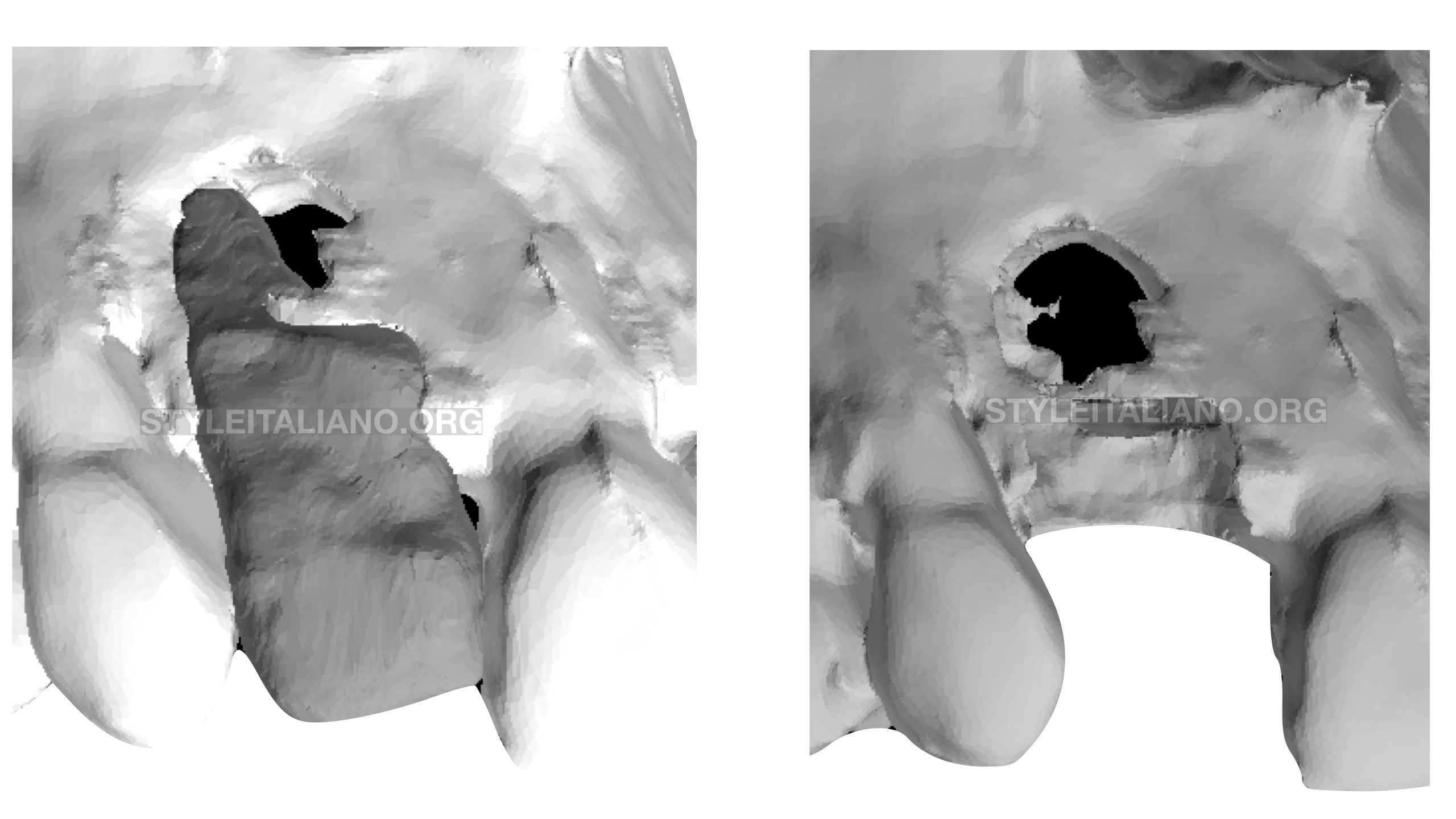
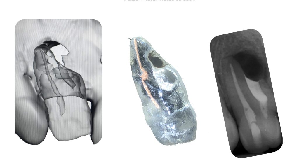
Fig. 3
The scanned volume derived from the CBCT software may allow generation of images in three planes that can be continuously scrolled through, thus allowing a three- dimensional understanding of the structure involved. They can also be converted into additional types of files.
the new types of files that can be derived from the CBCT scans is the stereo litography (STL) file. An STL file is a format used by stereoli- thography software to generate information needed to produce 3D plastic models using stereolithography machines or printers
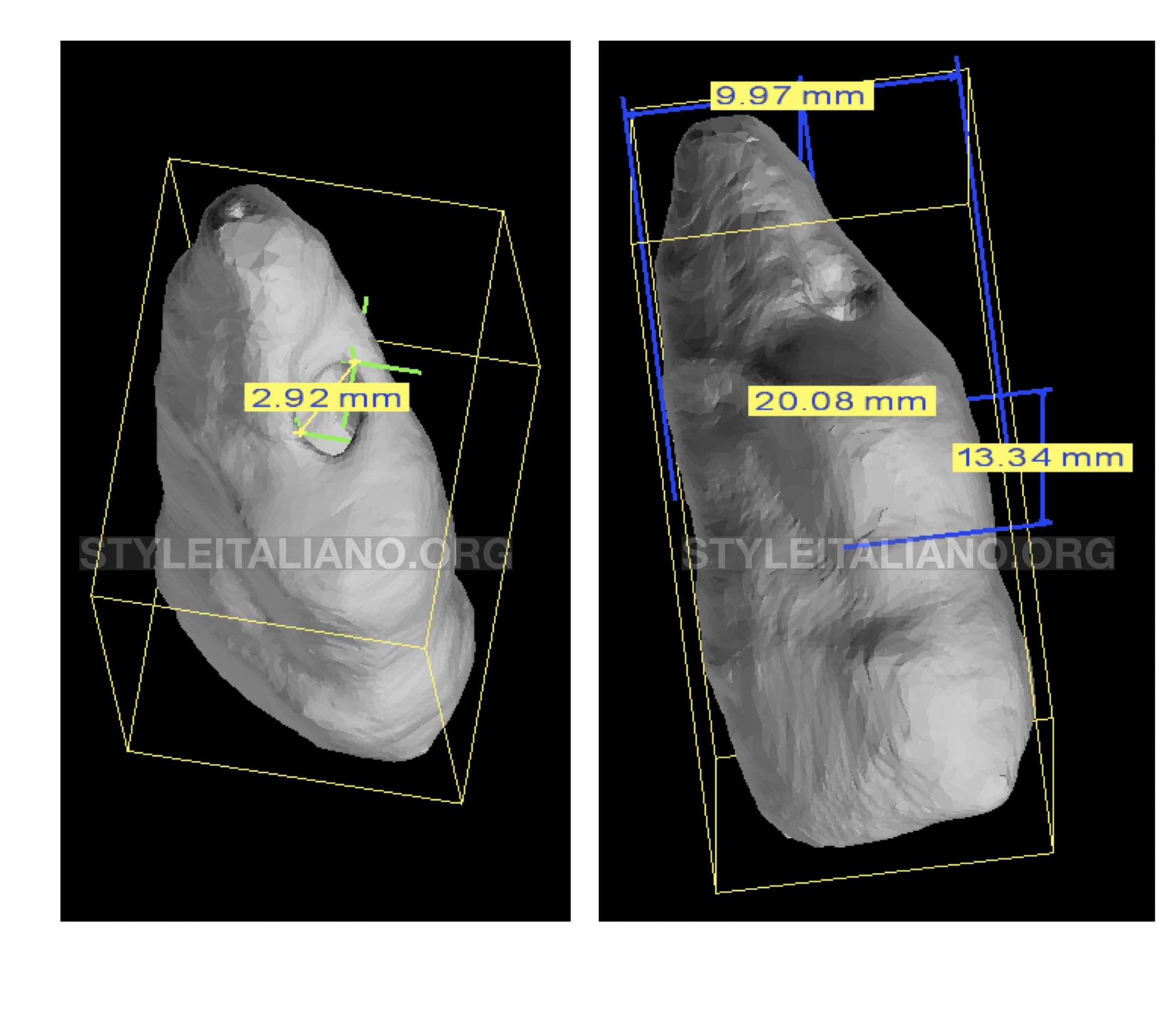
Fig. 4
Thanks to CBCT is also possible measure properly length an diameter of root canal anatomy
The CBCT data were used to produce precise 3D plastic models of the tooth. These models facilitated the treatment planning process and the trial of treatment approaches.
First visit: access cavity with a proper location in order to treat only the anatomy accessible with a conventional orthograde approach.
WL 19mm
Shaping:F2 gold
Filling: Warm vertical technique
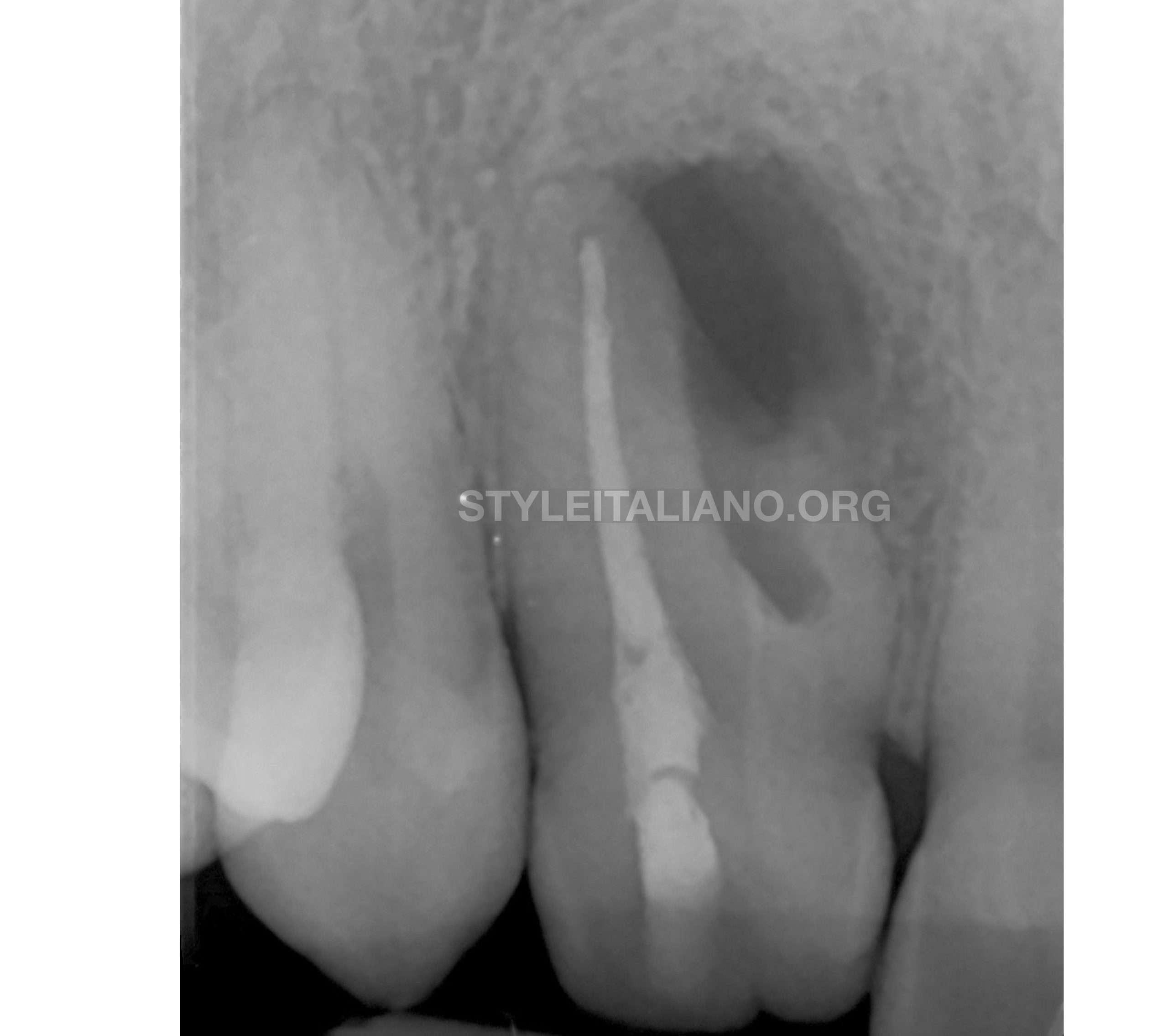
Fig. 5
Xray of RCT
Second visit: surgical approach.
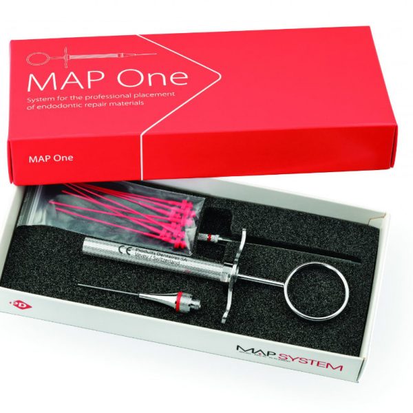
Cavity preparation with Spartan Obtura tips
Retrofilling with bioceramic putty
Res Membrane
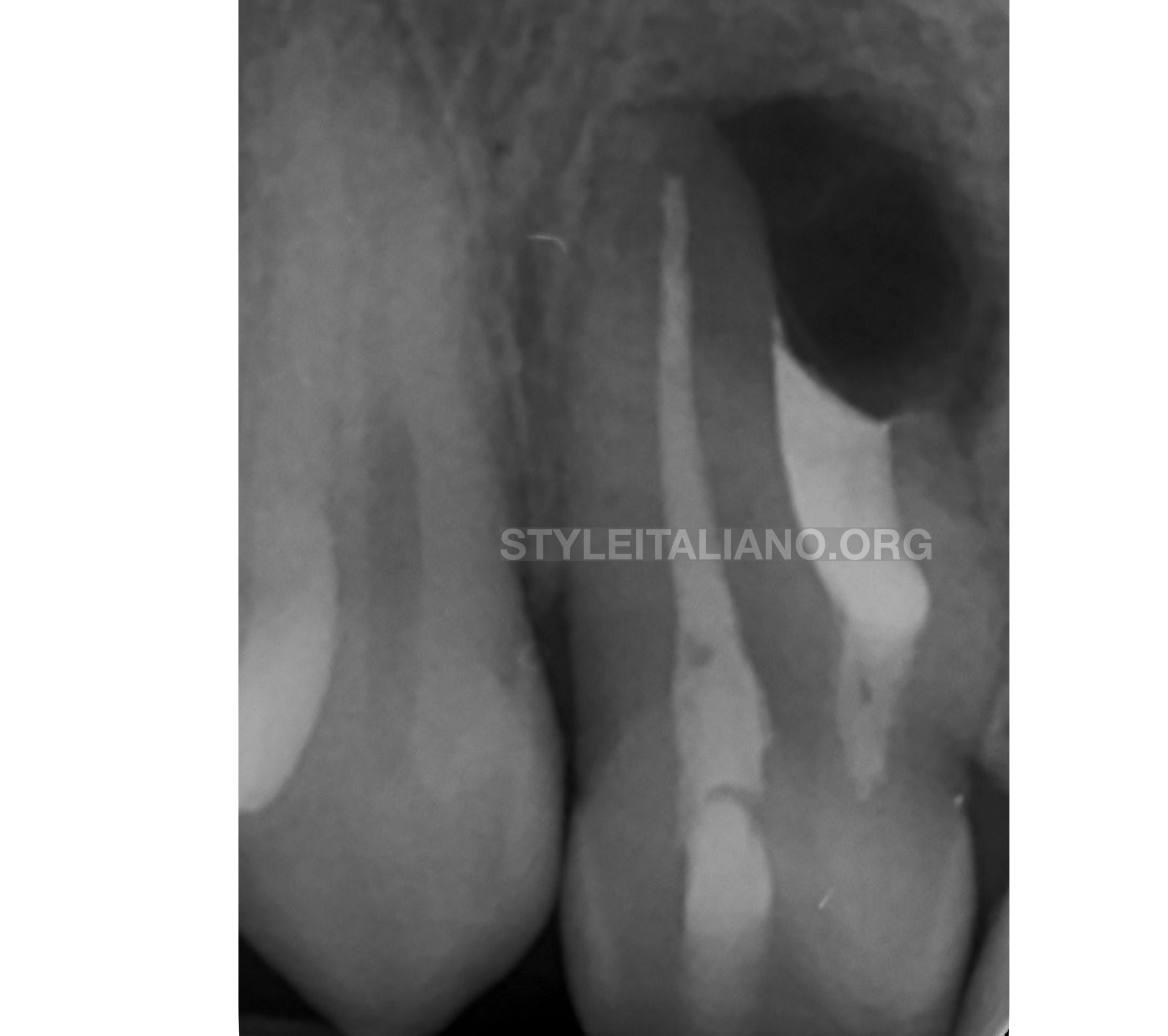
Fig. 6
Post op X-ray

Fig. 7
After 4 days seiches removal
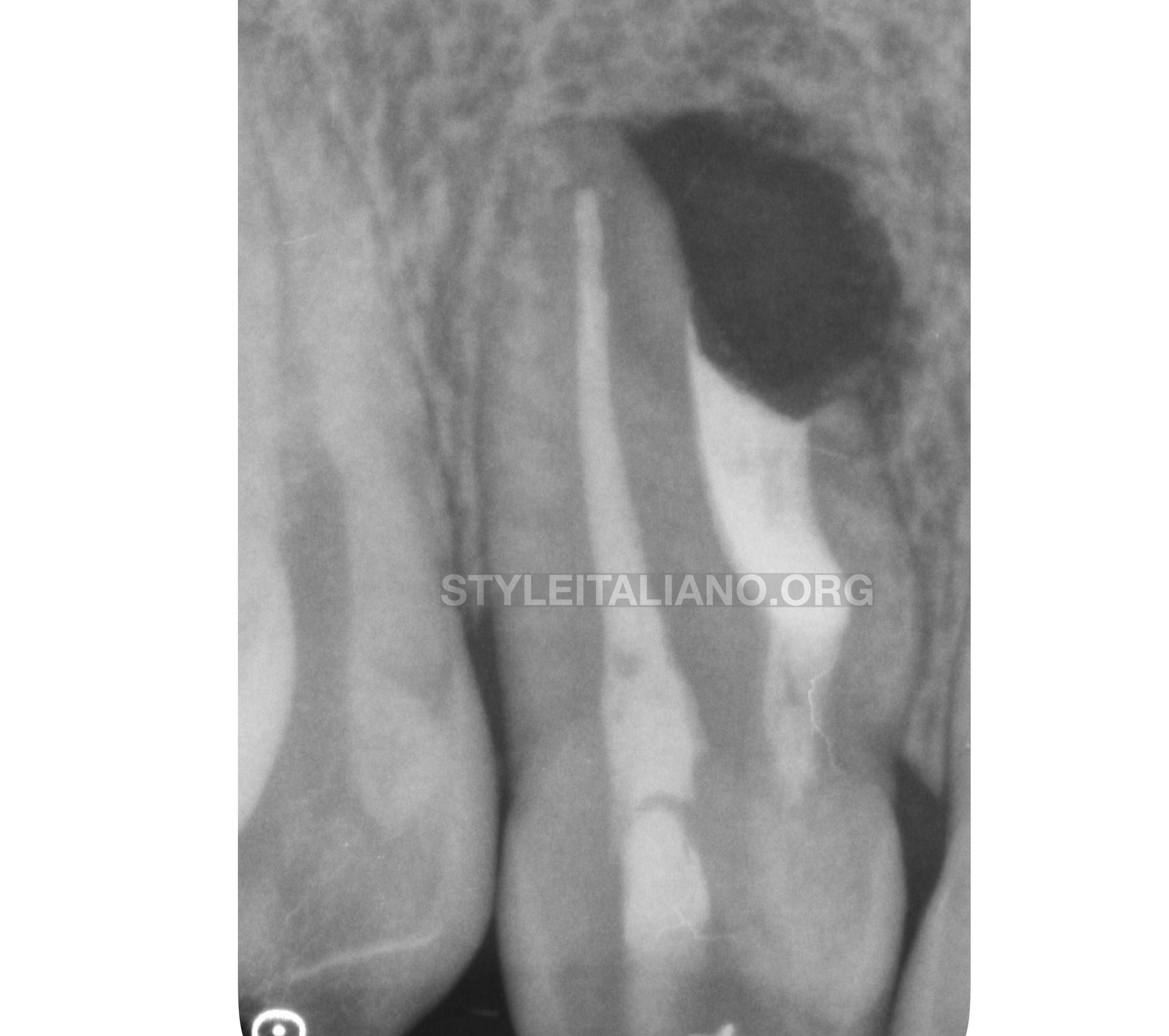
Fig. 8
Bone healing process after one month
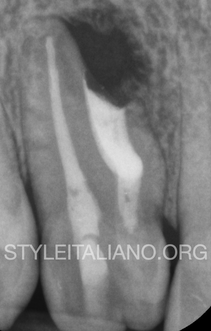
Fig. 9
5 months recall x-ray
Conclusions
Root canal treatment in teeth with dens invaginatus associated with apical pathosis often involves complicated procedures that require precise diagnosis, appropriate treatment planning and adequate implementation.
The printing of a transparent 3D plastic model of this tooth, with its complex invagination, provided an effective means for defining the correct approach, surgical and not surgical.
In complex cases, 3D models can facilitate treatment planning and is also useful for education purposes.
Bibliography
Management of a Type III Dens Invaginatus using a Combination Surgical and Non-surgical Endodontic Therapy: A Case Report
The journal of contemporary dental practice 10(5):E081-7 · September 2009
The diagnosis and conservative treatment of a complex type 3 dens invaginatus using cone beam computed tomography (CBCT) and 3D plastic models
A. Kfir, Y. Telishevsky-Strauss, A. Leitner & Z. Metzger