Management of a C-Shaped canal in second lower molar
18/11/2023
Fellow
Warning: Undefined variable $post in /home/styleendo/htdocs/styleitaliano-endodontics.org/wp-content/plugins/oxygen/component-framework/components/classes/code-block.class.php(133) : eval()'d code on line 2
Warning: Attempt to read property "ID" on null in /home/styleendo/htdocs/styleitaliano-endodontics.org/wp-content/plugins/oxygen/component-framework/components/classes/code-block.class.php(133) : eval()'d code on line 2
Anatomical variation of canals is well documented in the literature and C-shaped canal anatomy was first reported in literature by Cooke and Cox in 1979.
This anatomical variation mostly found in mandibular second molars with prevalence between 2.7–52% in different populations.
C-shaped canals have rarely been found in other teeth, maxillary first molars, mandibular first molars, third molars and mandibular premolars.
The most widely accepted theory for the formation of this unusual ribbon shaped canal is the failure of Hertwig’s epithelial.
Initial situation.
A 36-year-old woman came to our orthodontic referral clinic complaining of severe pain related to her mandibular second molar.
Pulp diagnosis: pulp necrosis, acute apical abscess.
Clinically a filtrated composite resin and sinus tract
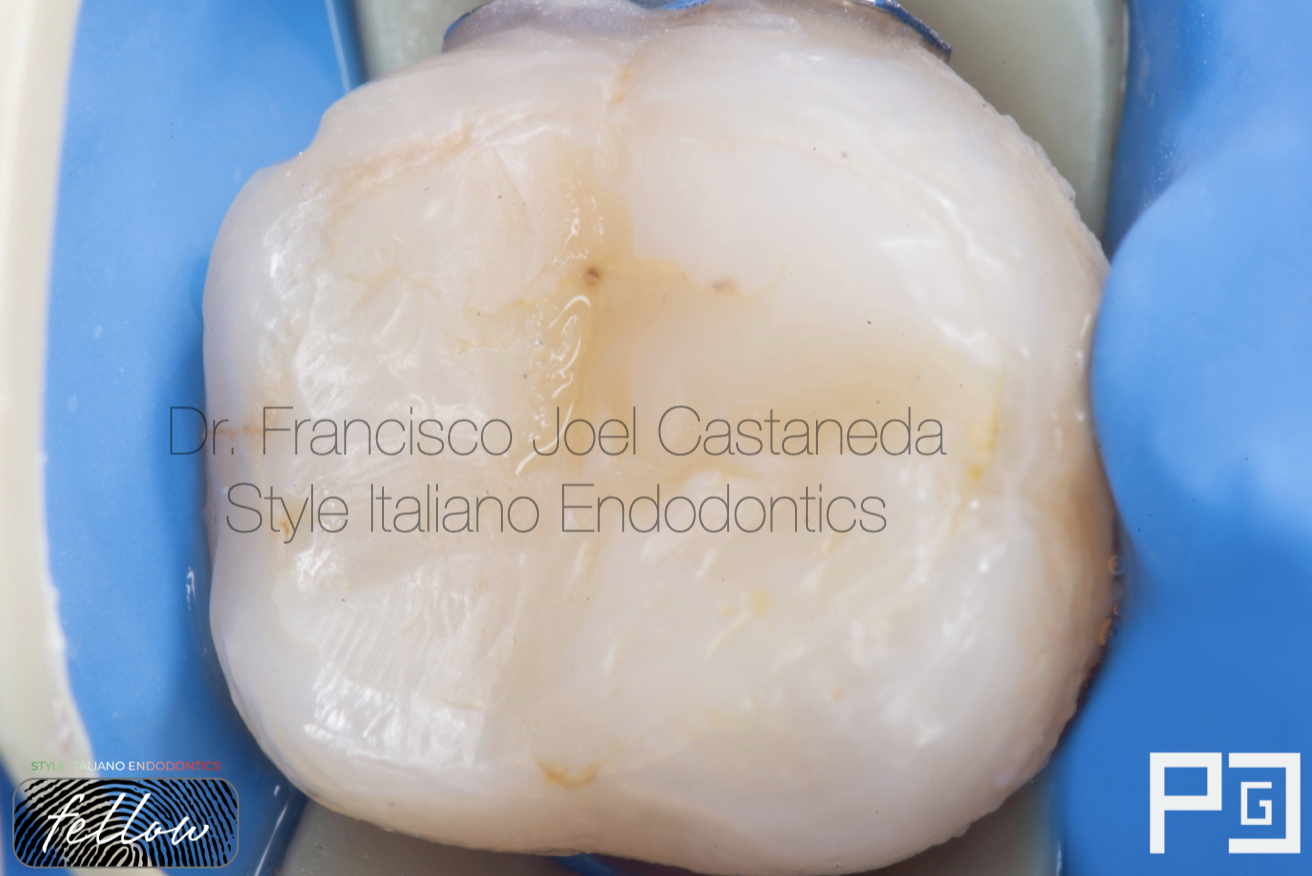
Fig. 1
Isolation of a single tooth with soft clamp, clinically a filtrated composite restoration.

Fig. 2
Pre-op X-Ray showed extend loss bone and conical root.
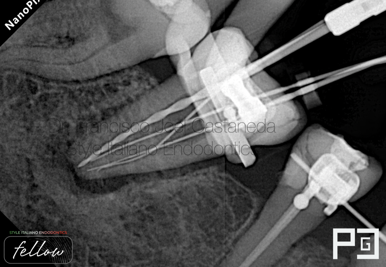
Fig. 3
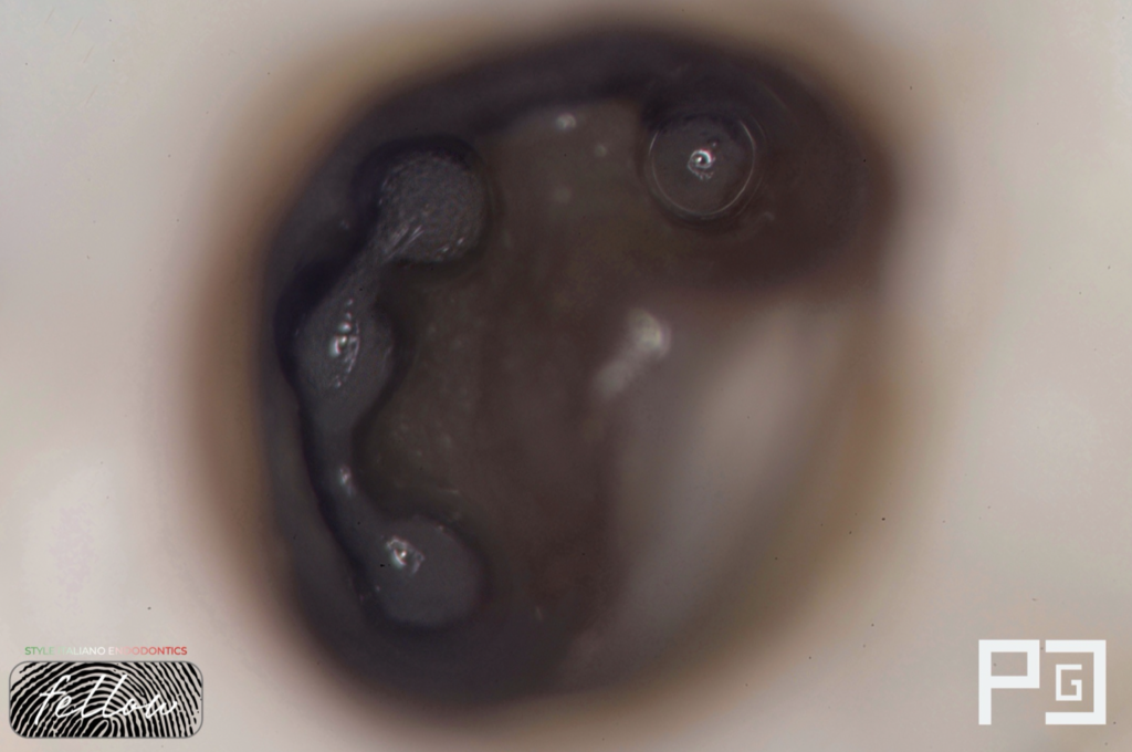
Working length and confirmation of electronic apex locator reading.
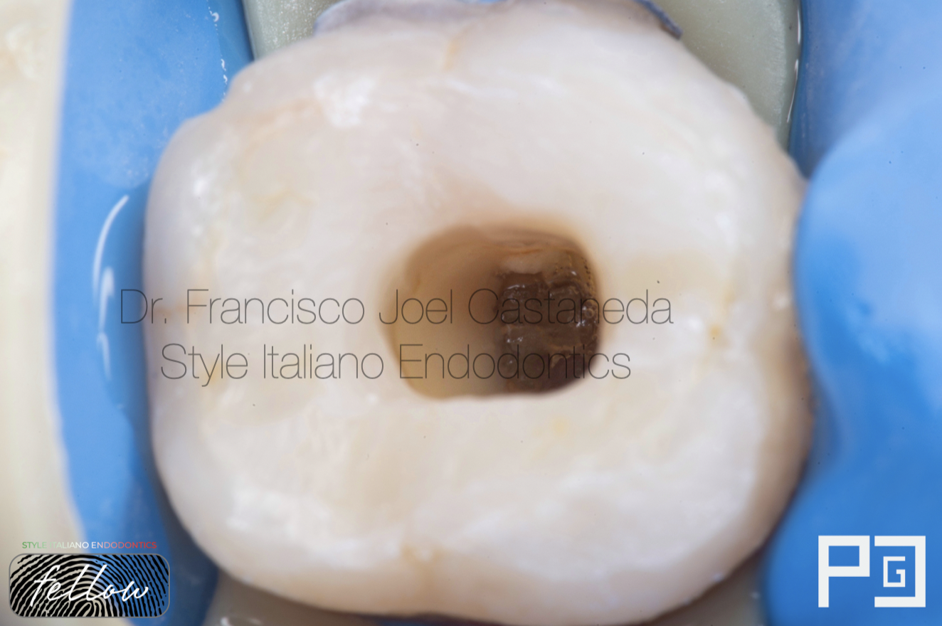
Fig. 4
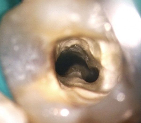
Access to the pulp chamber, location of the canals and discharge of pus via intracanal.
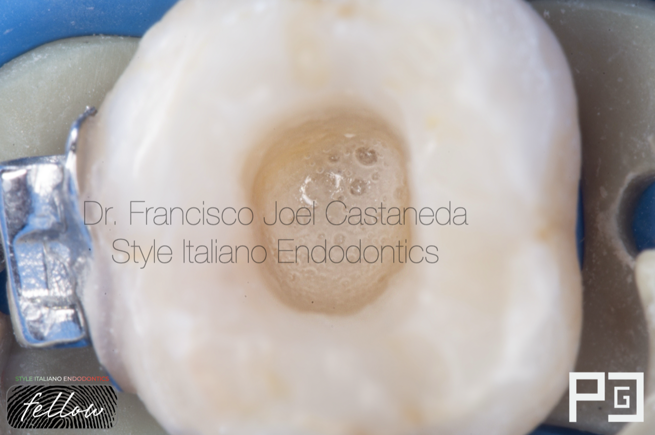
Fig. 5
Sodium Hypoclorite in action
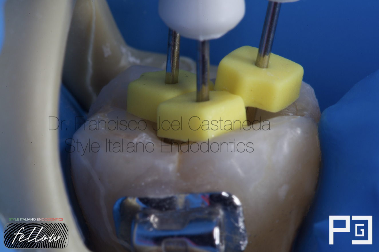
Fig. 6
Working length photograph.
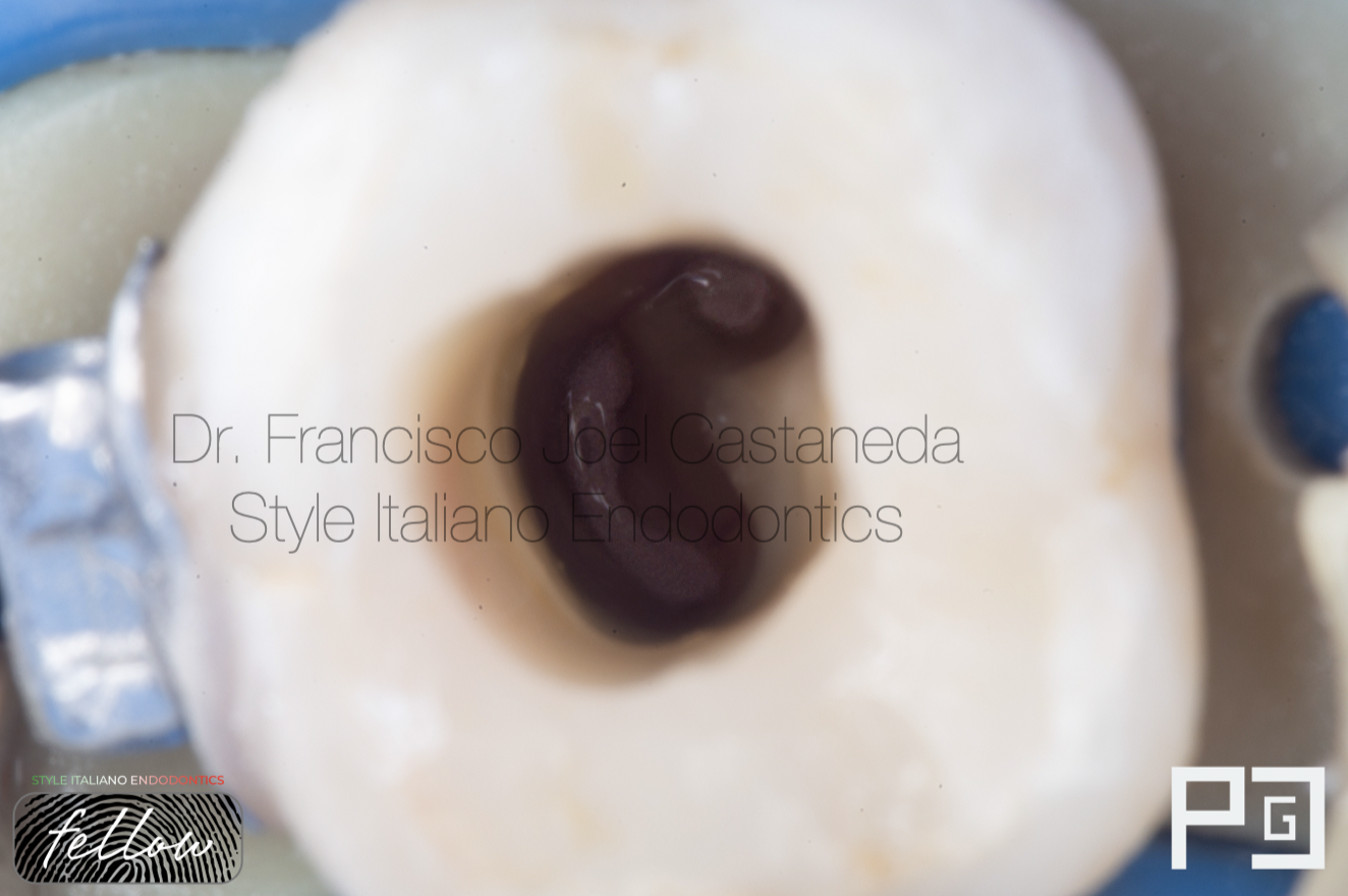
Fig. 7
After refining the access and locating all the canals, instrumentation and chemomechanical disinfection were performed with Dperfect MG3 Blue and RCS Rainbow 40.04 files.
C-shaped canal configuration
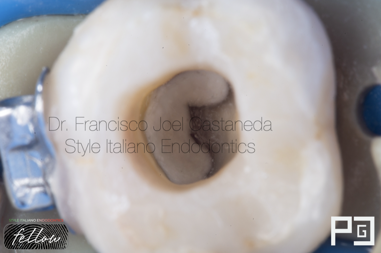
Fig. 8
Calcium hydroxide as intracanal medication
Calcium Hydroxide Video
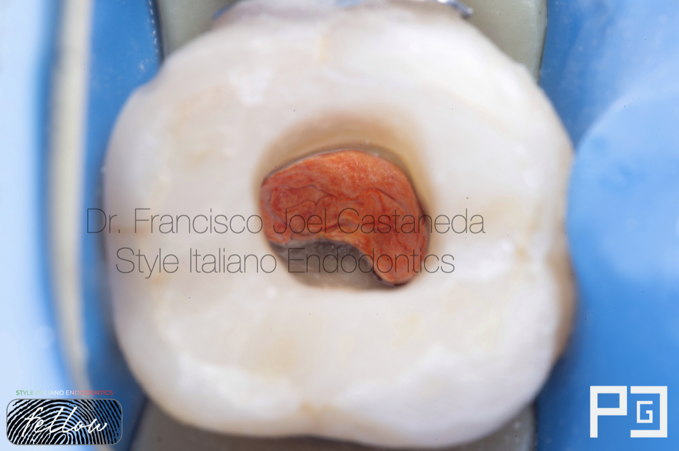
Fig. 9
We place Teflon tape on top of the calcium hydroxide followed by a temporary material by two weeks.
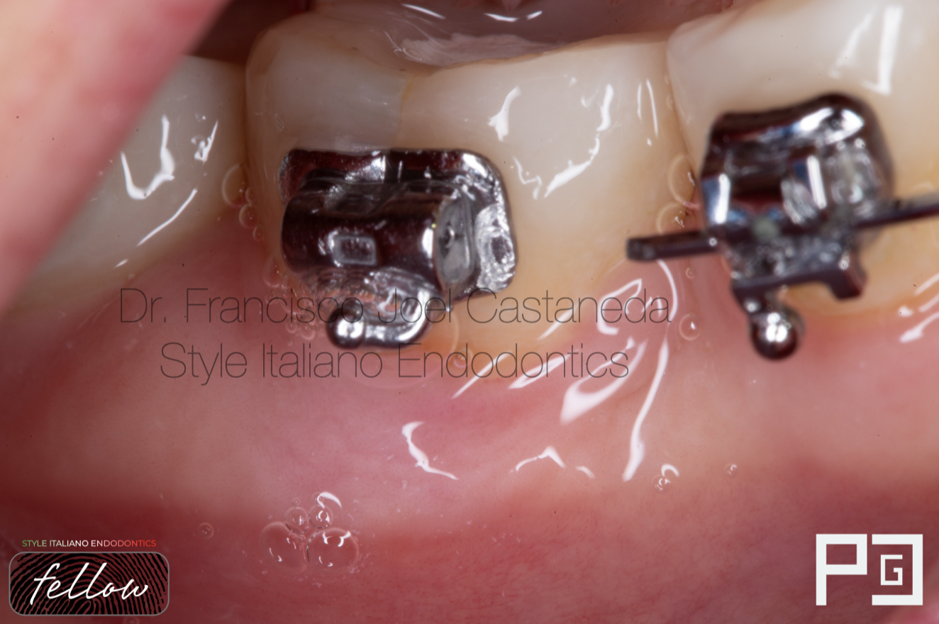
Fig. 10
After two weeks, we noticed the healing of the sinus tract and no symptoms.
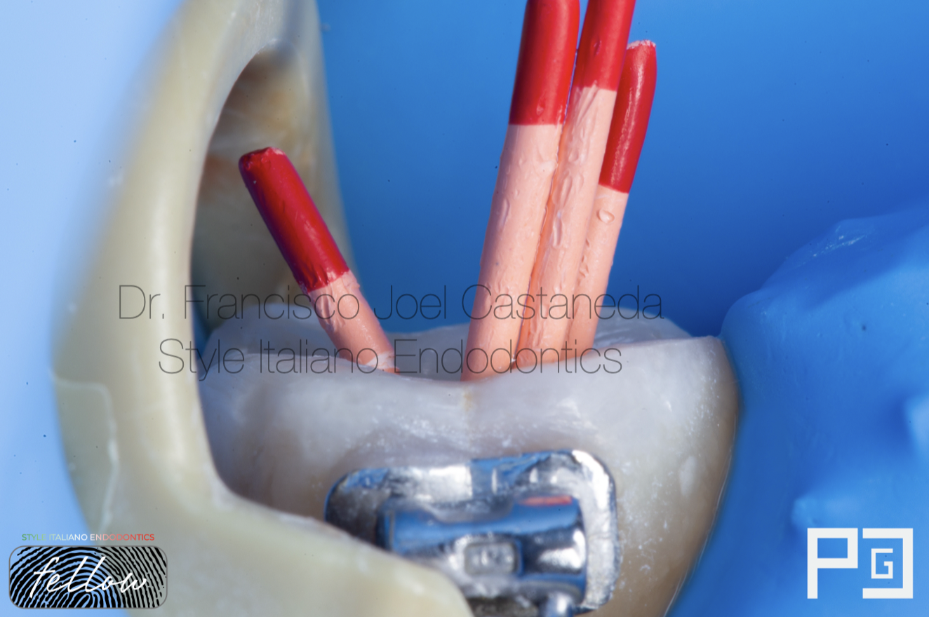
Fig. 11
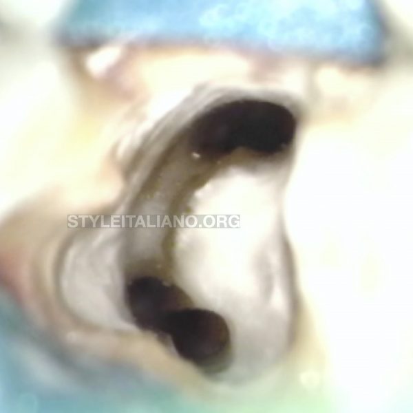
Cone fit photograph
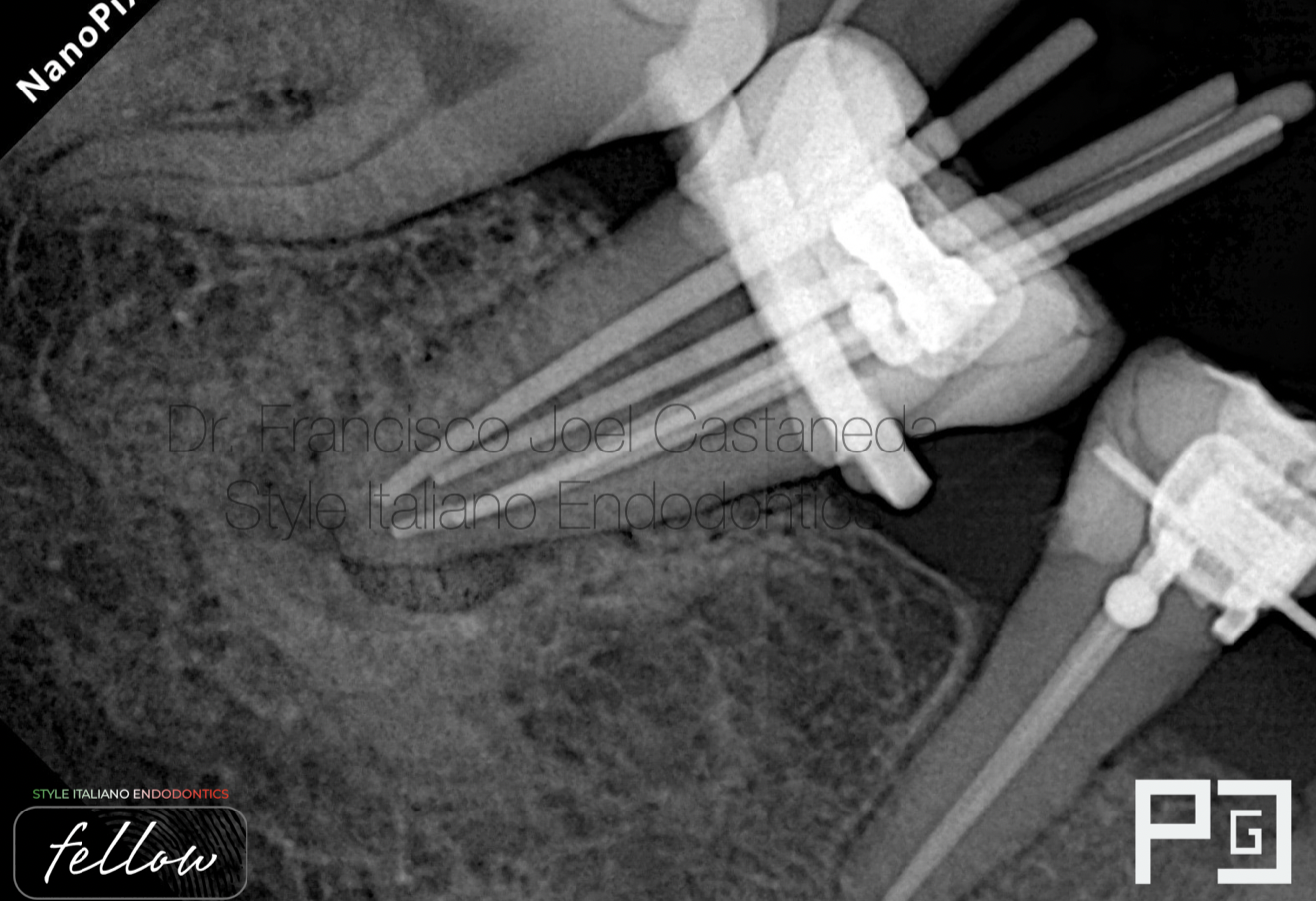
Fig. 12
Master cone fit
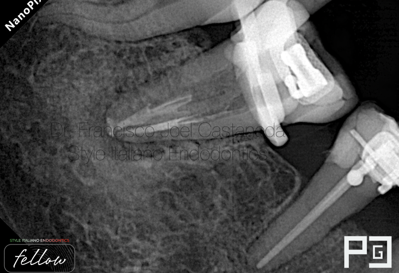
Fig. 13
Obturation by continuous wave technique, down pack step.
Down pack video
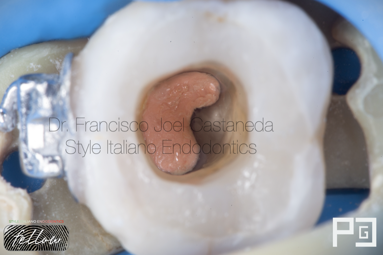
Fig. 14
Obturation done.
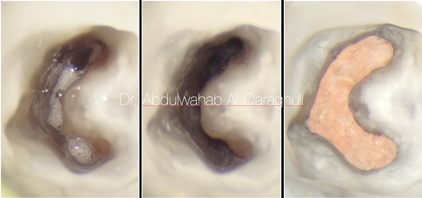
Clean the excess of gutapercha and sealer is a very important step in order to do a correct adhesive protocol step.
Clean chamber ( c-shape )
 : eval()'d code</b> on line <b>19</b><br />
)
Fig. 15
Sealing pulp chamber with flowable composite to prevent coronal leakage.
video of the pulp chamber
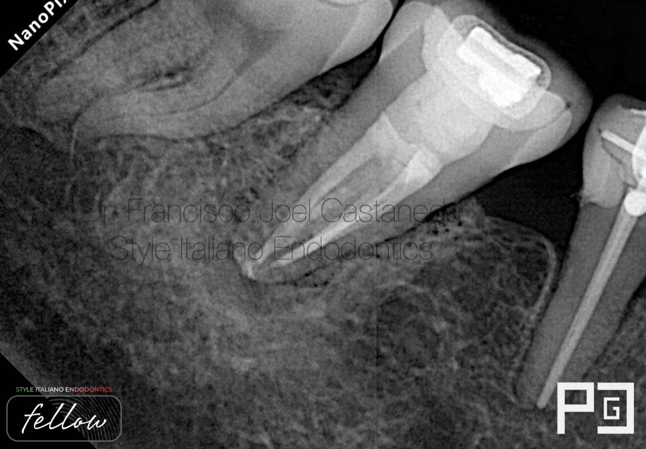
Fig. 16
Post-op X-Ray with the final temporary restoration
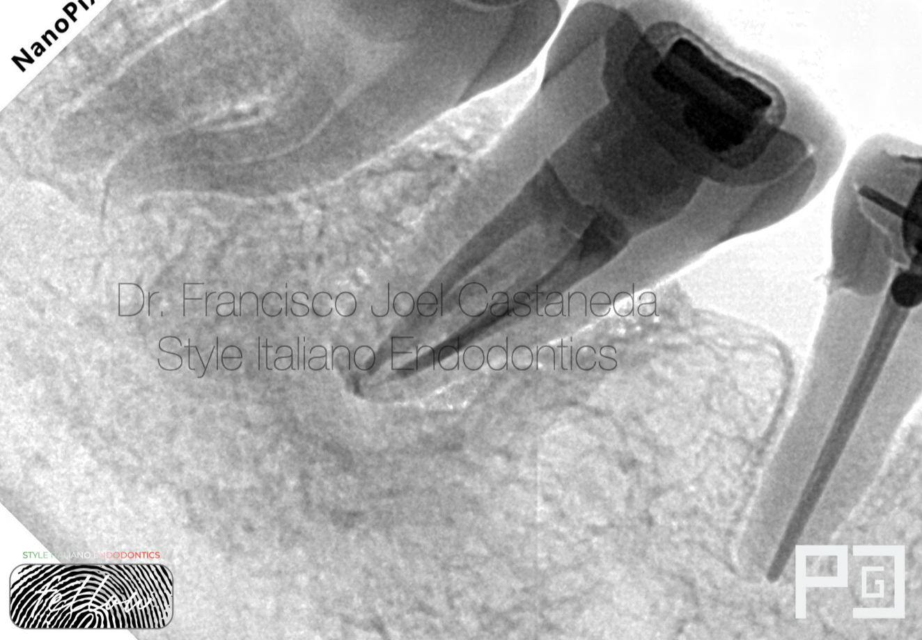
Fig. 17
Post-Op X-Ray inverted
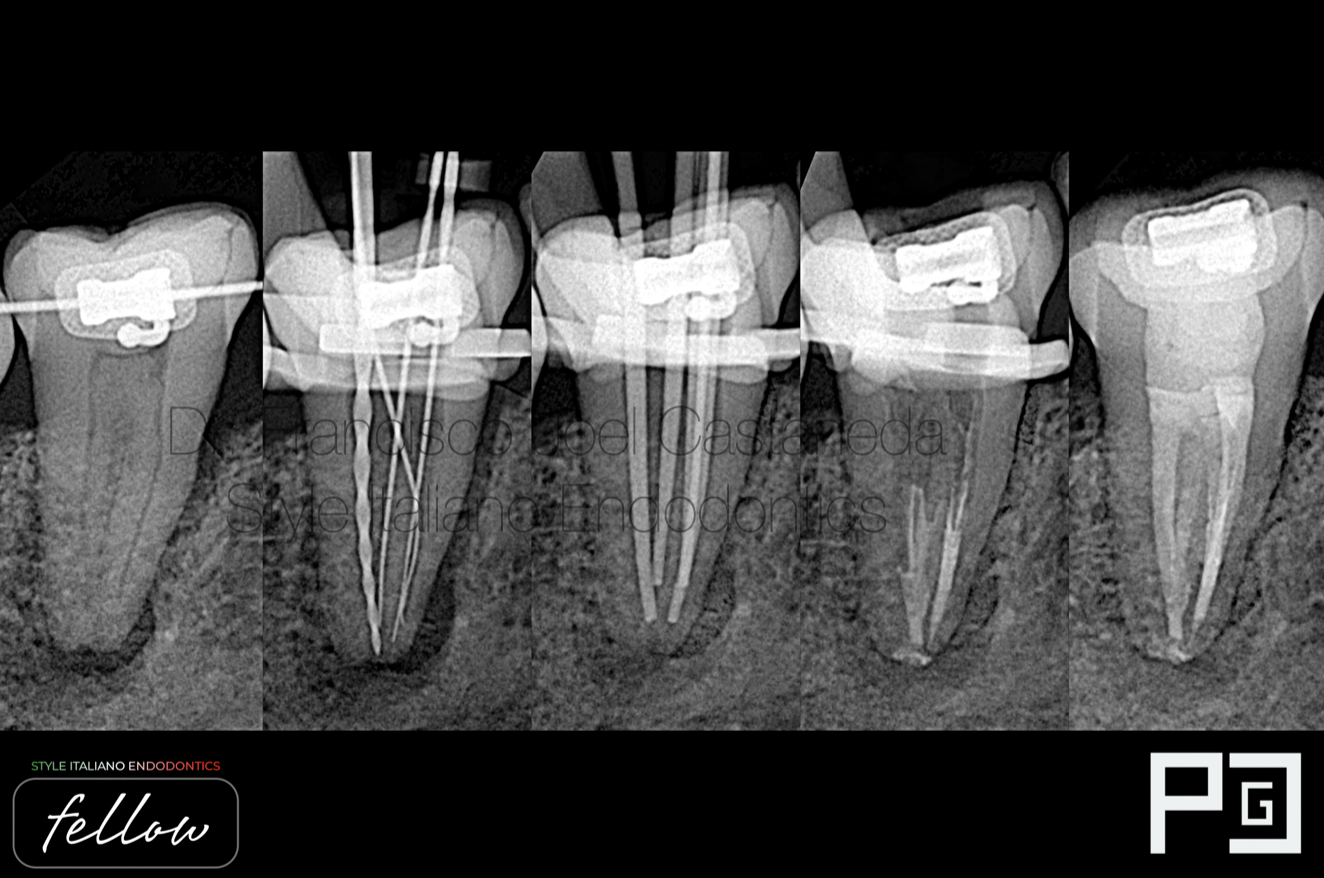
Fig. 18
Endo from A-Z

Fig. 19
Francisco Joel Castaneda Guerrero
Guadalajara, Jalisco - Mexico
2014-2018 dental surgeon by UDG (University of Guadalajara)
2021-2023 specialist in endodontics by UAG (University autonomy of Guadalajara)
2021- first place in artistic photography by AME (Mexican Association of Endodontics)
2022- first place in clinical photography, and second place in artistic photography by AME (Mexican Association of Endodontics)
Creator of ENDOGRAPHY
ENDOGRAPHY Courses (Endodontics and photography)
Conclusions
The prevalence of C-shaped canals is quite high in mandibular second molars. Understanding the anatomy and characteristics of this configuration is interesting in order to better manage cleaning, shaping, medication and obturation techniques.
Bibliography
Cooke HG, Cox FL (1979) C-shaped canal configurations in mandibular molars. J Am Dent Assoc 99:836–839
Keinan D, Nuni E, Slutzky-Goldberg I (2009) Is a C-shaped configuration possible in teeth other than mandibular molars? Quintessence Int 40:541–543Keinan D, Nuni E, Slutzky-Goldberg I (2009) Is a C-shaped configuration possible in teeth other than mandibular molars? Quintessence Int 40:541–543
Ladeira DB, Cruz AD, Freitas DQ, Almeida SM (2013) Prevalence of C-shaped root canal in a Brazilian subpopulation: a cone-beam computed tomography analysis. Braz Oral Res 28:39–45
Soo WK, Thong YL, Gutmann JL (2015) A comparison of four gutta-percha filling techniques in simulated C-shaped canals. Int Endod J 48:736–746
Vertucci FJ, Haddix EJ (2011) Tooth morphology and access cavity preparation. In: Cohens. Pathways of the Pulp, 10th edn. St Louis, Missouri, USA: Mosby-Year Book, Inc, pp 217.
Jafarzadeh H, Wu YN (2007) The C-shaped root canal configuration: a review. J Endod 33:517–523