Treatment of a C shaped canal of a mandibular second molar
02/12/2020
Marc Kaloustian
Warning: Undefined variable $post in /home/styleendo/htdocs/styleitaliano-endodontics.org/wp-content/plugins/oxygen/component-framework/components/classes/code-block.class.php(133) : eval()'d code on line 2
Warning: Attempt to read property "ID" on null in /home/styleendo/htdocs/styleitaliano-endodontics.org/wp-content/plugins/oxygen/component-framework/components/classes/code-block.class.php(133) : eval()'d code on line 2
Mandibular second molars are known to have a complex root canal anatomy with anastomosis, resulting in some difficulties during root canal treatment, especially for the complete debridement of organic tissues and bacteria during the shaping and cleaning process and the possibility of insuring a good 3-dimensional seal during obturation. The prevalence of such anatomy is variable and depends on the population investigated with a wide range from 2.7 to 52 % according to some authors. A patient was referred to the office for a root canal treatment of tooth #37. The patient was complaining of spontaneous acute pain in the entire lower jaw. A diagnosis of irreversible pulpitis was determined and the treatment was performed at the same session to relieve the symptoms.
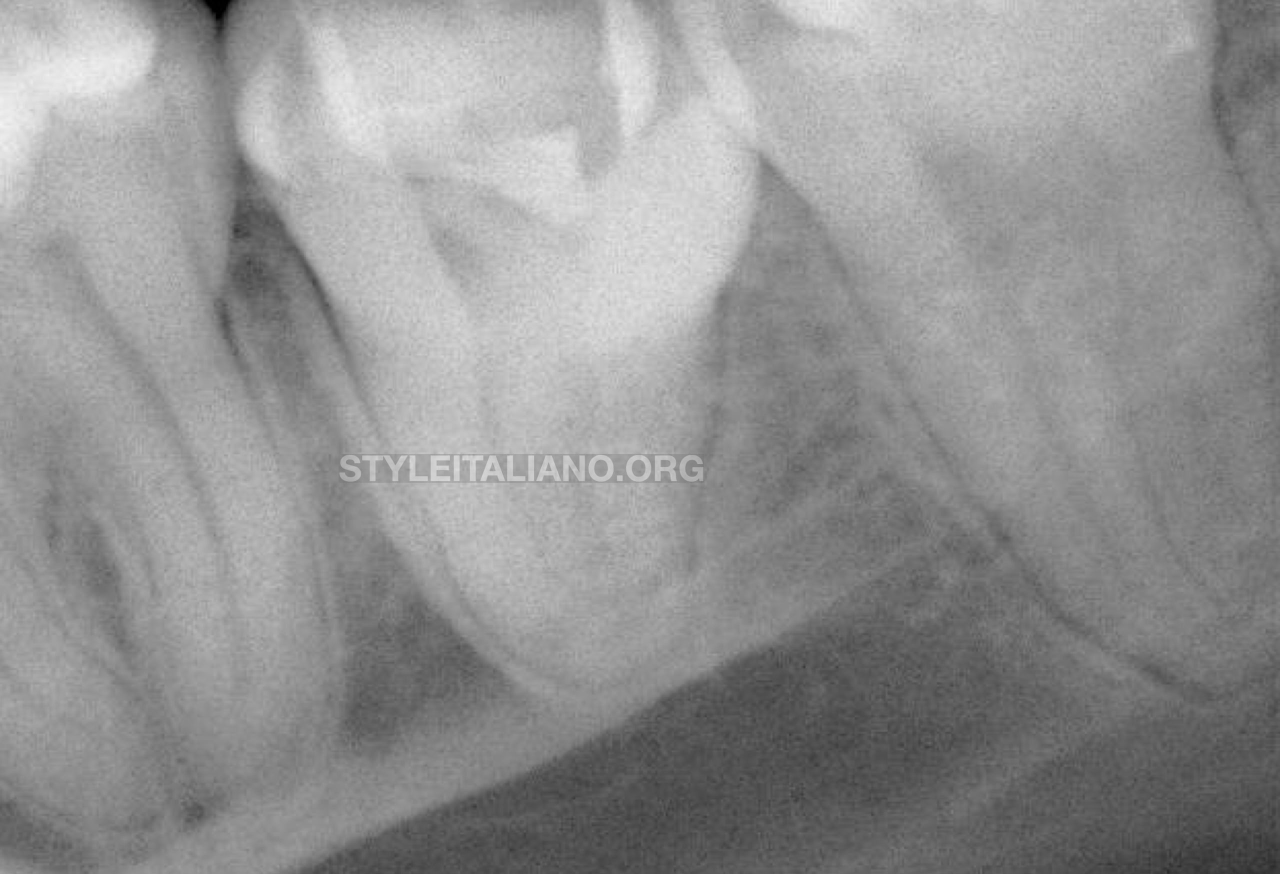
Fig. 1
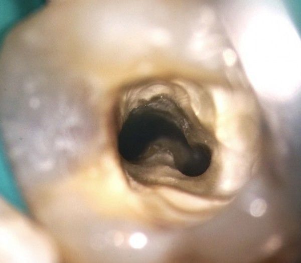
A clinical case report of an endodontic treatment on a mandibular second molar with a particular C shape configuration
Cavity was prepared with a long surgical bur #14 (Meisinger, CO, USA) and finished with an ultrasonic tip ET18 (Satelec, Acteon group, Merignac, France). A glide path was attempted as shown in the small footage using ProGlider (Dentsply Sirona, Balaigues, Switzerland), but it was impossible to reach the last 2 mm in any of the canals because of an abrupt curvature.
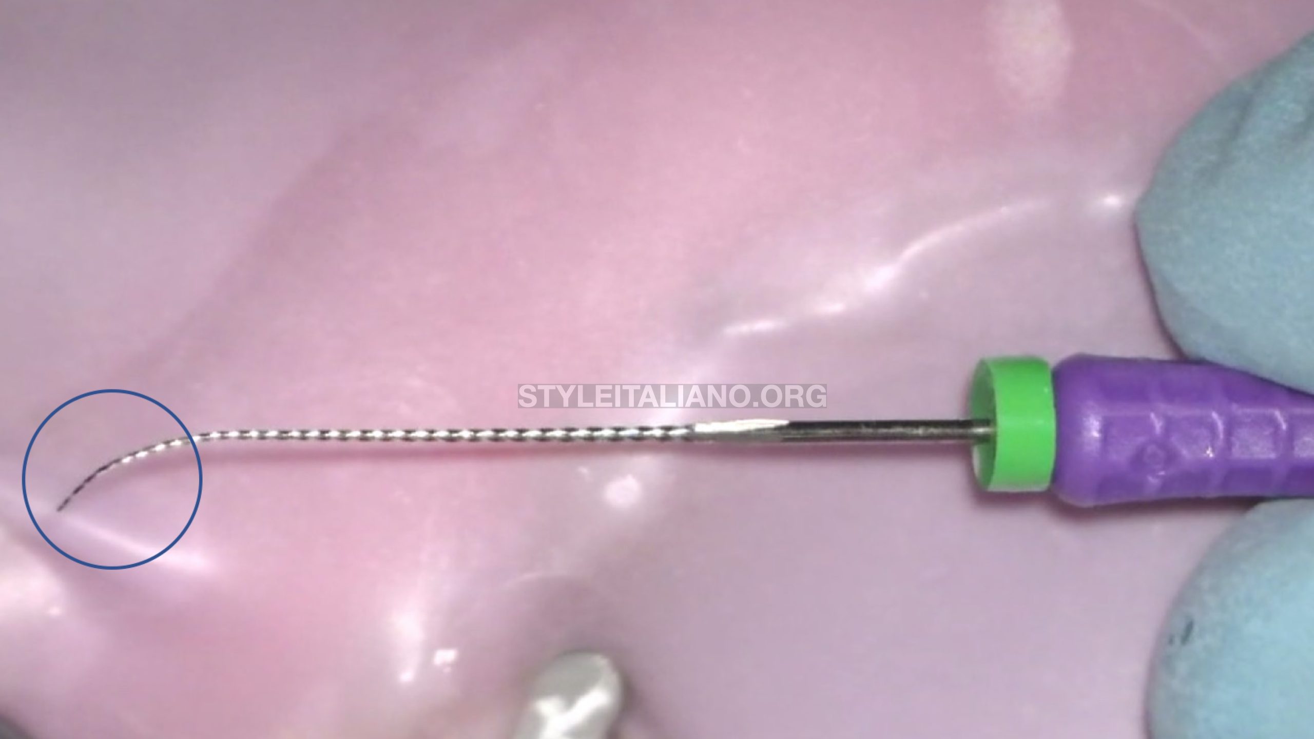
Fig. 2
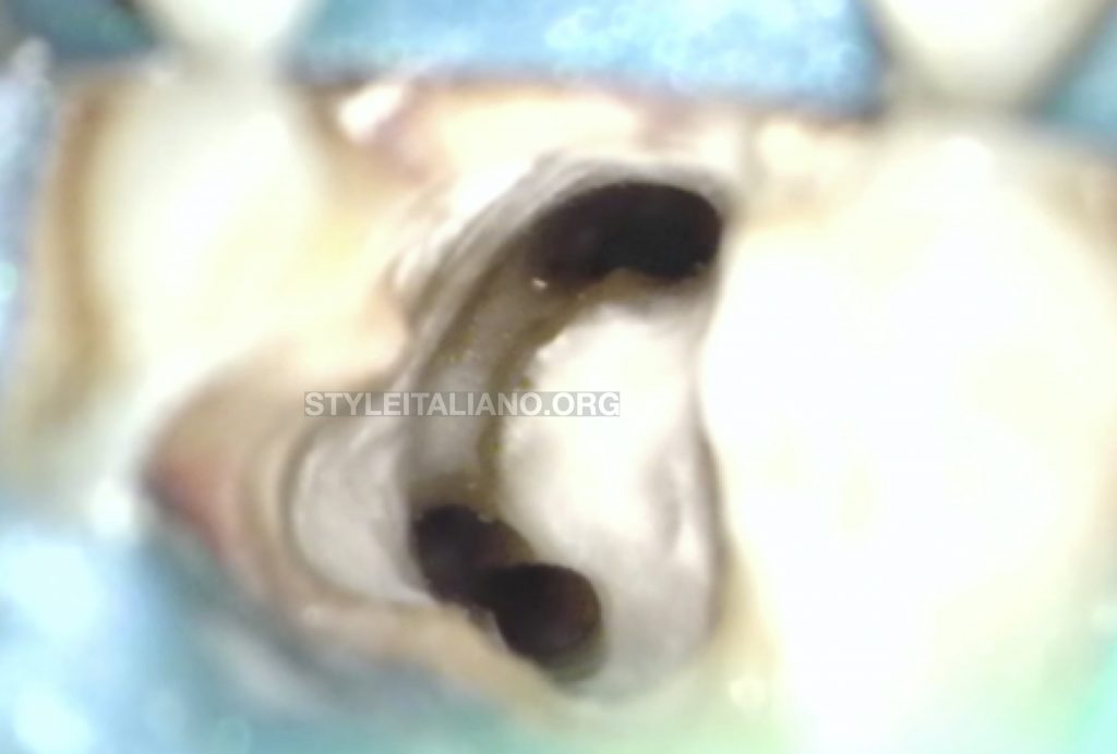
A number 10 K file (Dentsply Sirona, Balaigues, Swizerland) was precurved properly at its last 2 mm and penetrated carefully in the canals to prevent any ledges.
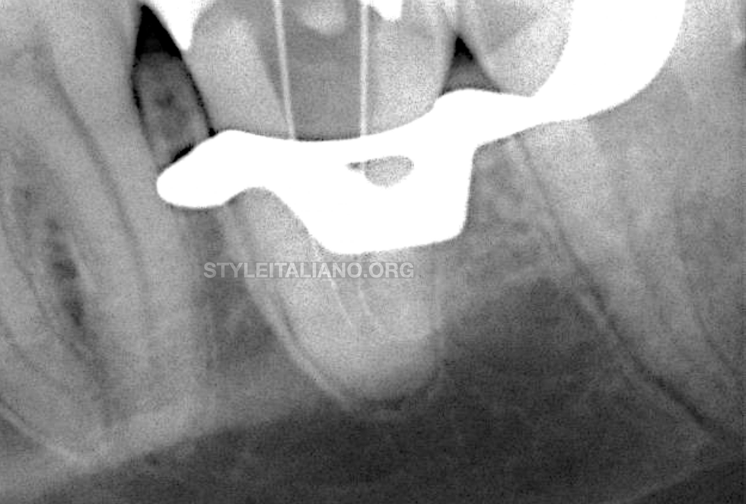
Fig. 3
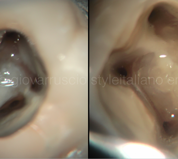
Once the the patency was obtained the file was not removed, an “in and out” motion was applied many times till the file was felt loose inside the canal. The canals were then shaped with Endostar E3 Azure (Poldent, Poland) 30/08 for preflaring 20/04, 20/06 for deep shaping.
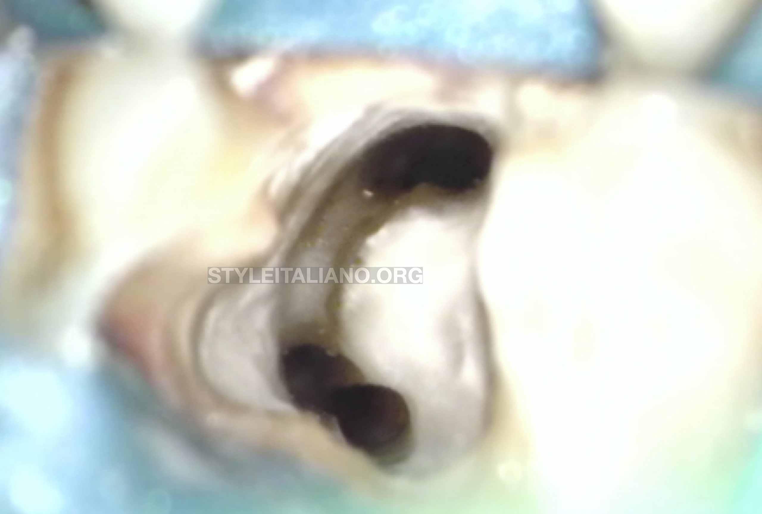
Fig. 4
A view of the mesial and distal orifices after shaping and cleaning
Mixing the Zinc Oxyde Eugenol type sealer EssenSeal
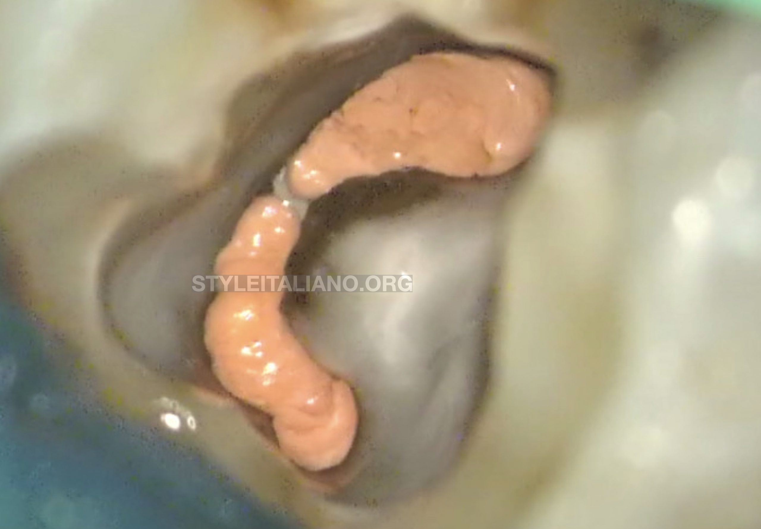
Fig. 5
Gutta percha was injected with the Obtura II gun and 23 G tip (Obtura Spartan Endodontics, Algonquin, IL,USA).
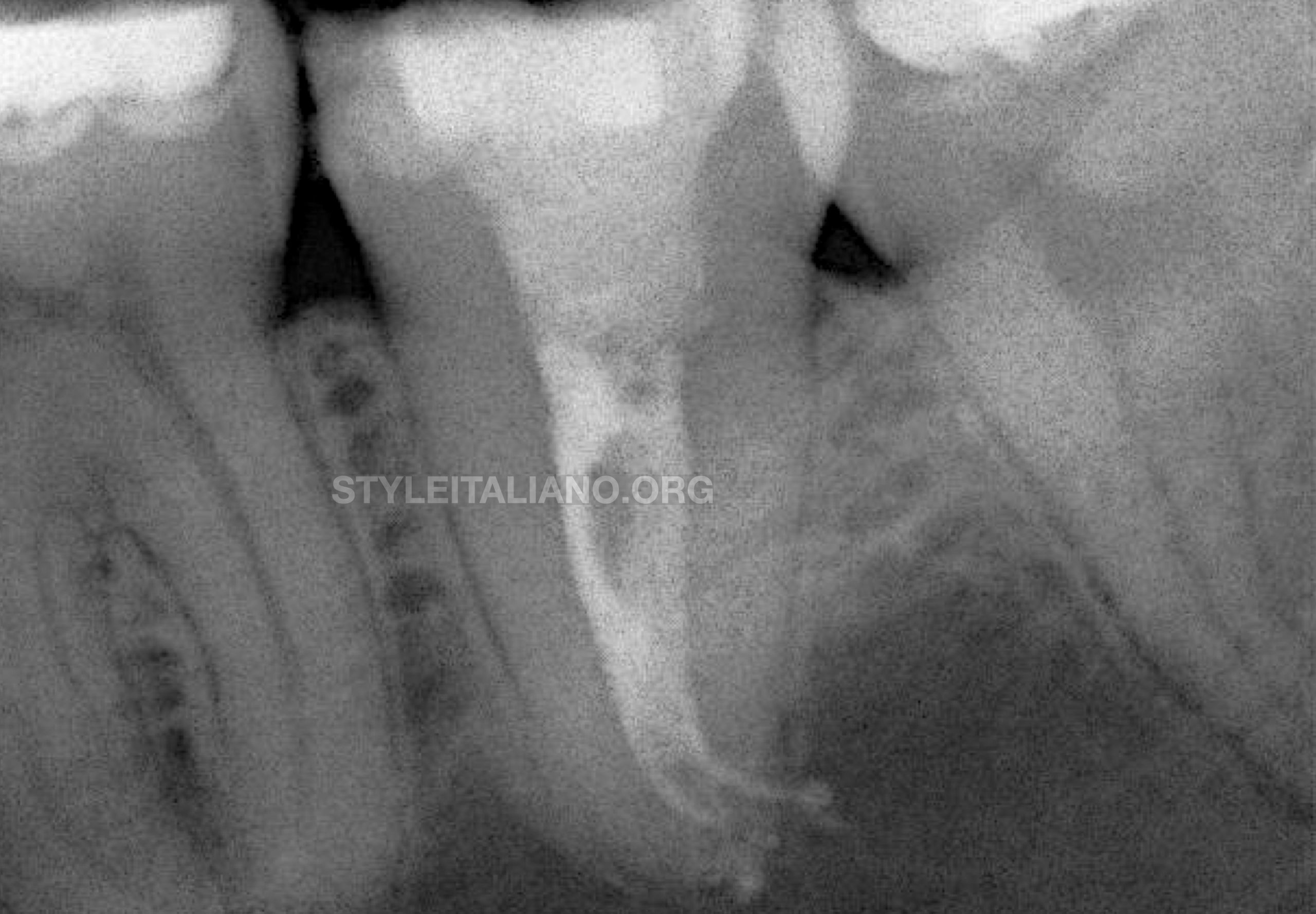
Fig. 6
Final Xray showing a type VI Vertucci 2-1-2 classification with large anastomosis in the middle part of the root.
Conclusions
Many classifications including the one of Melton et al. and Fan et al. were suggested to describe this particular ribbon shape anatomy, that can be basically a continuous or an interrupted system. Whereas, what is important for us clinicians is to adapt our treatment to each particular situation by observing carefully the periapical xray or sometimes the CBCT and to address clinically the entire system regardless of the type of C shape canal treated. Therefore, the respect of the original anatomy, the meticulous cleaning protocol including the activation and an adapted obturation technique are essential to succeed in such cases.
Bibliography
Fan B, Cheung GS, Fan M, Gutmann JL, Bian Z. C-shaped canal system in mandibular second molars: part I—anatomical features. J Endod 2004, 30:899–903
Melton DC, Krell KV, Fuller MW. Anatomical and histolog- ical features of C-shaped canals in mandibular second molars. J Endod 1991, 17:384–8.
Shemesh A, Levin A, Katzenell V, Itzhak JB, Levinson O, Avraham Z, Solomonov M. C-shaped canals-prevalence and root canal configuration by cone beam computed tomography evaluation in first and second mandibular molars-a cross-sectional study. Clin Oral Investig 2017 21(6):2039-44.
Srivastava S, Gaikwad RN, Alsalhi N, Alrogaibah NA. Cone-beam Computed Tomographic Analysis of C-shaped Canals and Radicular Grooves in Mandibular Premolars: Prevalence and Related Factors. J Contemp Dent Pract 2019 20(11):1350-4.