As endodontists we often encounter retreatment cases with procedural errors such as canal blockages, ledges, instrument fractures and perforations. The ledge is defined as an iatrogenic deviation from the original […]

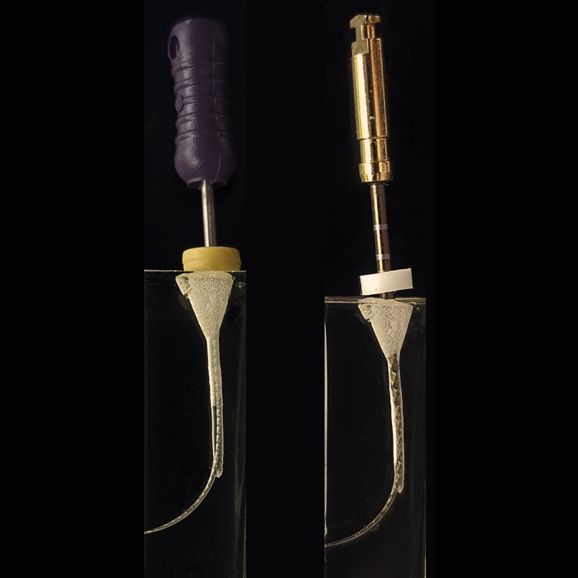 The endodontic ledge: causes and management
As endodontists we often encounter retreatment cases with procedural errors such as canal blockages, ledges, instrument fractures and perforations. The ledge is defined as an iatrogenic deviation from the original […]
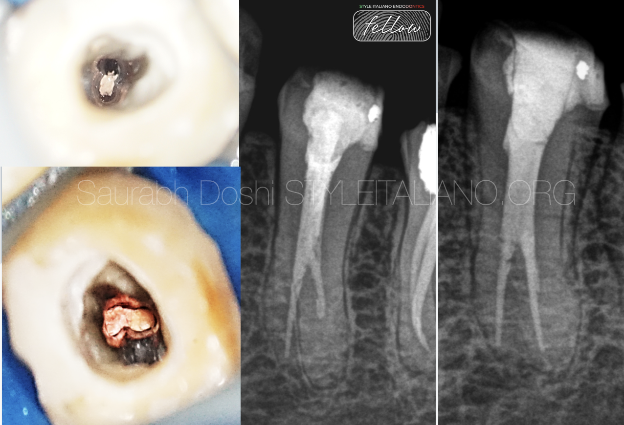
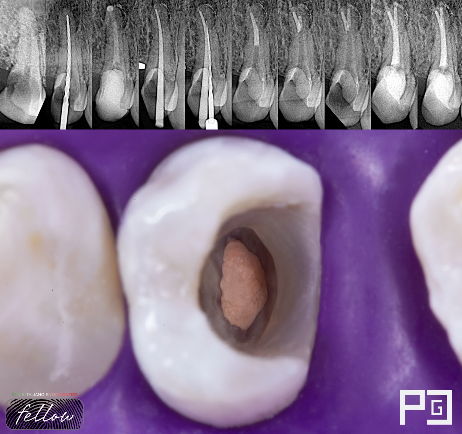
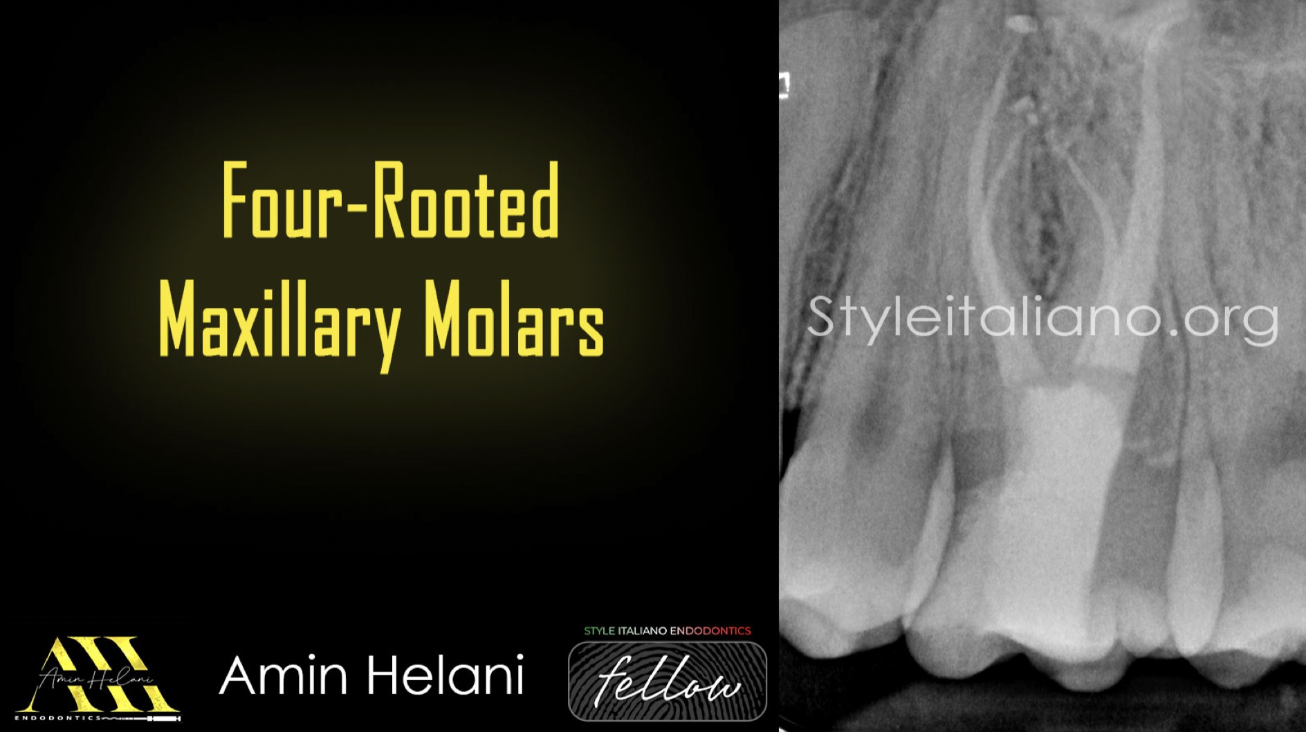 Four-Rooted Maxillary Molars
The maxillary first molar typically exhibits complex root canal anatomy, commonly presenting with three roots—two buccal and one palatal. However, anatomical variations, such as the presence of a fourth root, […]
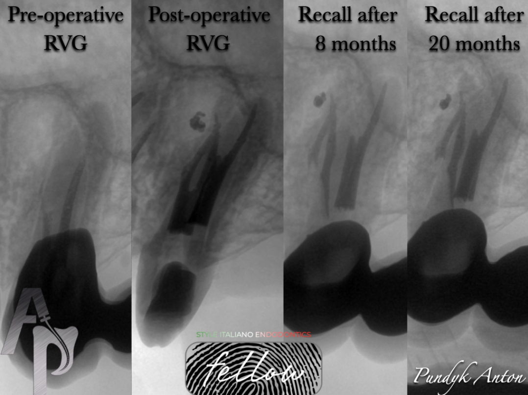
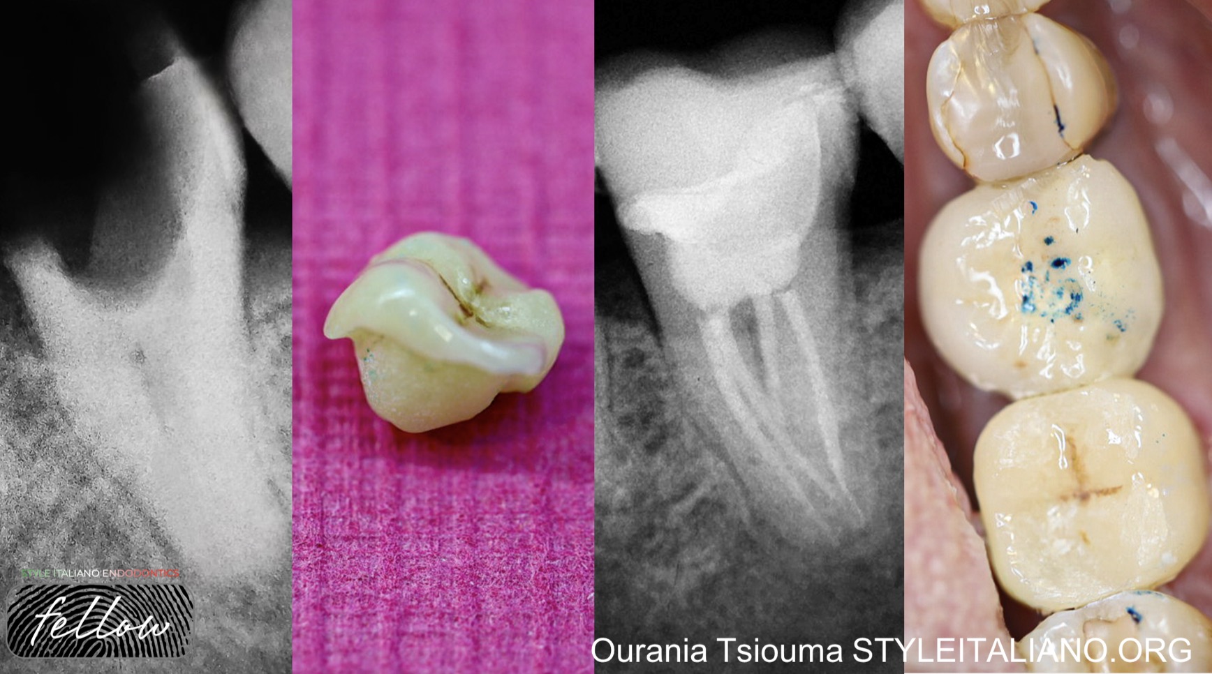
 Selective Root Canal Re-Treatement of Lower Left Pre-Molar
In retreatment cases , the clinician has to deal with many mishaps such as underfilled, missed and obstructed canals as also with iatrogenic damages as separated instruments and perforations. Underfilled […]
 Management of anatomy in a first tricky premolar (Deep Split)
The clinician must be aware of the possible anatomical variations of these teeth and their relationship to adjacent anatomical structures when planning and performing endodontic procedures.
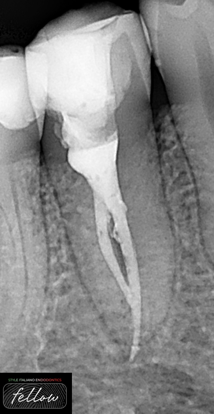 Retreatment case with two types of perforations
Root perforations were the second greatest cause of failure accounting for 9.62% of all unsuccessful cases. Seltzer et al. also attributed 3.52% of all endodontic failures to perforation. This perforation […]
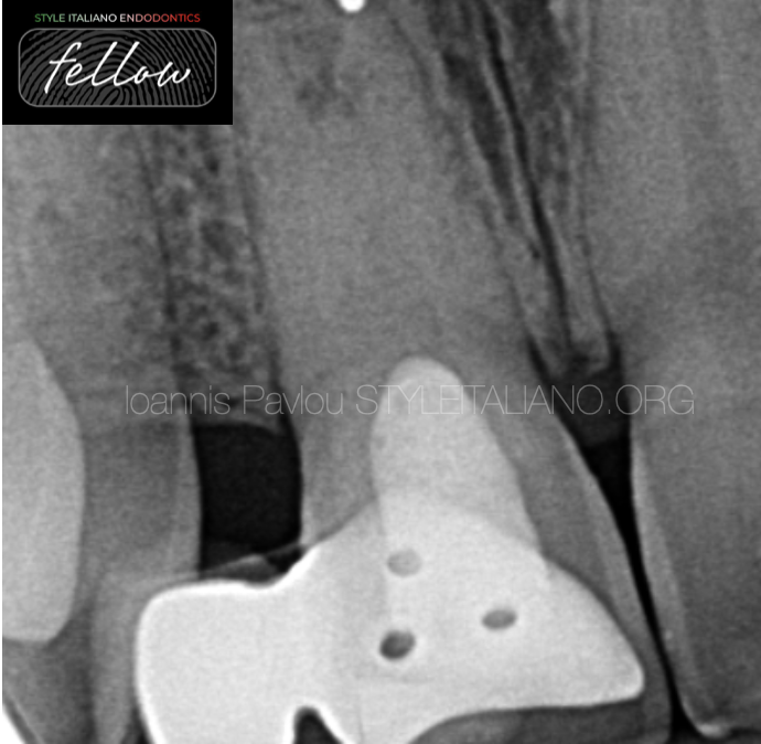
 Retreatment of teeth with a complex root system anatomy #1
Modern endodontic treatment is based on optimal cleaning,disinfection and high-quiality obturation of the root canal system.Anatomy is considered as a key factor in the healing process.That is why a clinician have to be aware of all normal,possible and abnormal variations in the teeth anatomy for achieving a maximum clinical success.In the article presents clinical example that demonstrate the complexity of endodontic retreatment of the maxillary left premolar
 Dental Trauma: Forever classic, Never boring
A 23 years old, female patient, was referred to my practice for an endodontic treatment on tooth 11. Patient complained of pain above the root and was concerned of the […]
 A story of a dilacerated premolar
Premolars are the most versatile teeth in regards of anatomy. One of the most challenging anatomical variations we have to face is dilaceration. Abrupt curvatures complicate the RCT especially when […]
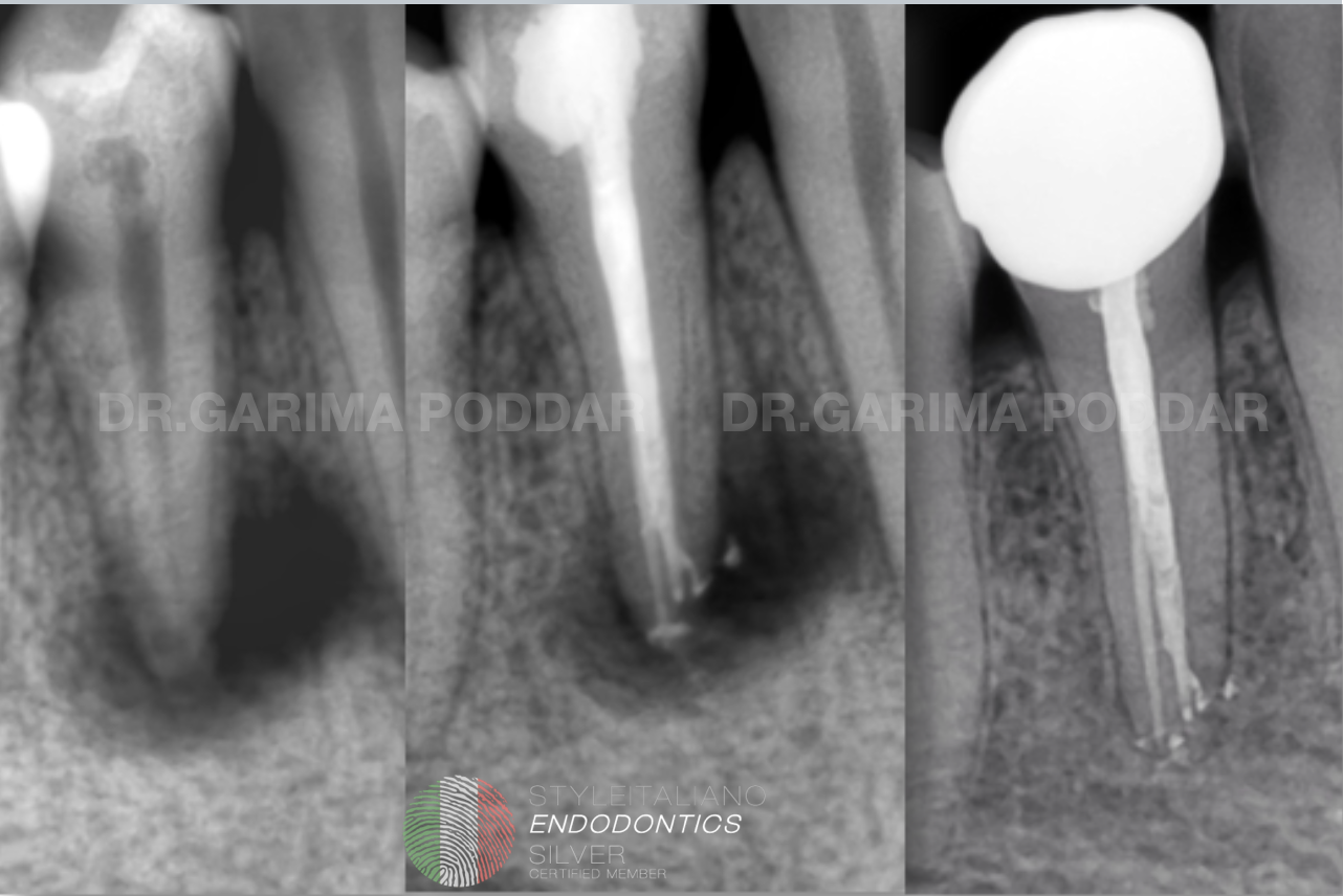 Mysterious pathology, fascinating anatomy & healing- a case of NSRCT of tooth 44
A periapical pathology, could be a result of caries, trauma, periodontally compromised oral cavity or even a thermal and chemical insult to the tooth. Effective cleaning, shaping and disinfection is the key to a successful endodontic treatment. A deep understanding of the anatomy of a root canal system, is of high value while performing a non-surgical root canal treatment. We often come across mandibular premolars with aberrant root canal anatomies. They often present with multiple canals, splits and accessory canals.
This article describes a case of NSRCT of a mandibular premolar with 1-2 configuration and it presented with a big peri-apical radiolucency.
 Management of Type II dens invaginatus
Dens invaginatus (DI) is one of the rare malformations of teeth which results from an infolding of the dental papilla during the development of teeth. This defect gives rise to a possible communication between the pulp and oral environment, thereby increasing the susceptibility to caries, pulpitis, and pulp necrosis.
 Zoning Technique in Endodontics
The Zoning Technique is an innovative approach in endodontics that aims to optimize root canal treatment by dividing the root canal system into distinct zones and applying specific instruments and techniques tailored […]
 A C-shaped story: endodontic management and endocrown rehabilitation
The C-shaped root canal configuration is an anatomical variation which is caused by the fusion of the mesial and distal roots either on the buccal or lingual root surface (1). […]