Management of Type II dens invaginatus
24/04/2024
José Conde Pais
Warning: Undefined variable $post in /home/styleendo/htdocs/styleitaliano-endodontics.org/wp-content/plugins/oxygen/component-framework/components/classes/code-block.class.php(133) : eval()'d code on line 2
Warning: Attempt to read property "ID" on null in /home/styleendo/htdocs/styleitaliano-endodontics.org/wp-content/plugins/oxygen/component-framework/components/classes/code-block.class.php(133) : eval()'d code on line 2
During the morphodifferentiation stage of development of teeth, various malformations can develop. Dens invaginatus (DI) is a rare malformation that results in deepening or invagination of the enamel organ into the dental papilla before the calcification of the dental hard tissue.It is frequently related to complicated crown and root canal morphology, proposing a challenge to the clinician in diagnosis as well as treatment. The reported occurrence ranges from 0.04% to 12%. .The teeth most affected are maxillary lateral incisors and bilateral occurrence is not uncommon and occurs in 43% of all cases (Grahnen et al. 1959).
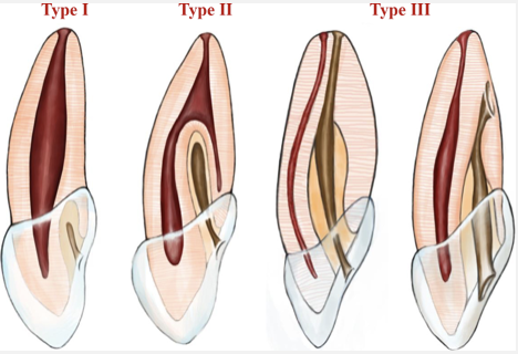
Fig. 1
The degree of malformation associated with DI was classified by Oehlers in 1957. According to Oehlers classification, Type I is characterized by enamel lined invagination, which remains confined to the coronal part of the tooth. Type II represents the extension of the invagination in the root extending beyond the cementoenamel junction, which ends in a blind sac. Accordingly, Type IIIa is described as invagination that extends throughout the root and communicates laterally with Periodontal Ligament (PDL) space, and Type IIIb represents invagination extending through the root and communicating the PDL space at the level of the apical foramen.
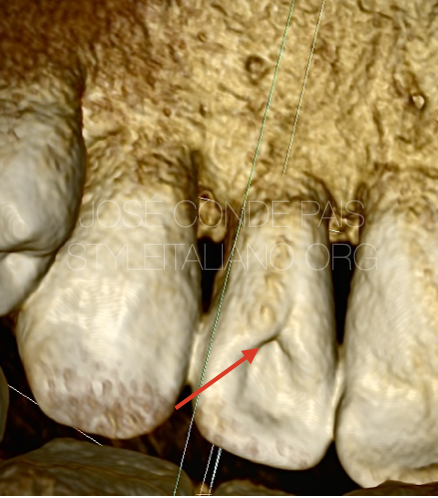
Fig. 2
A 35 years old male patient was referred from a general Practitioner to my clinic for management of 22 root canal treatment.
After a clinical and radiographic examination, we observe the following :
Tooth 22 presents Dens invaginatus type II in Oehlers classification, asymptomatic apical periodontitis and physiological probing.
We decide to perform a non-surgical approach with Mta apical plug under surgical microscope. The patient was informed of the pros and cons of the treatment and signed the informed consent.
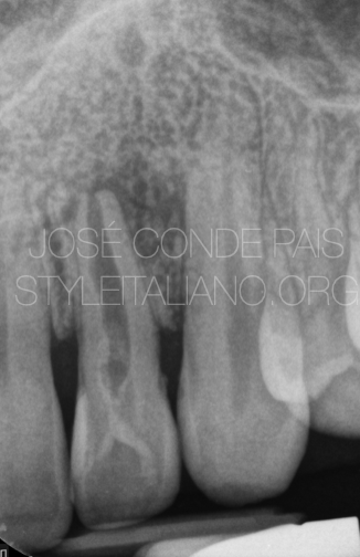
Fig. 3
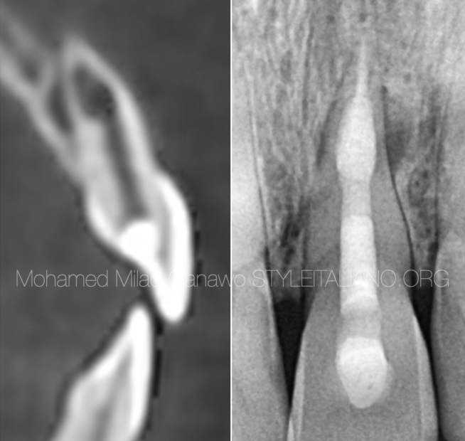
Preoperative X- Ray
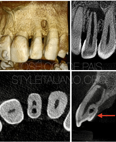
Fig. 4
CBCT Slides
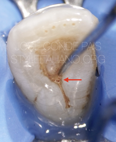
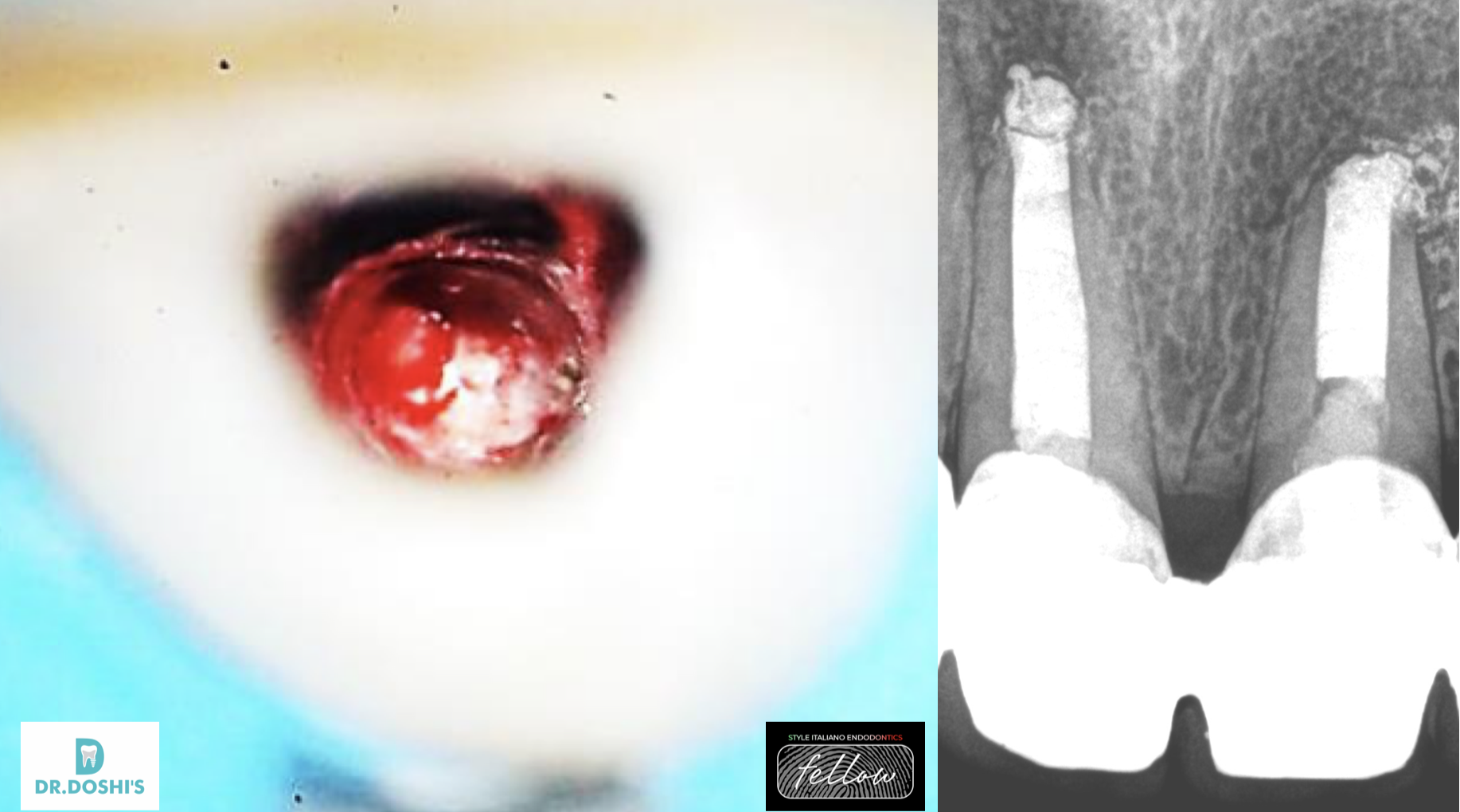
Fig. 5
Preoperative Clinical image, where we can observe the extraoral communication of the dens invaginatus
Fig. 6
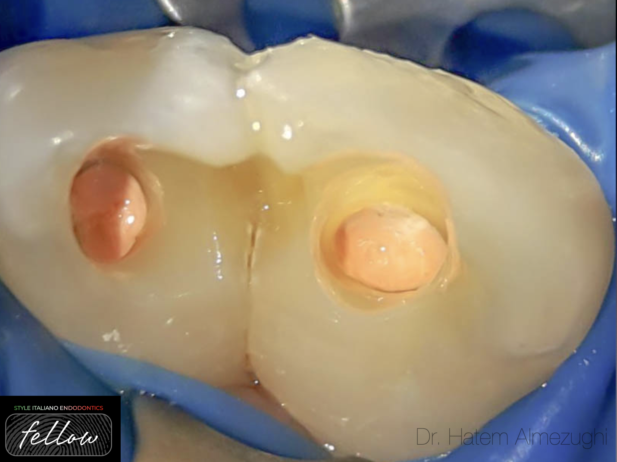
In this image we can see the dens in dente and the canal .
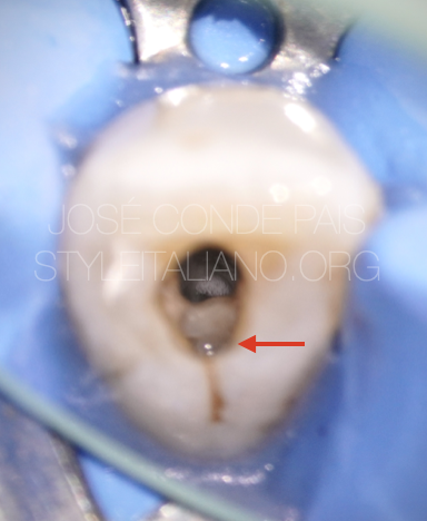
Fig. 7
Image after Chemomechanical Preparation where we can see 2 apical foramen.
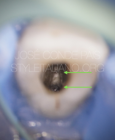
Fig. 8
Mta Apical plug

Fig. 9
Back Fill
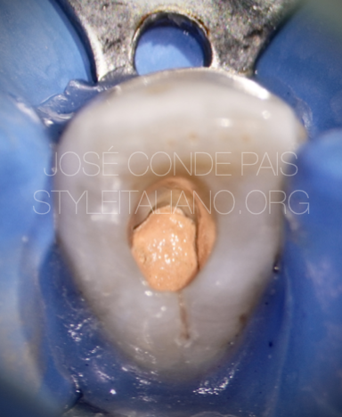
Fig. 10
Final X-Ray
Video of the procedure
Conclusions
Dens invaginatus is a dental malformation that with proper diagnosis, treatment and tools has a predictable prognosis.
Bibliography
1. Hülsmann M. Dens invaginatus: Aetiology, classification, prevalence, diagnosis, and treatment considerations. Int Endod J. 1997;30:79–90.
2. Akers HF, Henderson CM, Foley MA. Diagnosis and management of a maxillary lateral incisor exhibiting dens invaginatus and dens evaginatus. Aust Endod J. 2014;40:32–8.
3. Kirzioǧlu Z, Ceyhan D. The prevalence of anterior teeth with dens invaginatus in the western Mediterranean region of Turkey. Int Endod J. 2009;42:727–34.
4.OEHLERS FA (1957a) Dens invaginatus. I. Variations of the invagina- tion process and associated anterior crown forms. Oral Surgery, Oral Medicine and Oral Pathology 10, 1204–18.
5.OEHLERS FA (1957b) Dens invaginatus. II. Associated posterior crown forms and pathogenesis. Oral Surgery, Oral Medicine and Oral Pathology 10, 1302–16.
6.Ali A, Arslan H, Jethani B. Conservative management of Type II dens invaginatus with guided endodontic approach: A case series. J Conserv Dent. 2019 Sep-Oct;22(5):503-508