Management of internal root resorption in a maxillary central incisor
05/08/2021
Mohamed Milad Ganawo
Warning: Undefined variable $post in /home/styleendo/htdocs/styleitaliano-endodontics.org/wp-content/plugins/oxygen/component-framework/components/classes/code-block.class.php(133) : eval()'d code on line 2
Warning: Attempt to read property "ID" on null in /home/styleendo/htdocs/styleitaliano-endodontics.org/wp-content/plugins/oxygen/component-framework/components/classes/code-block.class.php(133) : eval()'d code on line 2
Root resorption is a particular physiologic or a pathologic pulpal disease associated with loss of dentin, cementum, and/or bone. This resorption may occur following different types of injuries, including chemical, mechanical or thermal injury. Furthermore, it can be cla classified as internal or external root resorption.
Internal root resorption (IRR), is a rare condition that commonly referred to as intracanal resorption, resulting in dystrophy of the pulp with destruction of the hard tissues. Majority of the cases remain asymptomatic and are often incidentally detected during radiographic evaluation. Once detected, it should be treated as soon as possible to limit its progression. Moreover, the use of Cone-Beam Computed Tomography(CBCT) provides a 3-dimensional appreciation of the resorption lesion with axial, coronal, parasagittal views of the anatomy that provide cross-sectional views which clearly determine the size and the location of the resorption with high sensitivity and an excellent specificity. This is a case of internal resorption started with pulp and led to loss of dentin and may be cementum. In such cases I believe that role of CBCT is CRUCIAL to visualize missing information in 2D x-ray
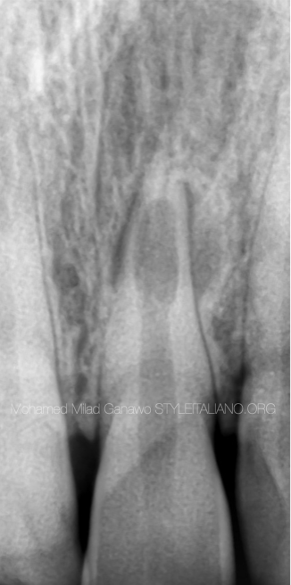
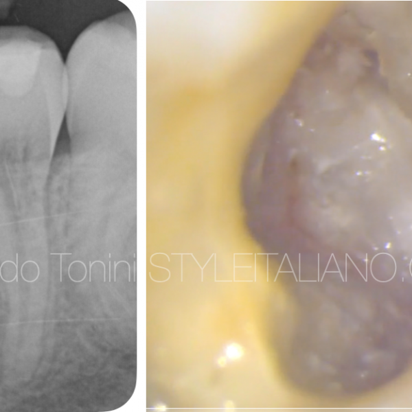
Fig. 1
A 25-year-old female patient came to the dental office with pain related to upper anteriors,history was taken and it turned out that she had trauma 5 years ago.
Initial radiograph was taken then CBCT was requested .
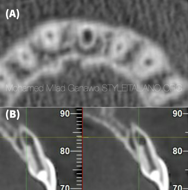
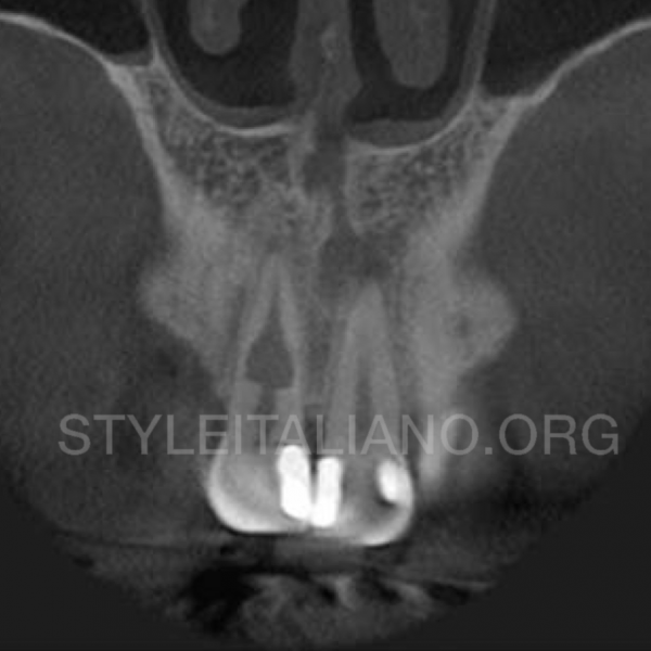
Fig. 2
A) CBCT image showing axial section view of thooth 21 with an internal resorptive defect.
B) CBCT image showing sagittal view of tooth 21 with an internal resorptive defect.
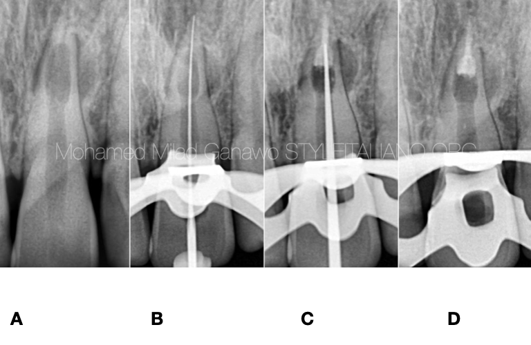
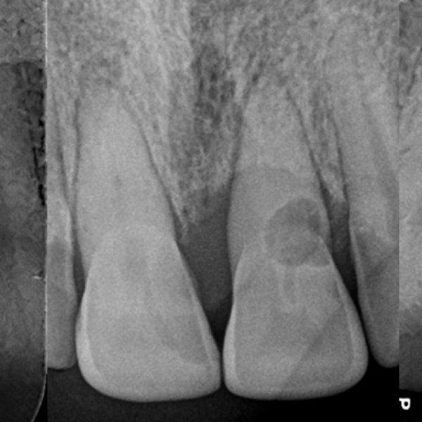
Fig. 3
This figure shows some steps of therapy
A) initial radiograph revealed internal root resorption at apical part
B) taking working length and confirm it
C) MTA apical plug also you can notice manual plugger
D) shows MTA plug done .
MTA = mineral trioxide aggregate
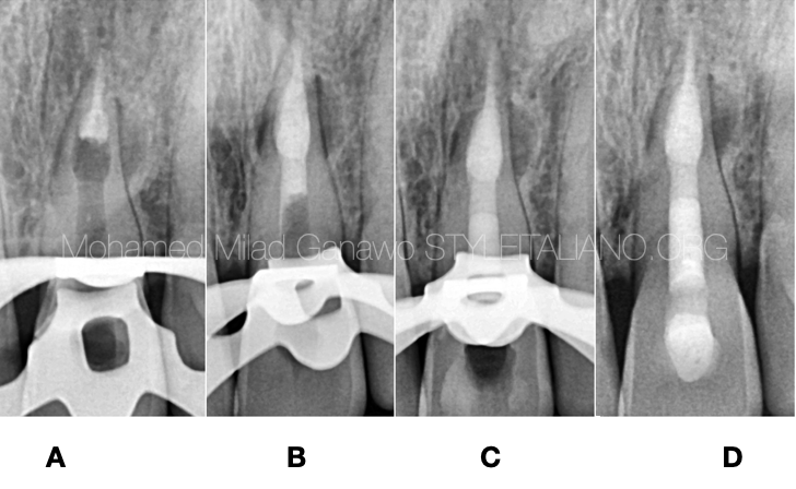
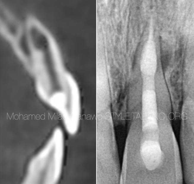
Fig. 4
This figure shows the rest of therapy steps:
A and B: MTA plug
C: back filling of gutta-percha
D: final radiograph including permanent restoration.
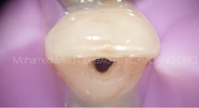
Fig. 5
intra oral picture shows complete isolation followed by secondary isolation & access cavity
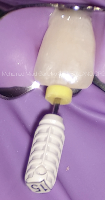
Fig. 6
step of introducing 15K file to size up the situation and working length .
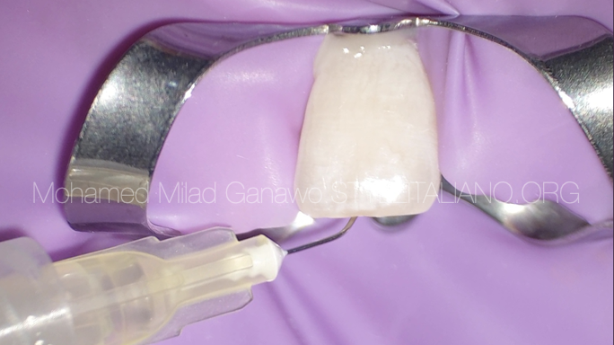
Fig. 7
Intra oral photo shows chemical debridement with sodium hypochlorite solution in the canal and the resorption lacuna
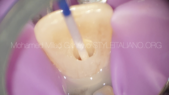
Fig. 8
The root canal has been dried with sterile paper points
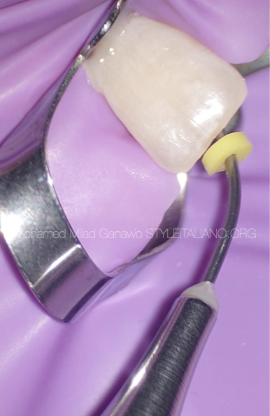
Fig. 9
The MTA is placed with a dedicated carrier
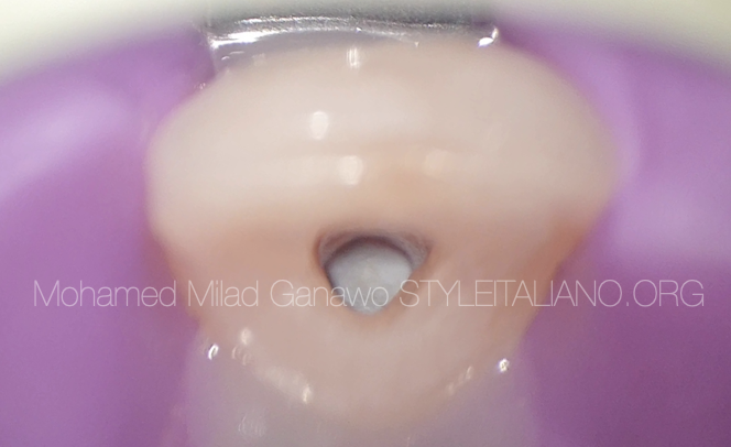
Fig. 10
The microscopic picture shows the first step of apical obturation with MTA
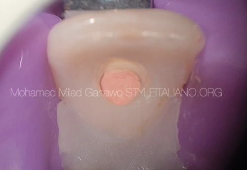
Fig. 11
Subsequently the root canal is filled with warm gutta percha
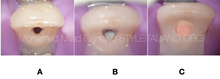
Fig. 12
The intra oral pictures show
A: access cavity
B: MTA plug
C: GP filling
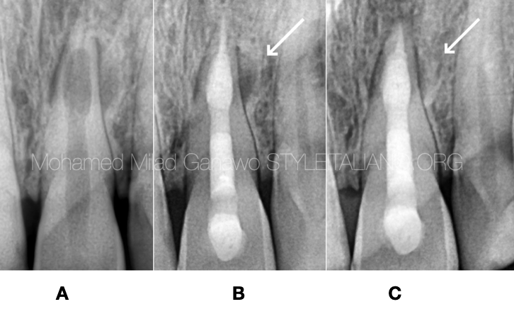
Fig. 13
A: Pre-operative radiograph
B: Post-operative radiograph
C: seven-months follow-up showing healing
Conclusions
Internal inflammatory root resorption is a particular pulpal disease, which can be diagnosed by clinical and radiographic examinations. Nowadays, the diagnosis of internal root resorption is significantly improved by the three-dimensional imaging as CBCT that provides superior diagnostic accuracy resulted in an improved management of the resorptive defects and a better outcome of conservative therapy.
Bibliography
Ne RF, Witherspoon DE, Gutmann JL: Tooth resorption. Quintessence Int. 1999, 30:9-25.
Fuss Z, Tsesis I, Lin S: Root resorption- diagnosis, classification and treatment choices based on stimulation factors. Dent Traumatol. 2003, 19:175-182.
C.Estrela,M.R.Bueno,A.H.G.deAlencaretal.,“Method to evaluate inflammatory root resorption by using cone beam computed tomography,” Journal of Endodontics,vol.35,no.11, pp.1491–1497,2009.
Patel S, Ricucci D, Durak C, Tay F. Internal root resorption: a review. J Endod. 2010 Jul;36(7):1107-21.
Nilsson E, Bonte E, Bayet F, Lasfargues JJ. Management of internal root resorption on permanent teeth. Int J Dent. 2013;2013:929486.
Mehra N, Yadav M, Kaushik M, Roshni R. Clinical Management of Root Resorption: A Report of Three Cases. Cureus. 2018