
Gemination or fusion
07/10/2023
Fellow
Warning: Undefined variable $post in /home/styleendo/htdocs/styleitaliano-endodontics.org/wp-content/plugins/oxygen/component-framework/components/classes/code-block.class.php(133) : eval()'d code on line 2
Warning: Attempt to read property "ID" on null in /home/styleendo/htdocs/styleitaliano-endodontics.org/wp-content/plugins/oxygen/component-framework/components/classes/code-block.class.php(133) : eval()'d code on line 2
Dental anomaly is a condition in which one or more teeth deviate from the normal in form, function, or position.
And it includes abnormalities in teeth number, size, shape and structure
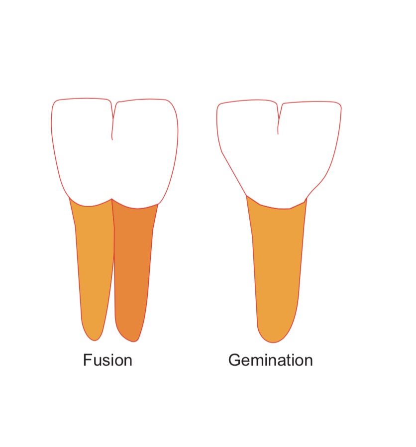
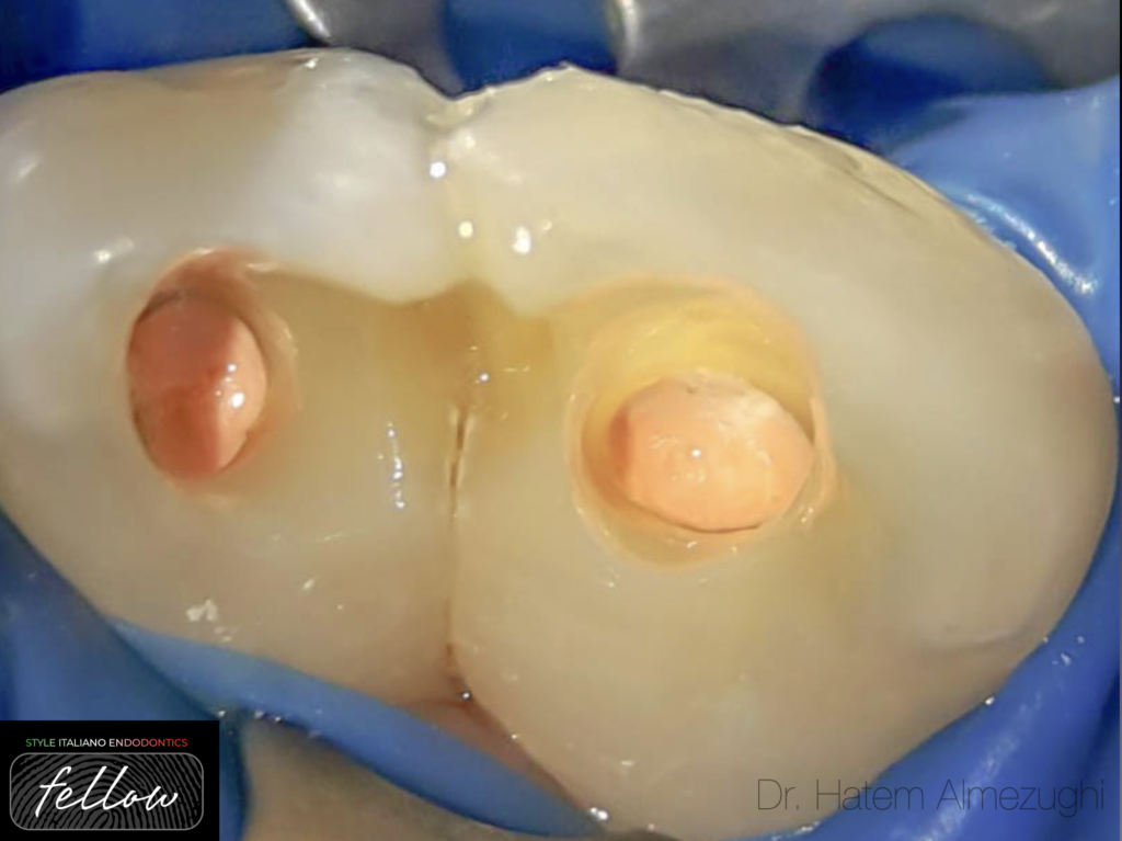
Fig. 1
FUSION
Union of two normally separated teeth germs, may be complete or incomplete.
Fusion can also be the union of a normal tooth bud to a supernumerary tooth germ.
The number of teeth is fewer if the anomalous tooth is counted as one tooth
Fusion can occur in deciduous or permanent teeth, its more common in anterior teeth depending on the stage of odontogenesis.
GEMINATION
Is a dental phenomenon that appears to be two teeth developed from one.
there's is one main crown with a cleft in it that, within the incisal third of the crown, the number of the teeth in the arch will be normal.
In the case of fusion there are usually two separate root canals, whereas in gemination there is usually one large common root canal
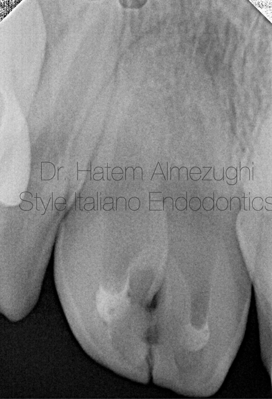
Fig. 2
This figure shows inter proximal caries between two fused teeth as pointed with one sided green arrow, and the diameter of orifices as shown in orange rectangle , diameter of canal coronally as shown in green, and apically there's radiolucency
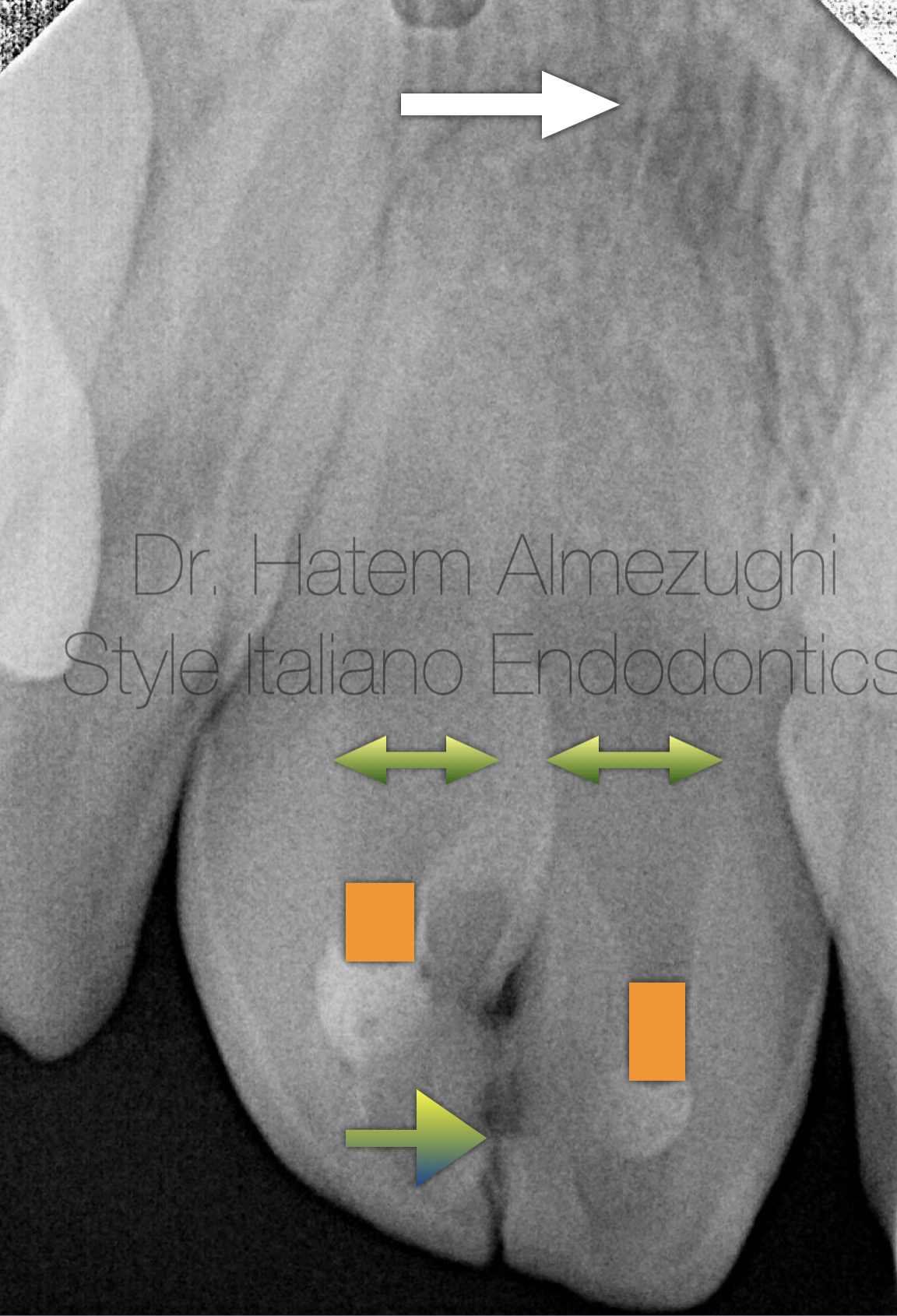
Fig. 3
TREATMENT
- multidisciplinary approach
- endodontic : root canal therapy,
- surgical : divide the tooth into two teeth
- orthodontic treatment : reshape and restore teeth with appropriate material
The image show the axial view of the CBCT
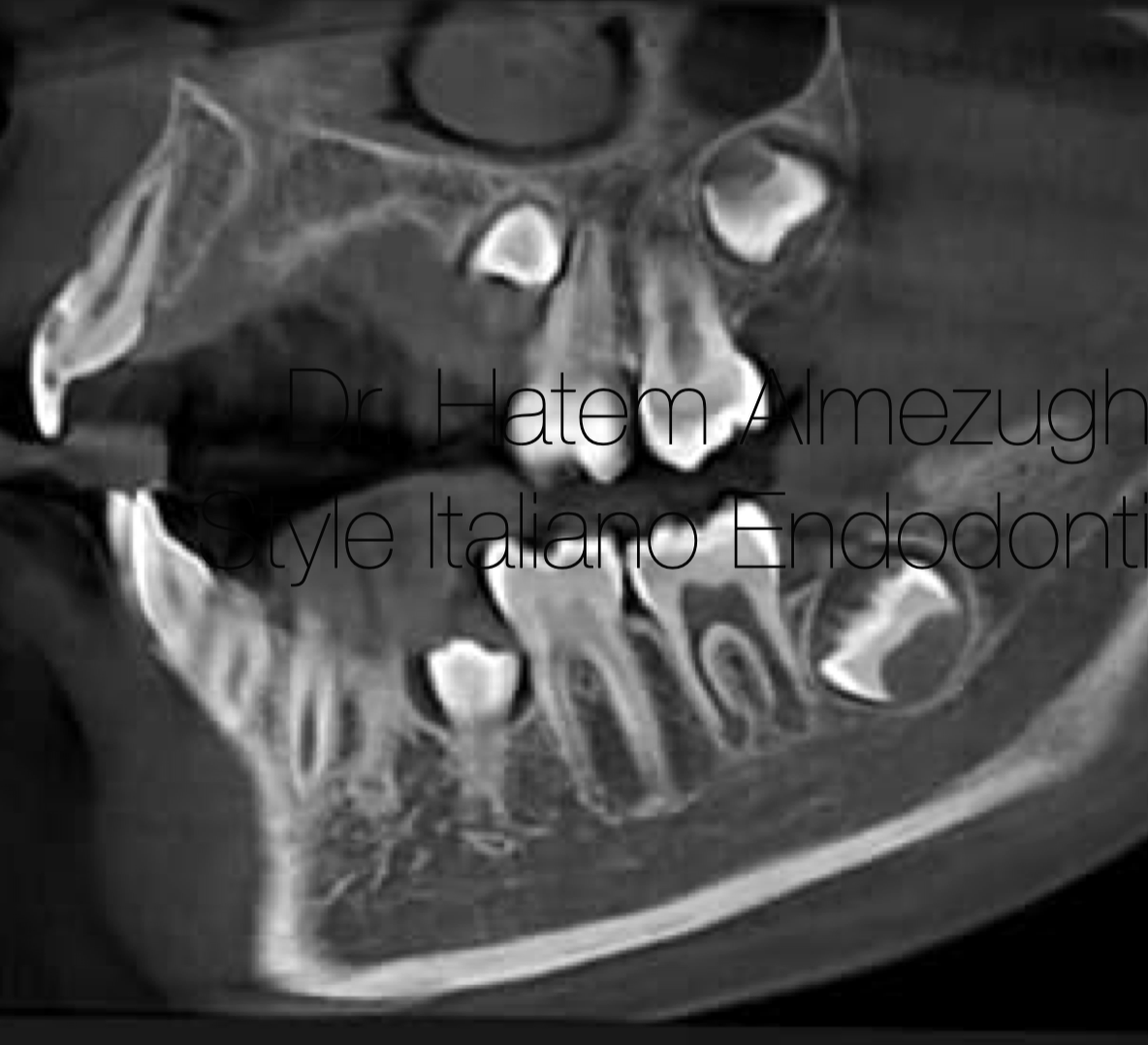
Fig. 4
Sagital View
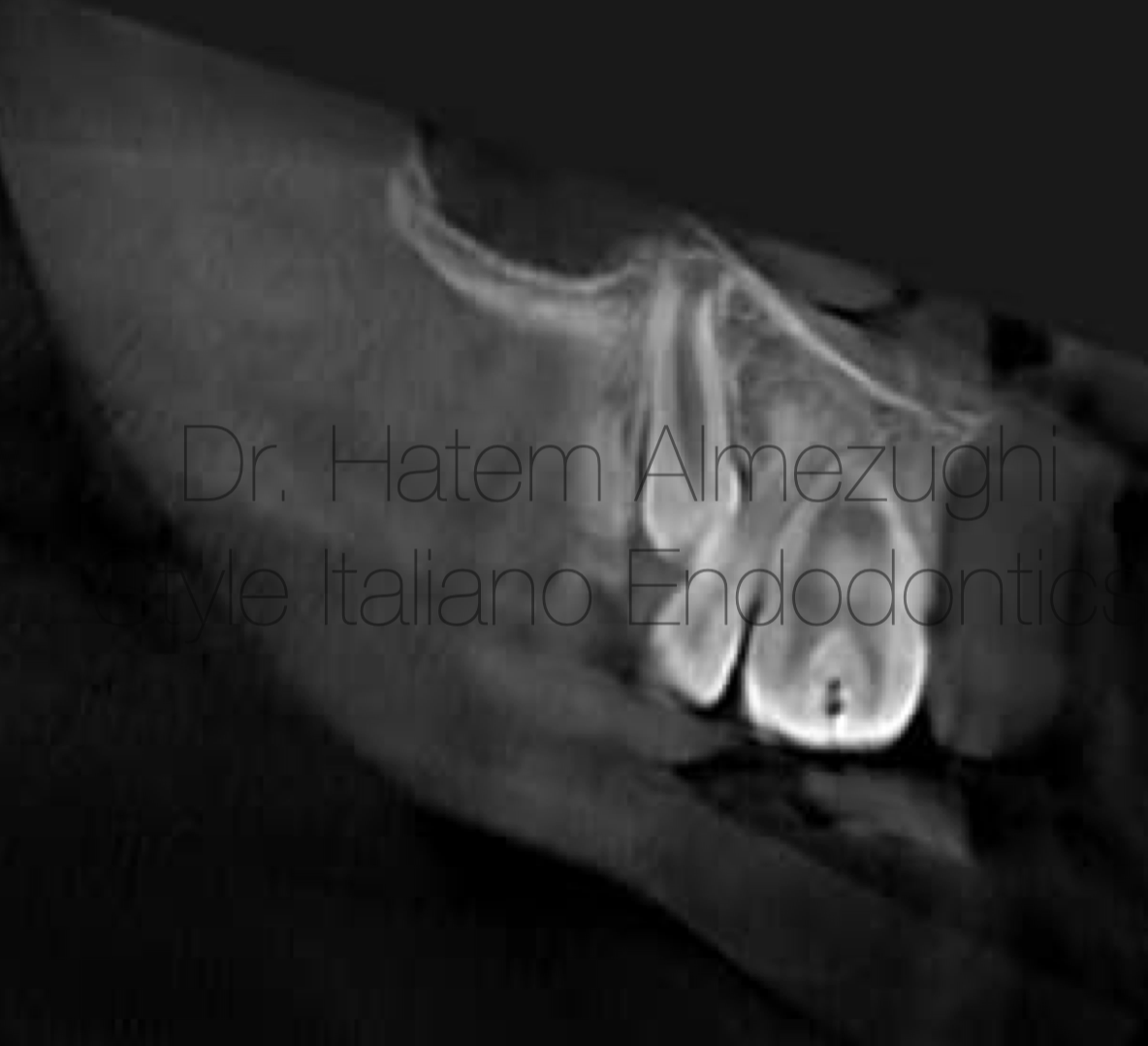
Fig. 5
Coronal View
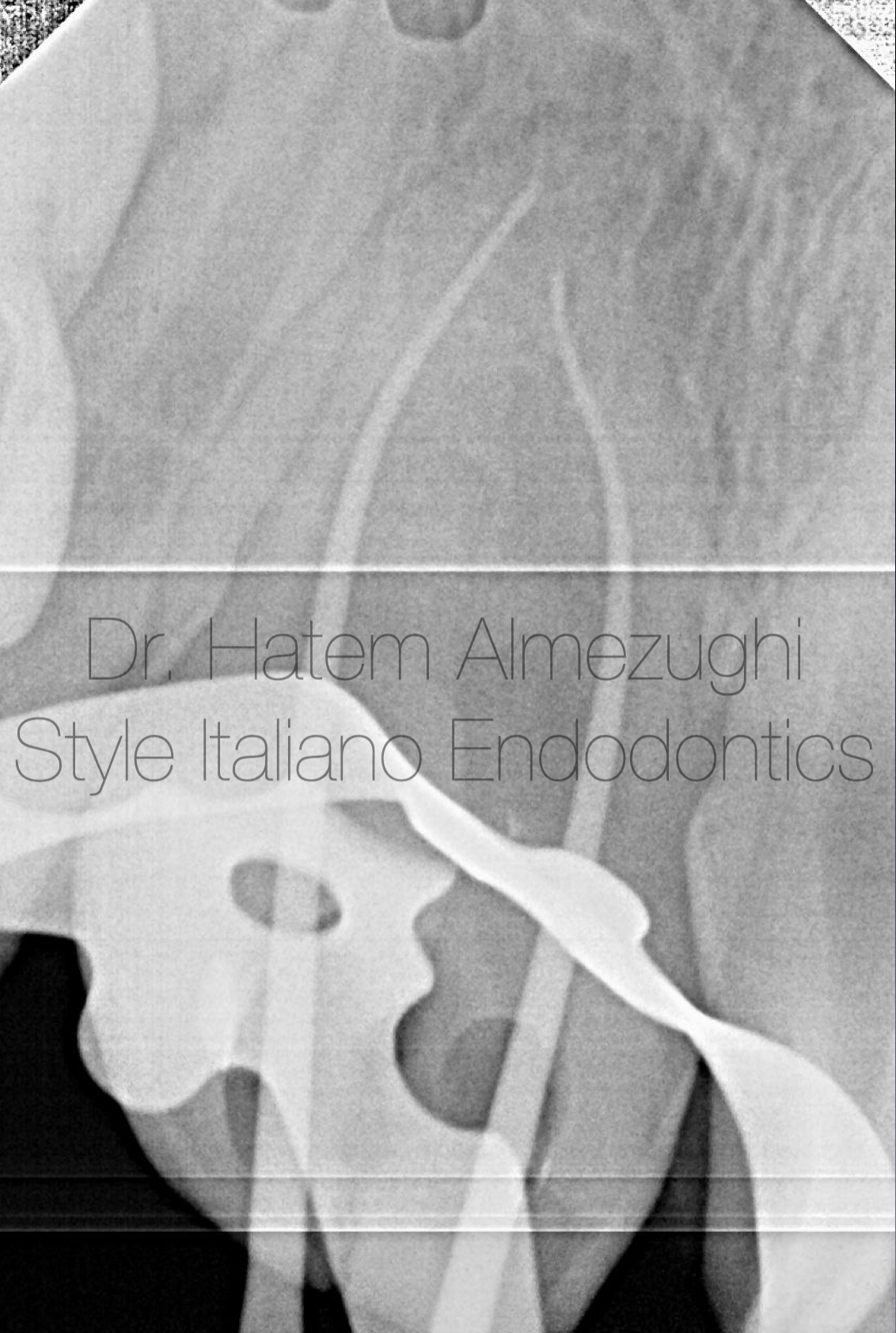
Fig. 6
This figure show cone fit after taking working length with eal which gave reading 25mm for right central and 24mm for left central
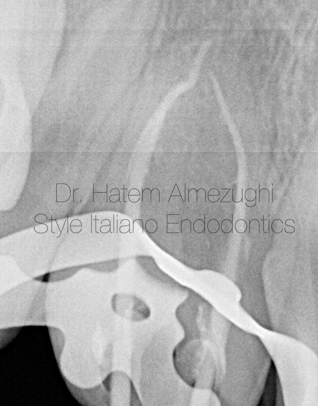
Fig. 7
Coating gps with sealer and sizing up the situation before obturation
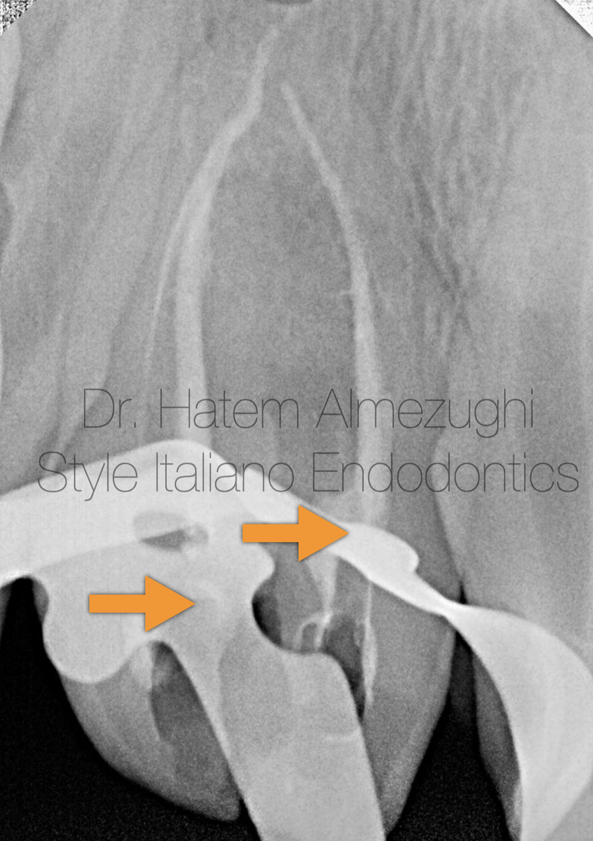
Fig. 8
Searing off gp to the orifice level or a little bit beyond as a first step to visualise the scene to go for next phase.
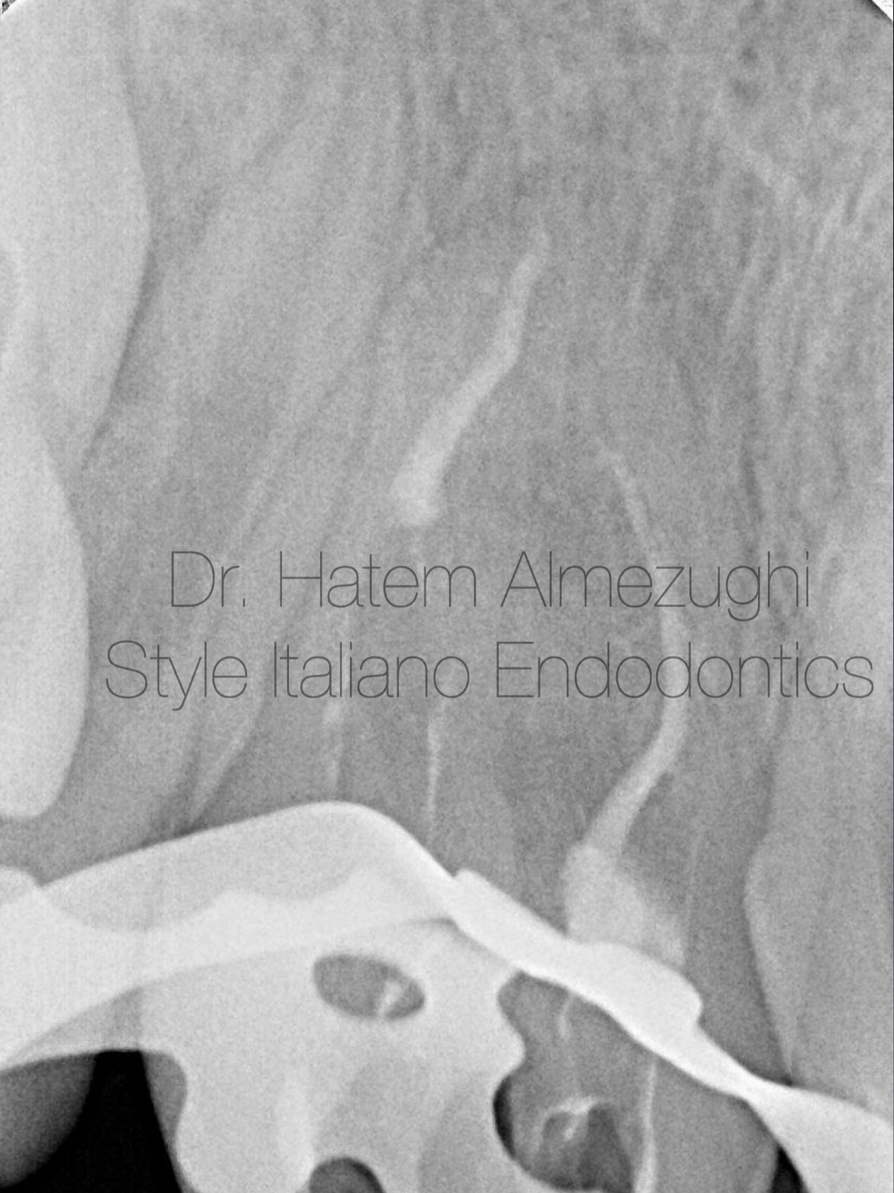
Fig. 9
Now after down packing there`s one more to go up to guarantee no voids left , to get ready for squirting
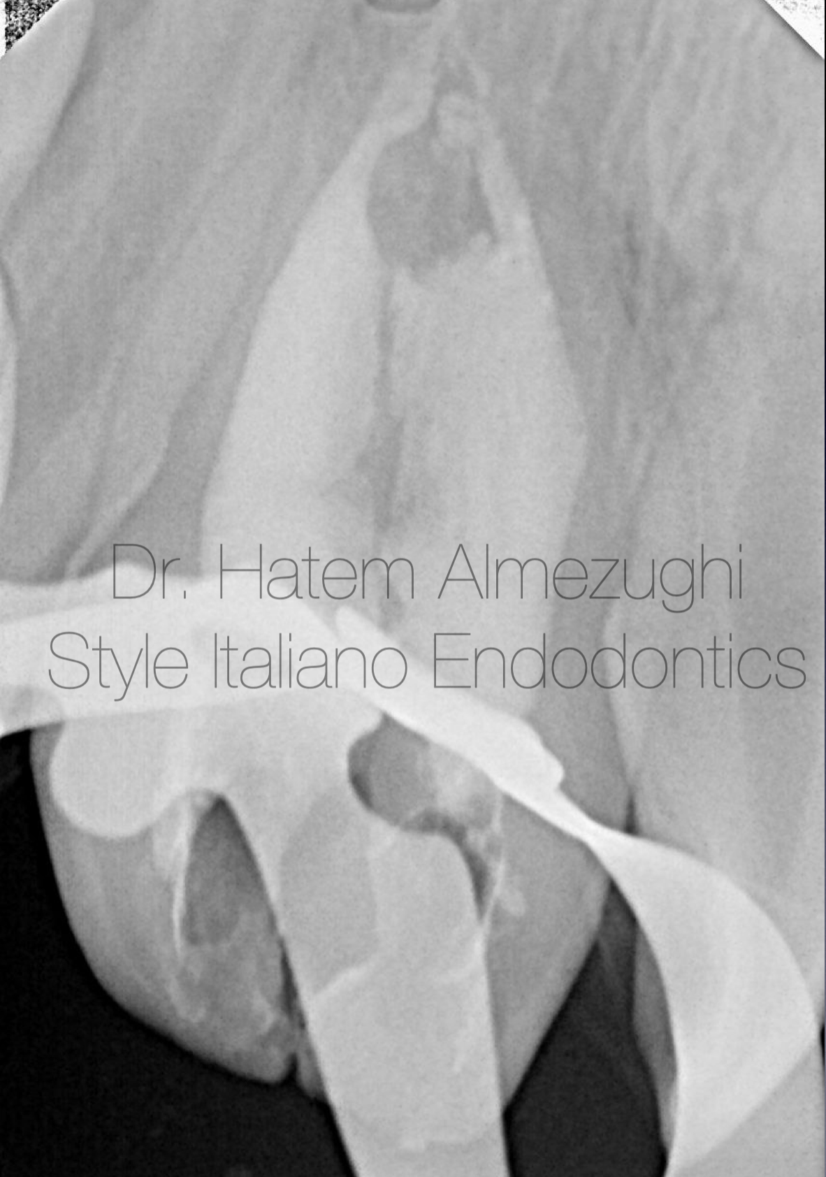
Fig. 10
Squirting done as a last phase of obturation , now tooth ready for permanent core.
now clealy it’s a “fusion” case after noticing the union of two teeth with two separate root canals
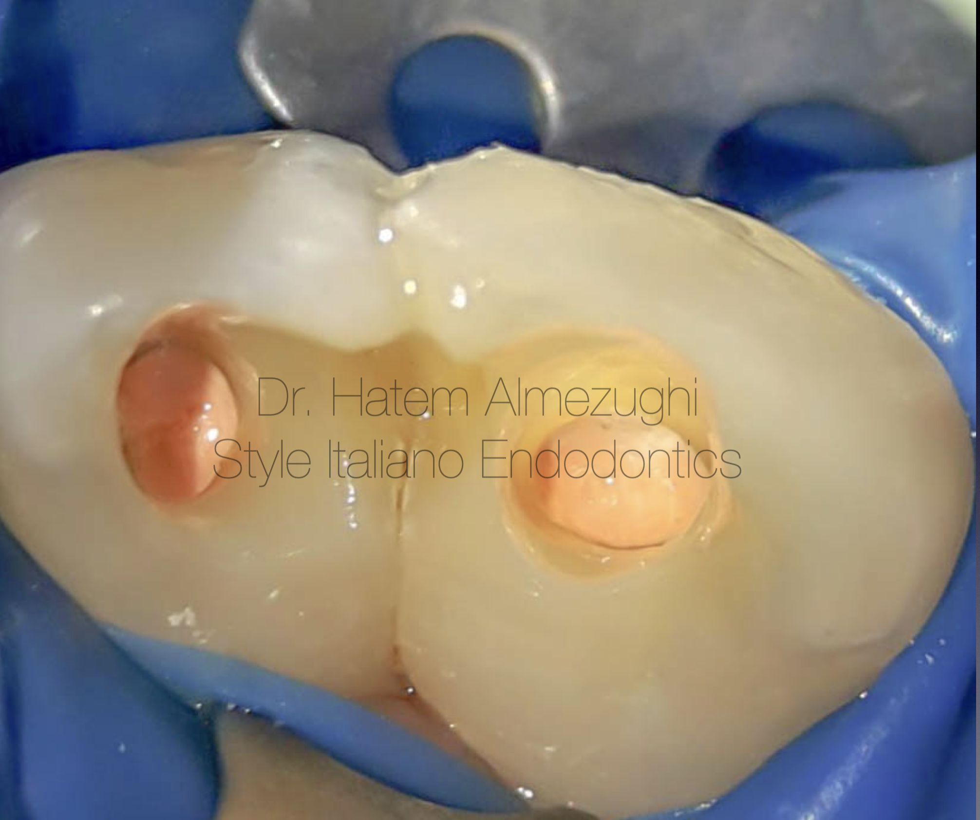
Fig. 11
Intra oral palatal view
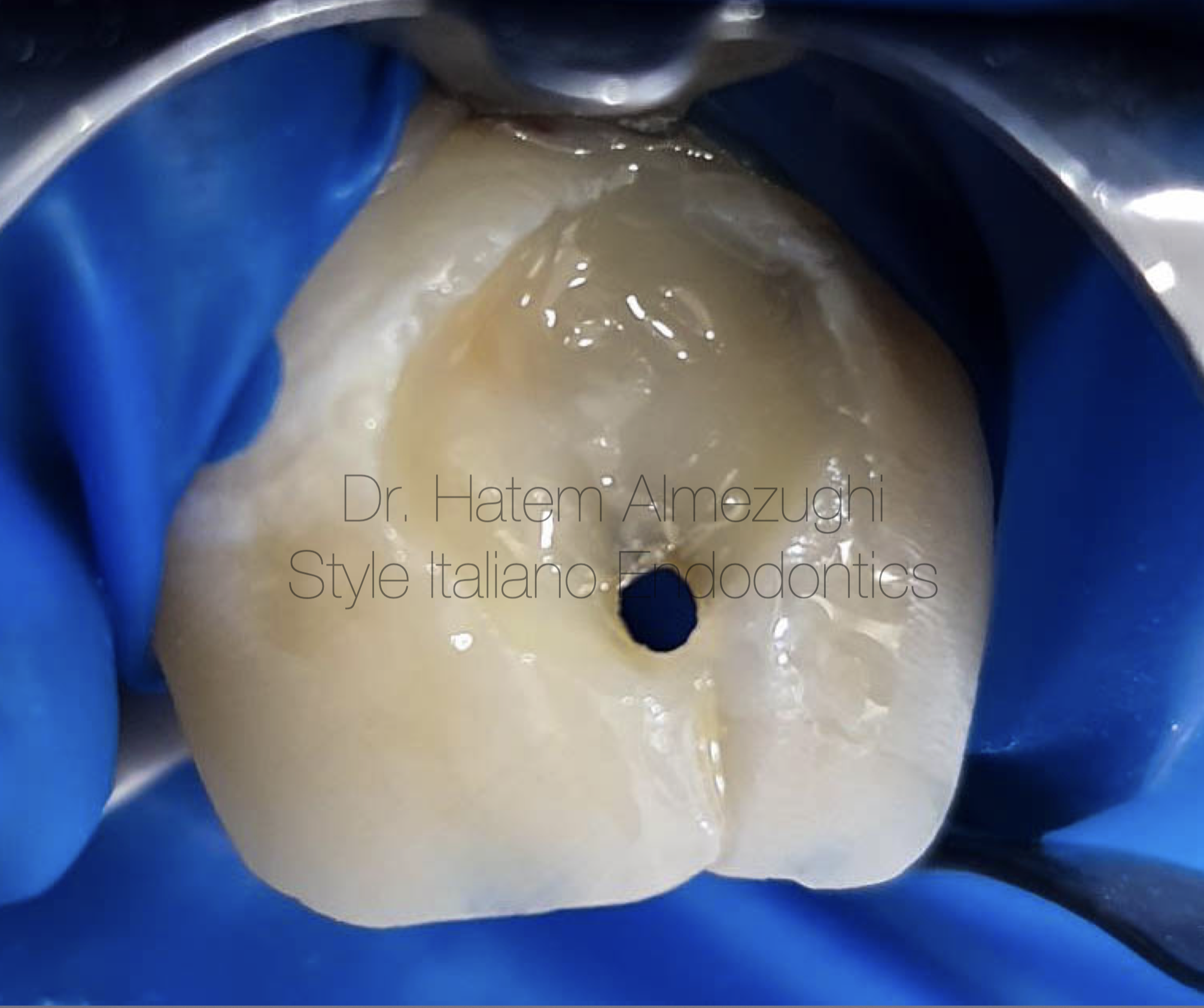
Fig. 12
Intra oral labial view
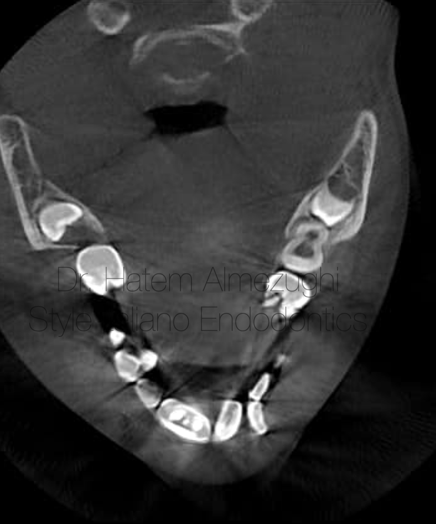
Fig. 13
Post operative axial section
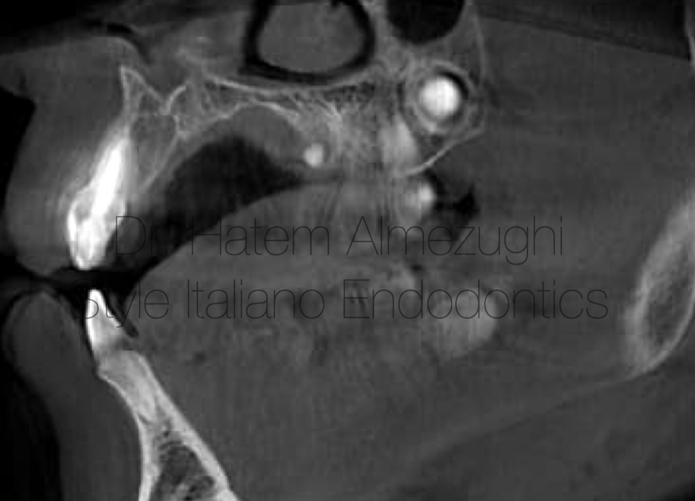
Fig. 14
Post operative saggital section
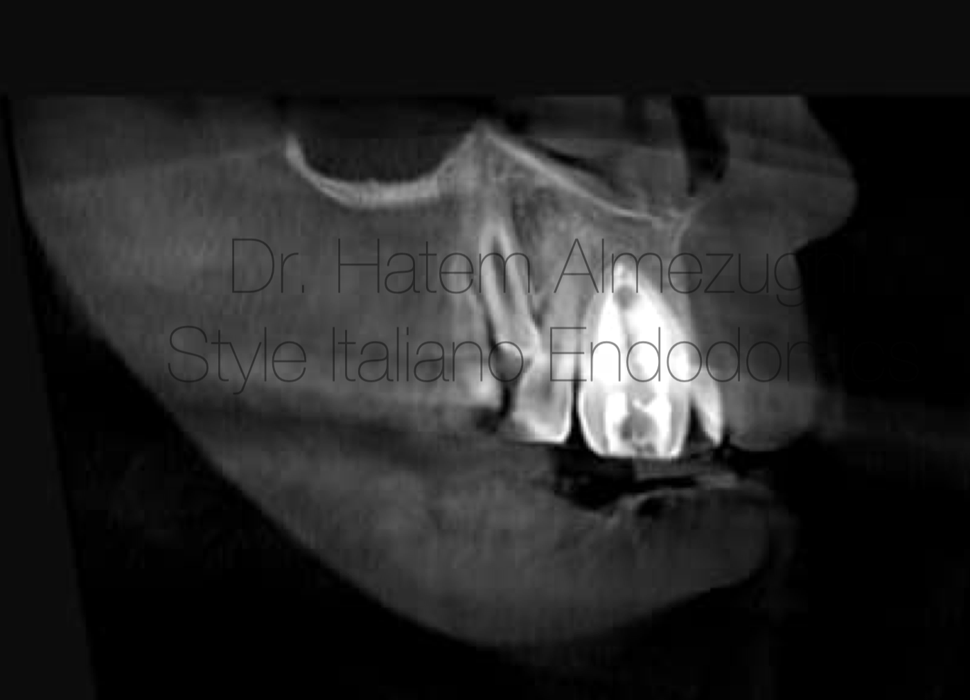
Fig. 15
Post operative coronal section

Fig. 16
Conclusions
Diagnosis and management of teeth anamolies has always been challenging for the clinicians. careful examination by clinical and radiographic methods provides early diagnosis and intervention.
Bibliography
M. O. Folayan, M. Alade, A. Adeniyi, M. El Tantawi, and T. L. Finlayson, “Association between developmental dental anomalies, early childhood caries and oral hygiene status of 3-5-year-old children in Ile-Ife, Nigeria,” BMC Oral Health, vol. 20, no. 1, 2020
R. S. Cunha, A. Junaid, and I. Mello, “Unilateral fusion of a supernumerary tooth to a maxillary permanent lateral incisor: a report of a rare case,” Journal of Endodontics, vol. 41, no. 3, pp. 420–423, 2015.
N. Tewari and R. K. Pandey, “Bilateral fusion in primary mandibular teeth: a report of two cases,” Journal of Indian Society of Pedodontics and Preventive Dentistry, vol. 29, no. 1, pp. 50–52, 2011.
H. Açıkel, S. İbiş, and E. Şen Tunç, “Primary fused teeth and findings in permanent dentition,” Medical Principles and Practice, vol. 27, no. 2, pp. 129–132, 2018.
