How To deal with Splits " Different Scenarios "
03/07/2023
Fellow
Warning: Undefined variable $post in /home/styleendo/htdocs/styleitaliano-endodontics.org/wp-content/plugins/oxygen/component-framework/components/classes/code-block.class.php(133) : eval()'d code on line 2
Warning: Attempt to read property "ID" on null in /home/styleendo/htdocs/styleitaliano-endodontics.org/wp-content/plugins/oxygen/component-framework/components/classes/code-block.class.php(133) : eval()'d code on line 2
Failure to locate and disinfect all the root canals may be a major reason for endodontic failure. The first Step to reach the success of root canal therapy is to have a good knowledge of the root canal morphology in order to get a successful Root Canal Treatment Slowey has suggested that mandibular first premolars , often called as " Endodontist's Enigma " may present the greatest difficulty of all teeth to perform a successful endodontic treatment due to their unpredictable anatomy with a wide variety of morphological rarities - - A recently published systematic literature review by Kottoor et al. summarized that “ lower bicuspids commonly have a single root (97%), with the mandibular first premolars being twice more likely to present with two canals (23.55%) than second premolars (12.64%)The aim of this article is to report diagnosis and endodontic treatment of a mandibular premolars with two or three canals.
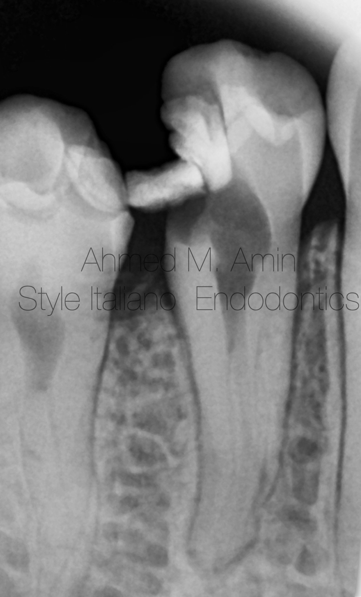
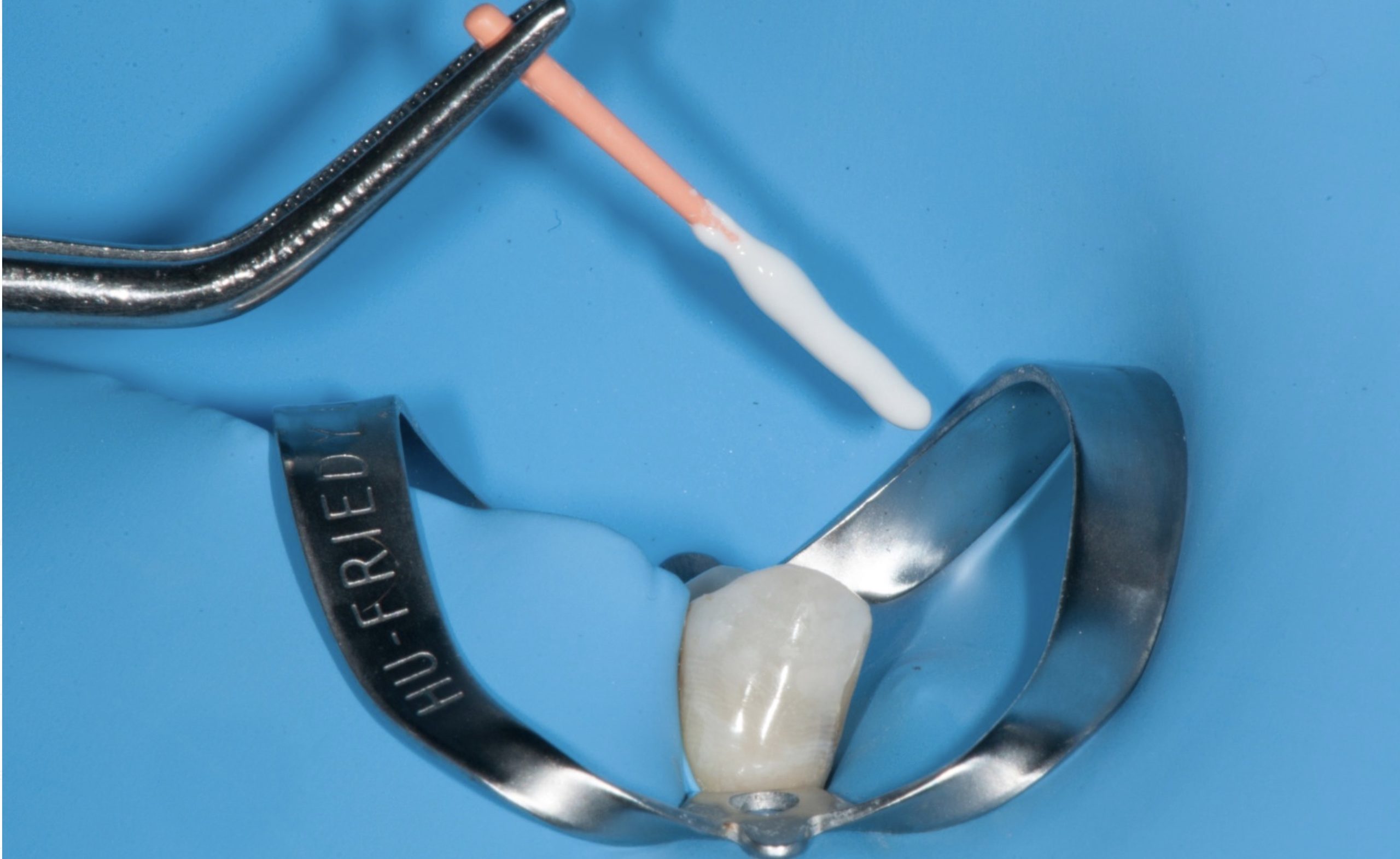
Fig. 1
A young female patient came to my clinic complaining from a severe pain related to lower right area , she had pain for about 1 month , the previous dentist suggested that there was a calcification, and he was facing a blockage , then she was addressed to my clinic The Pre-Operative Xray Shows an enlargement of the periodontal ligament Space and disappearing of the main canal in the mid root that confirming a Split related to tooth No. 44
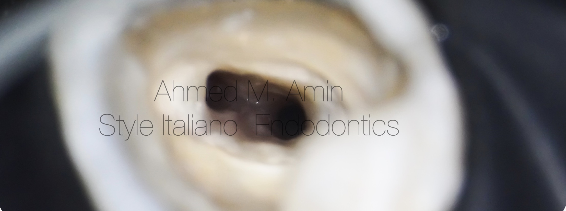
Fig. 2
Access Cavity after Pre-Endo buildup & Refinement
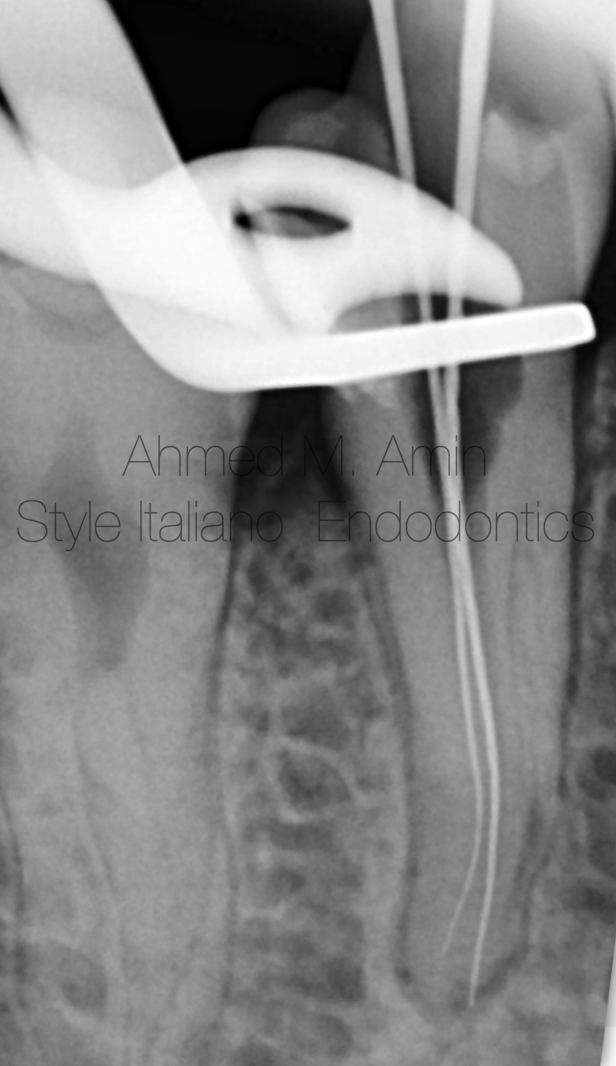
Fig. 3
Scouting of the main canal " Lingual " was easy but the difficulty was in how to catch and scout the buccal canal so I made some refinement of the access cavity in Bucco-lingual direction then i pre-curved the smallest St. St. manual file " 8k " and made the file sliding up & down alongside the lingual wall without rotation to avoid any changes in the tip direction and made its pre-curved tip toward the buccal wall until I could get a catch
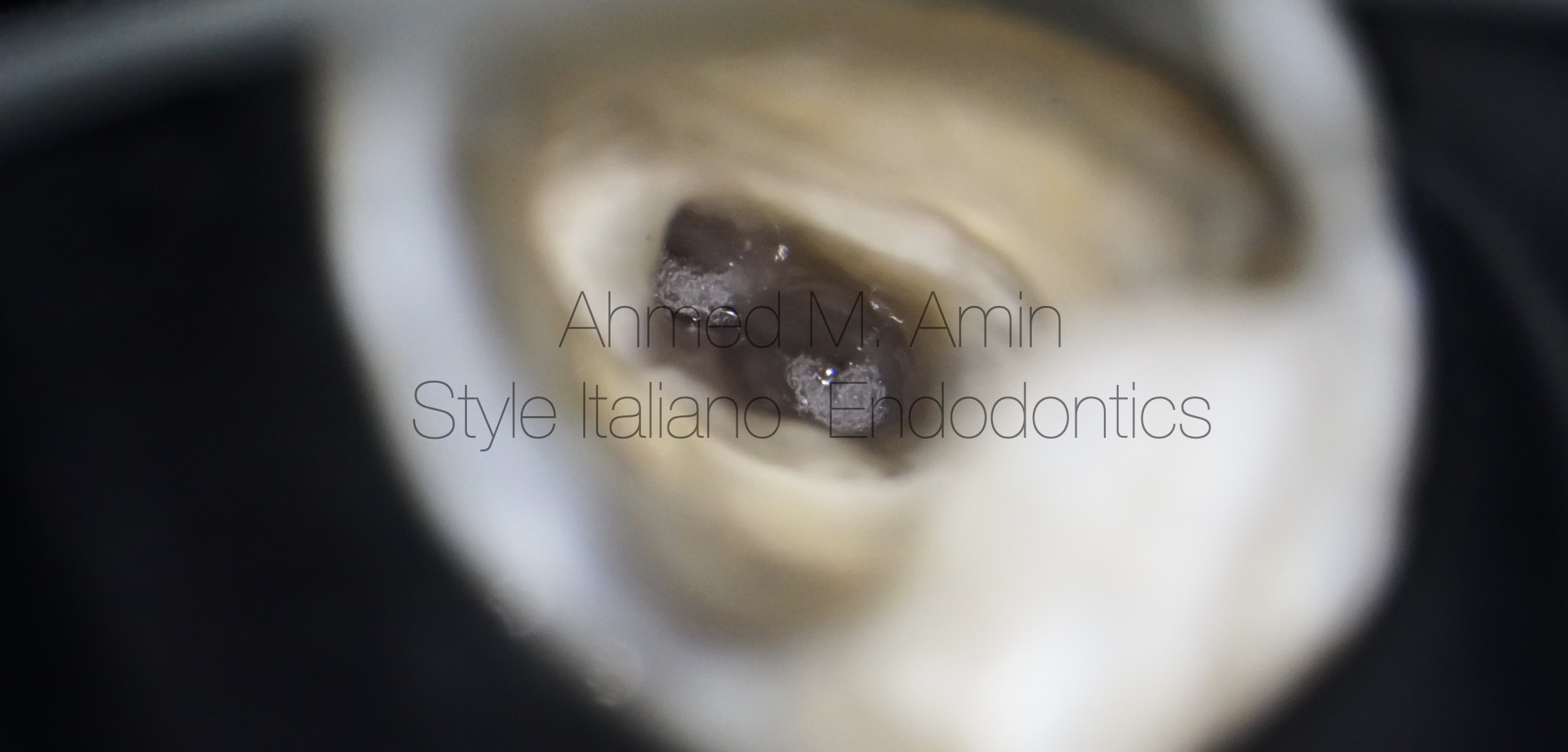
Fig. 4
This Pic shows the effervescence action of irrigation while heating
Irrigation Protocol was 5.25 % Naocl , 17 % EDTA Solution and Alcohol heated by FastPack & Activated by Ultrasonic " Ultra X device "
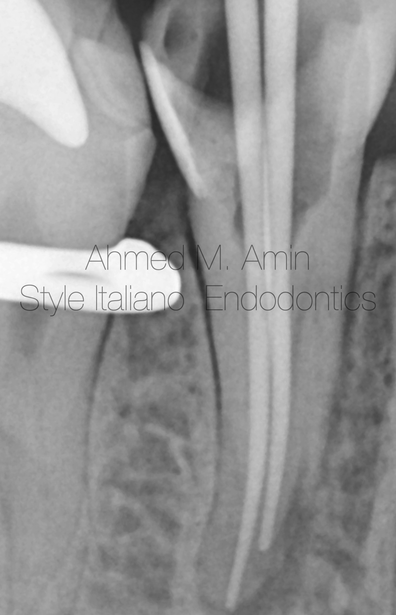
Fig. 5
-I shaped the root canals up to 30.04 with Controlled memory files
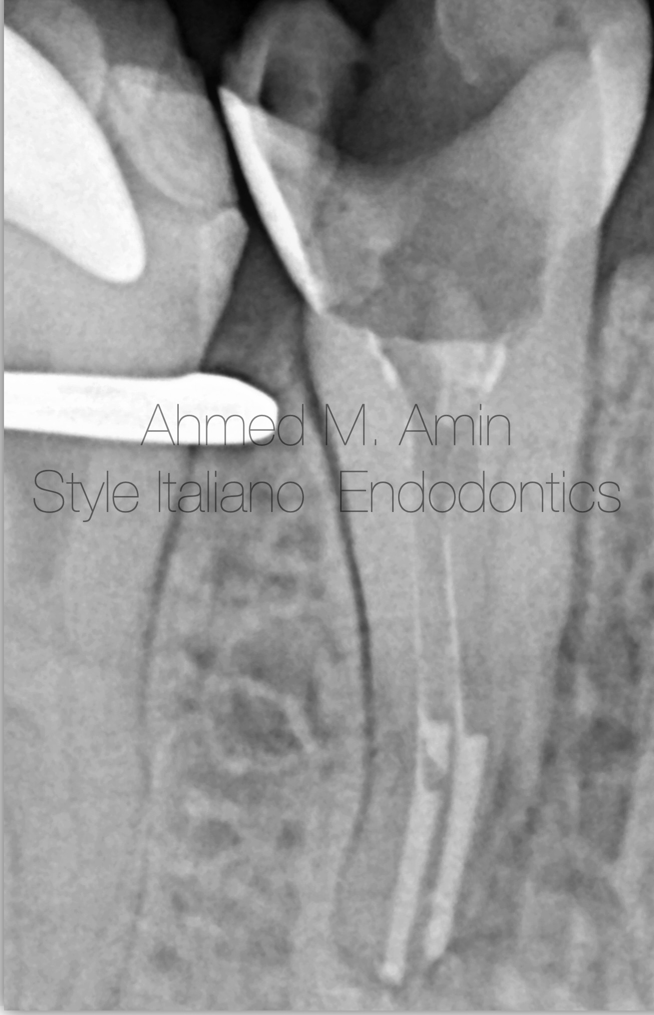
Fig. 6
I cut the gutta-percha 4-5 mm apical from the apex by down packing technique using FastPack device
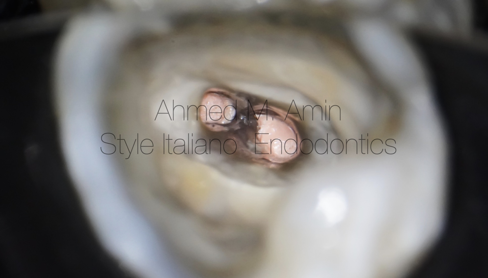
Fig. 7
Obturation of the buccal & Lingual canal at the Split level
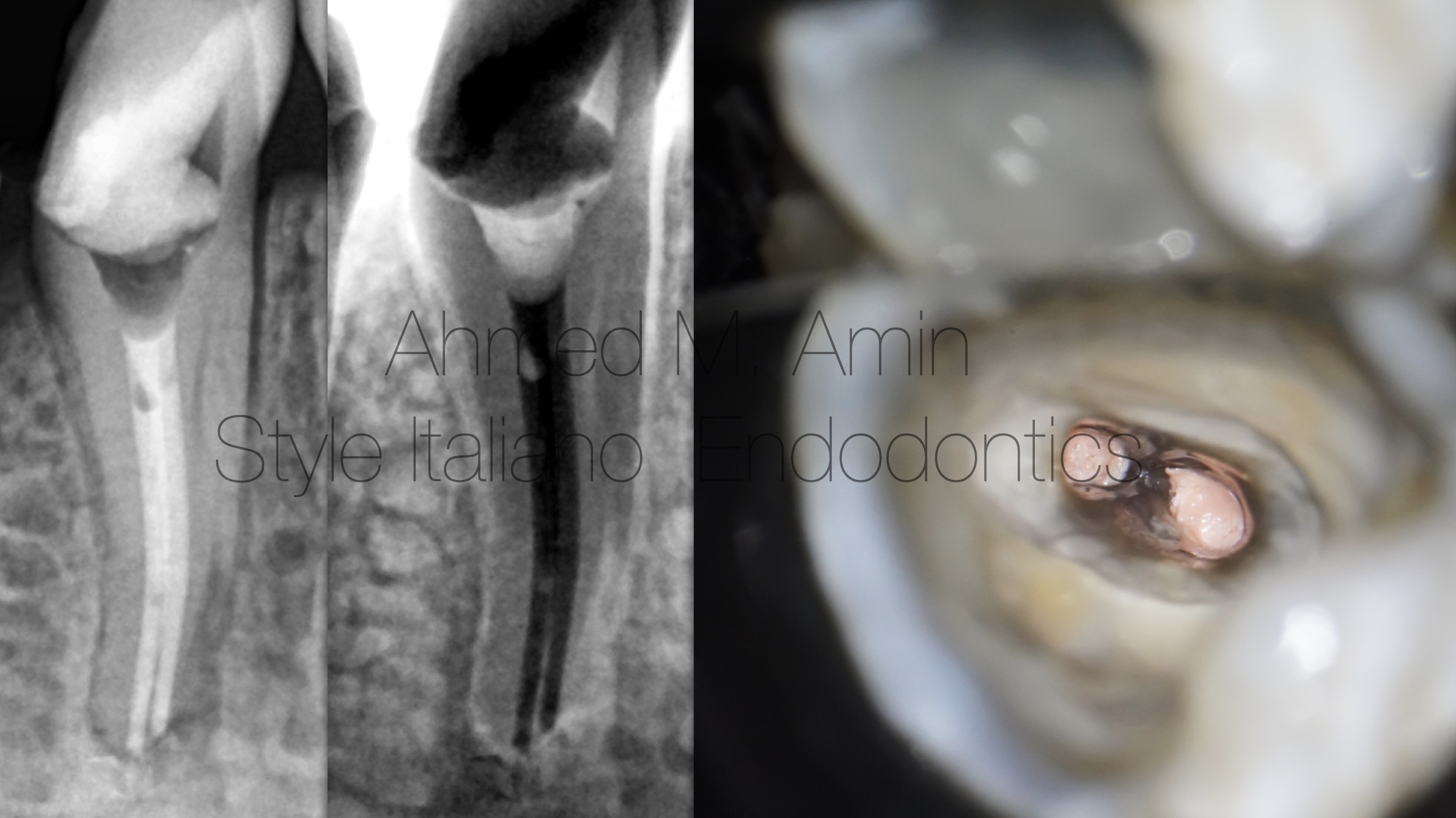
Fig. 8
Back-filing of the whole canal Space by thermoplastisized GP Using Fastfill Device
Then the case was refereed back to the previous dentist for completion of the final restoration
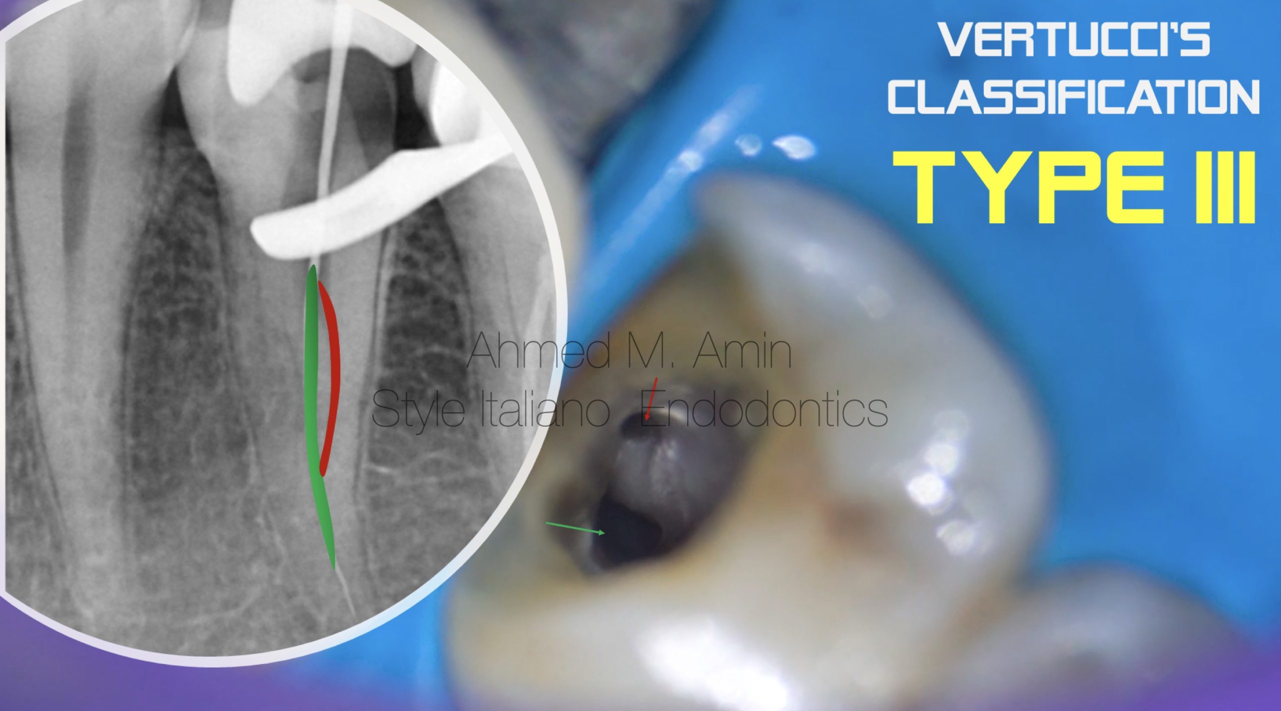
Fig. 9
Tooth No. 35 with Coronal Split into 2 canals
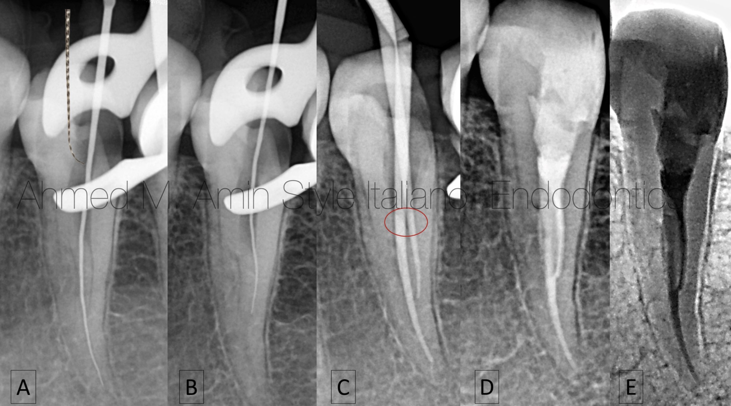
Fig. 10
A- Trials to Catch the buccal canal
B- Catching & Scouting the Canal
C- Check Master cone Fitting
D&E - obturation by WVC Tech.
This video shows us how to obturate the canals at the split level in tooth No. 35
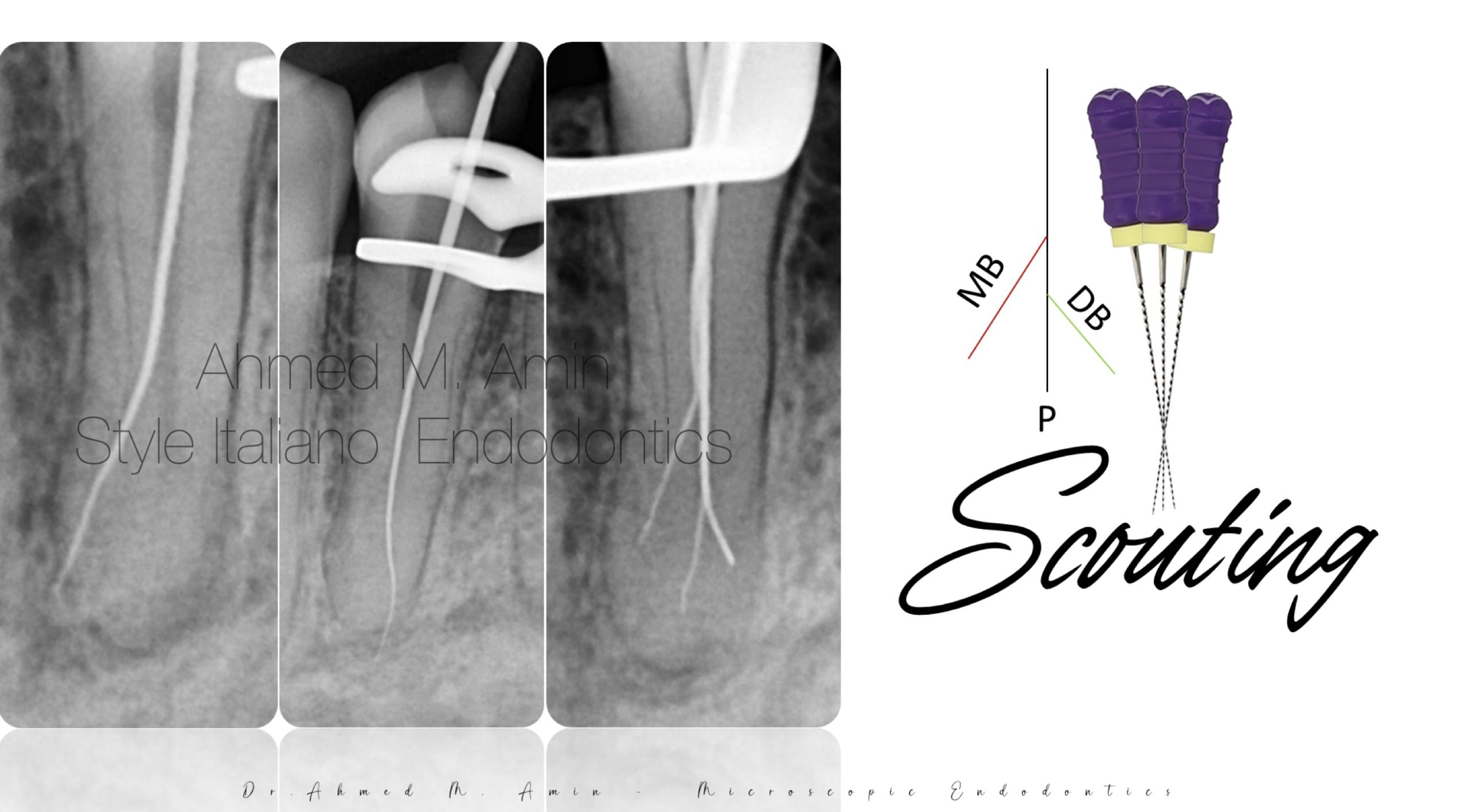
Fig. 11
Other Scenario of Diagnosis & Management of Mandibular 1st Premolar with three canals
During taking a working length Xray , I revealed that we hade a fast break phenomenon apically that suggests a Split in tooth No. 34
Then I tried to scout in more than one direction in order to catch other canals
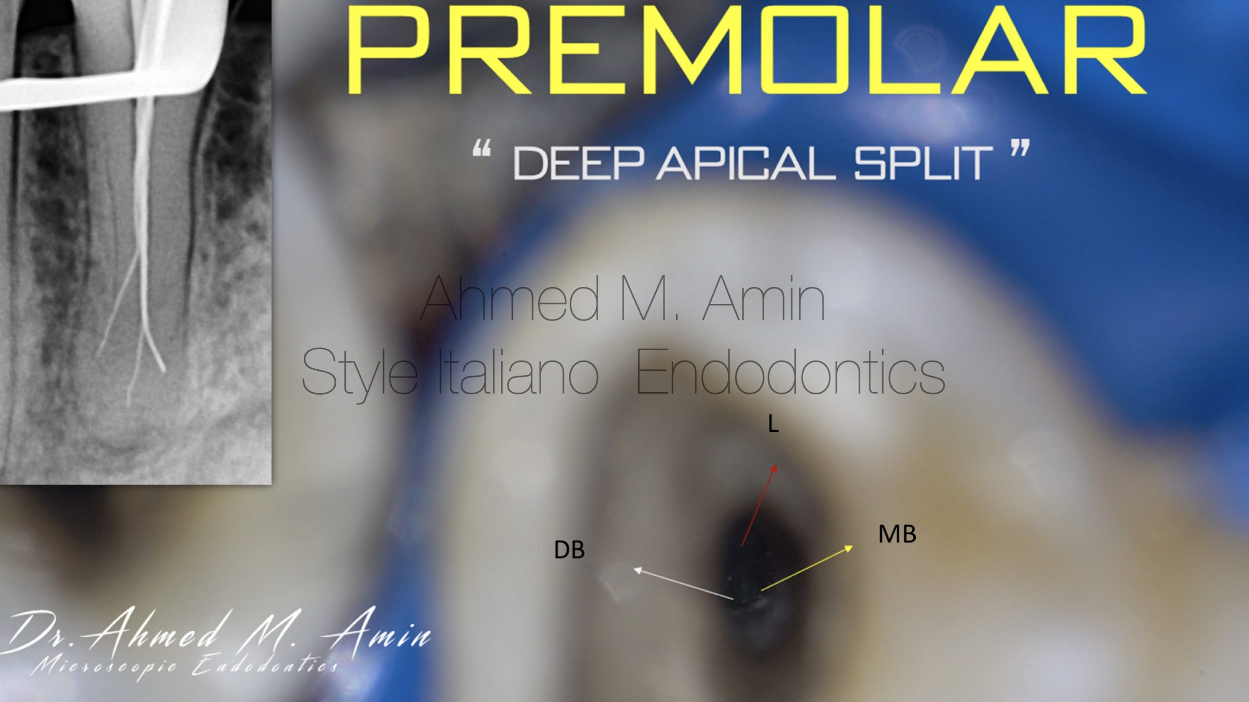
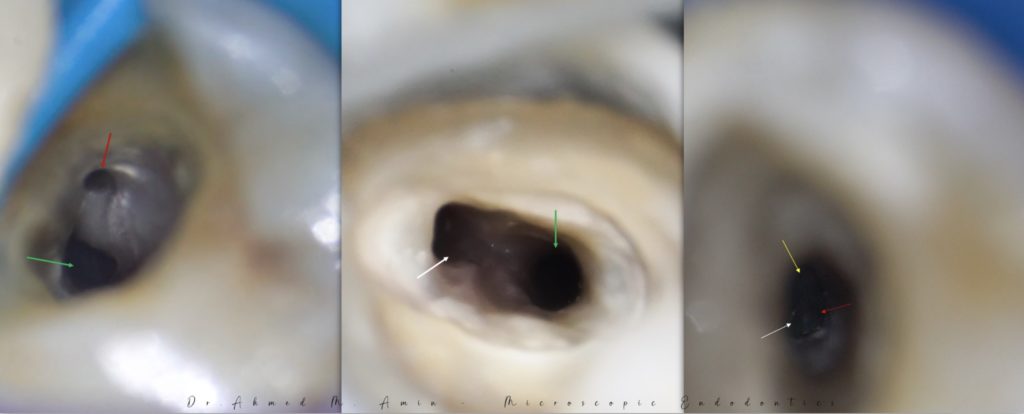
Fig. 12
Clinical Pic Shows 3 Canals Apically " L , MB & DB "
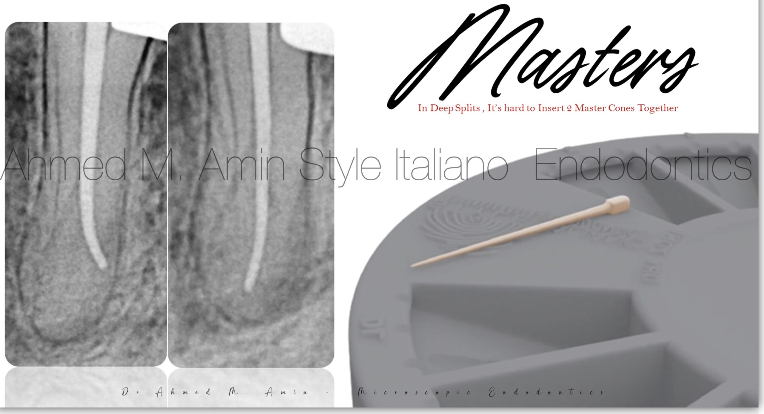
Fig. 13
All canals were prepared up to 25.04 using CM Files
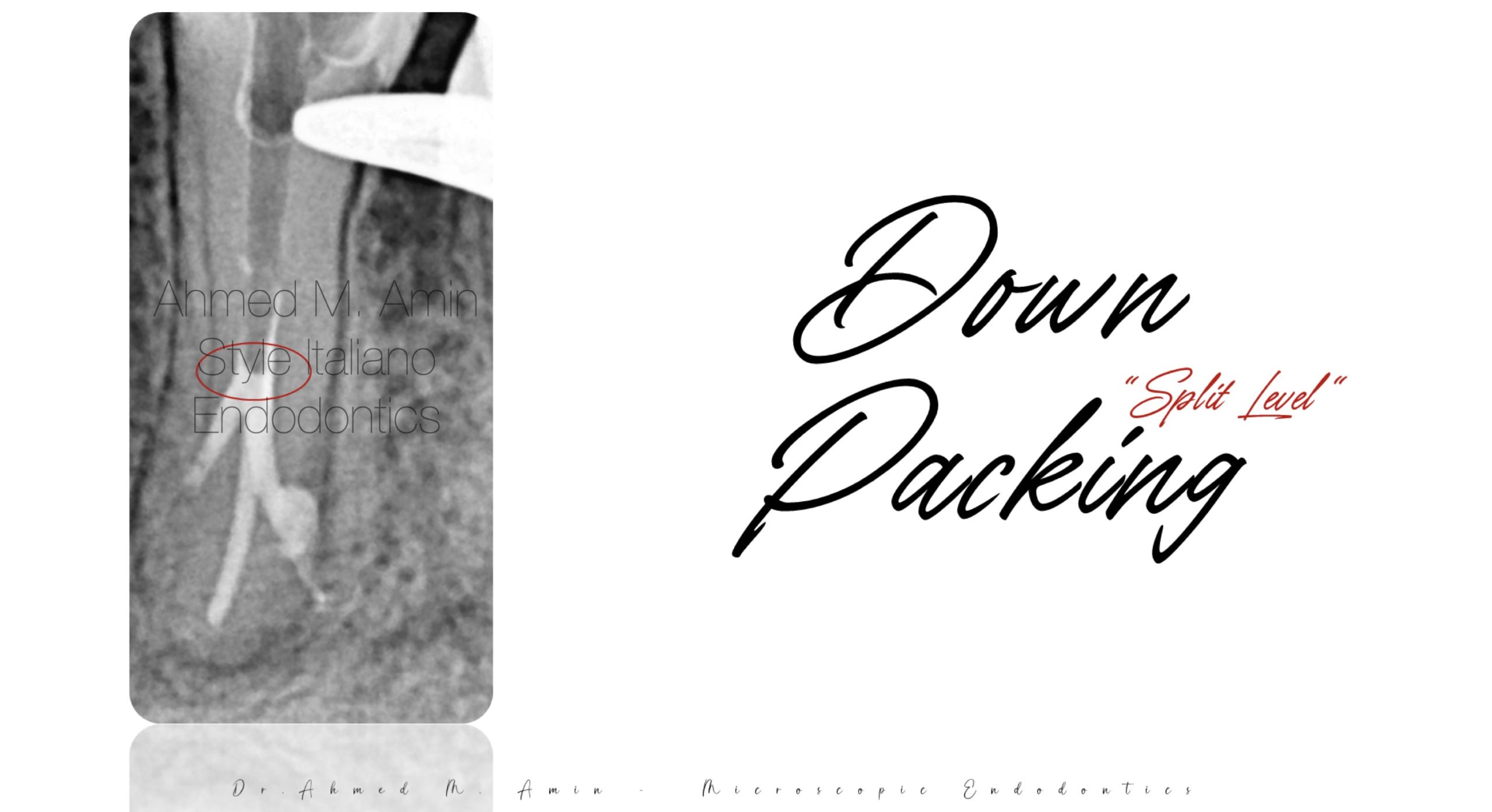
Fig. 14
I put each Master cone Separately and cut it at the split level using fastback device
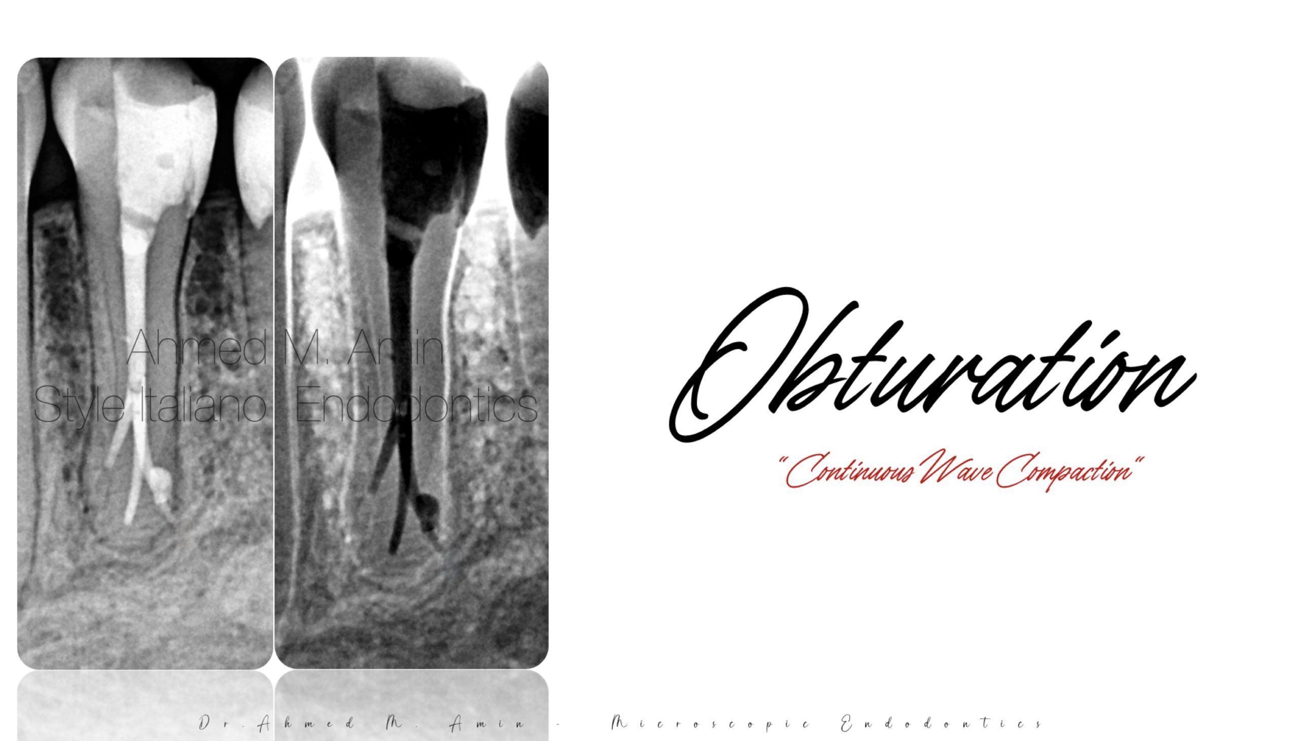
Fig. 15
Obturation by WVC Technique Using Fast-fill device , then restored the tooth with resin composite
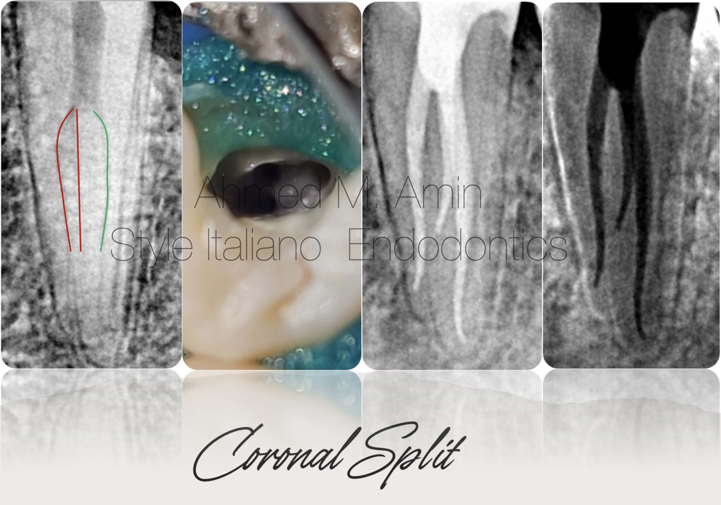
Fig. 16
1st premolar “ tooth no. 34 “ also has a division in the coronal 1/3 into three canals and managed by the same protocol that we discussed before
Conclusions
The Outcome of endodontic treatment can be jeopardized by a lack of anatomical knowledge and a poor attention to the Preoperative Xray Never underestimate the case you deal with especially the mandibular premolars A careful examination of the pre-operative x-ray can help the clinician in identifying such anatomic peculiarities in order to select the strategy and the correct instruments before starting the root canal therapy.
Bibliography
1- Albuquerque D, Kottoor J, Hammo M. Endodontic and clinical considerations in the management of variable anatomy in mandibular premolars: a literature review. Biomed Res Int. 2014;2014:512574. doi: 10.1155/2014/512574. Epub 2014 May 8. PMID: 24895584; PMCID: PMC4034431.
2- Kararia N, Chaudhary A, Kararia V. Mandibular left first premolar with two roots: A morphological oddity. Contemp Clin Dent. 2012 Apr;3(2):234-6. doi: 10.4103/0976-237X.96840. PMID: 22919233; PMCID: PMC3425116.
3- Arnaldo Castellucci, Endodontics text book, Access Cavity and Endodontic Anatomy P.244.
4- Vertucci FJ. Root canal morphology of mandibular premolars. J Am Dent Assoc
1978;97:47-50.
5- England MC Jr, Hartwell GR, Lance JR. Detection and treatment of multiple canals in mandibular premolars. J Endod 1991;17:174 – 8.