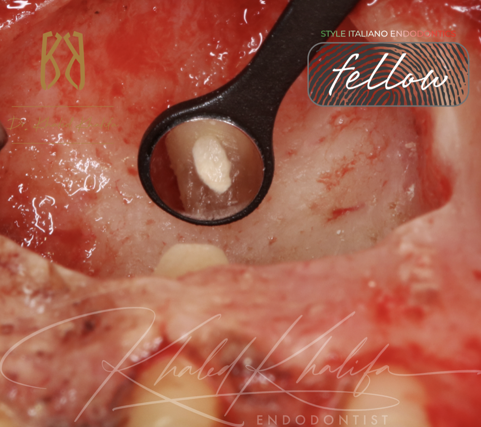
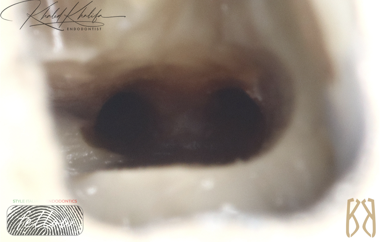
ECR External Repair should be opted for over default RCT

 Minimally Invasive Management of External Cervical Resorption Avoiding Root Canal Treatment
ECR External Repair should be opted for over default RCT
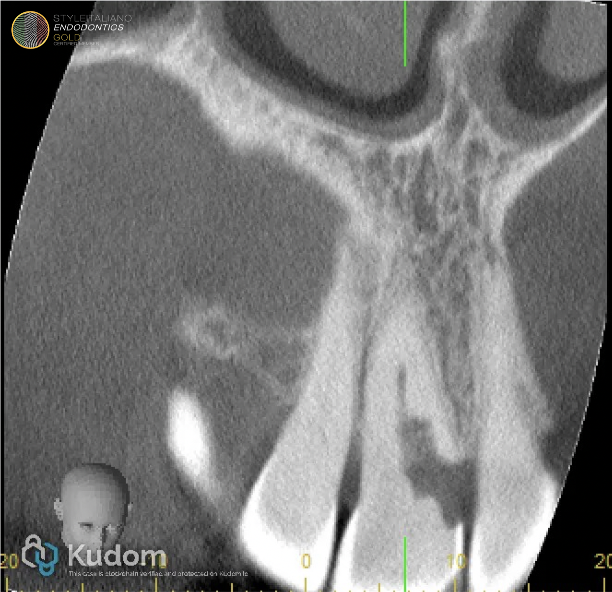
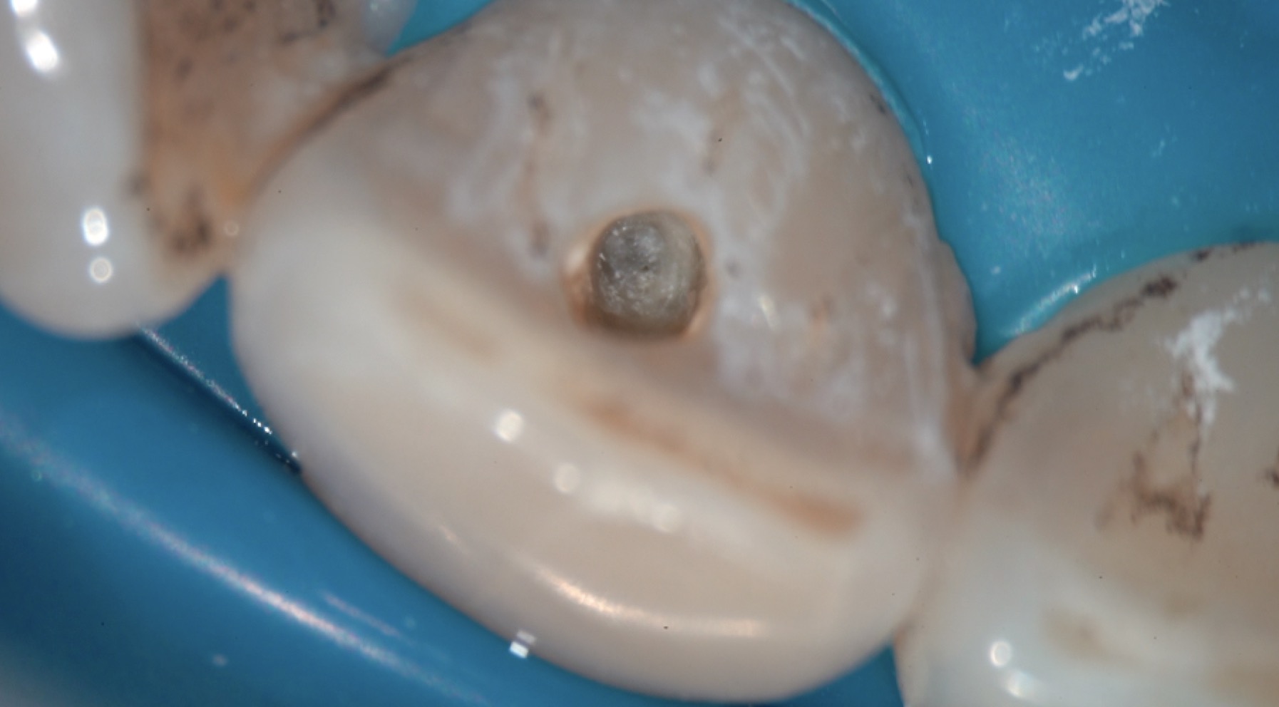
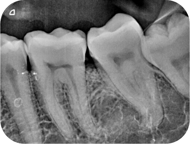
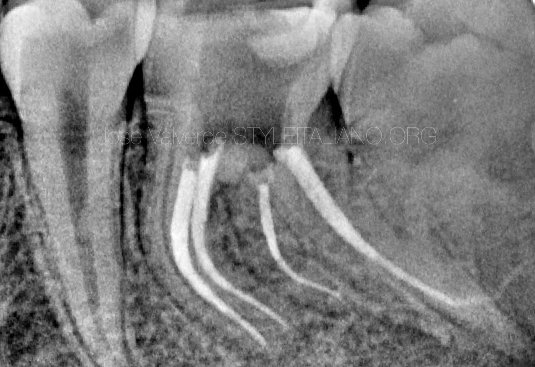
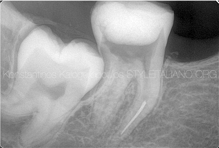
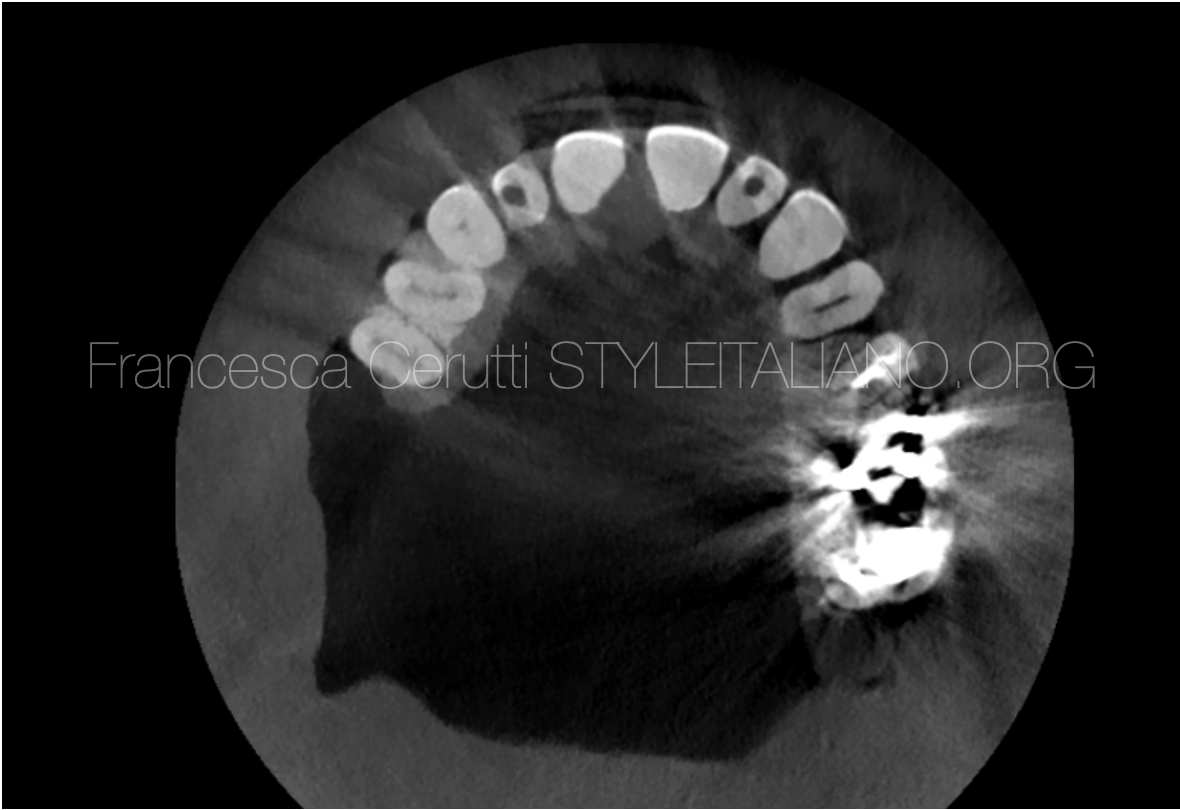
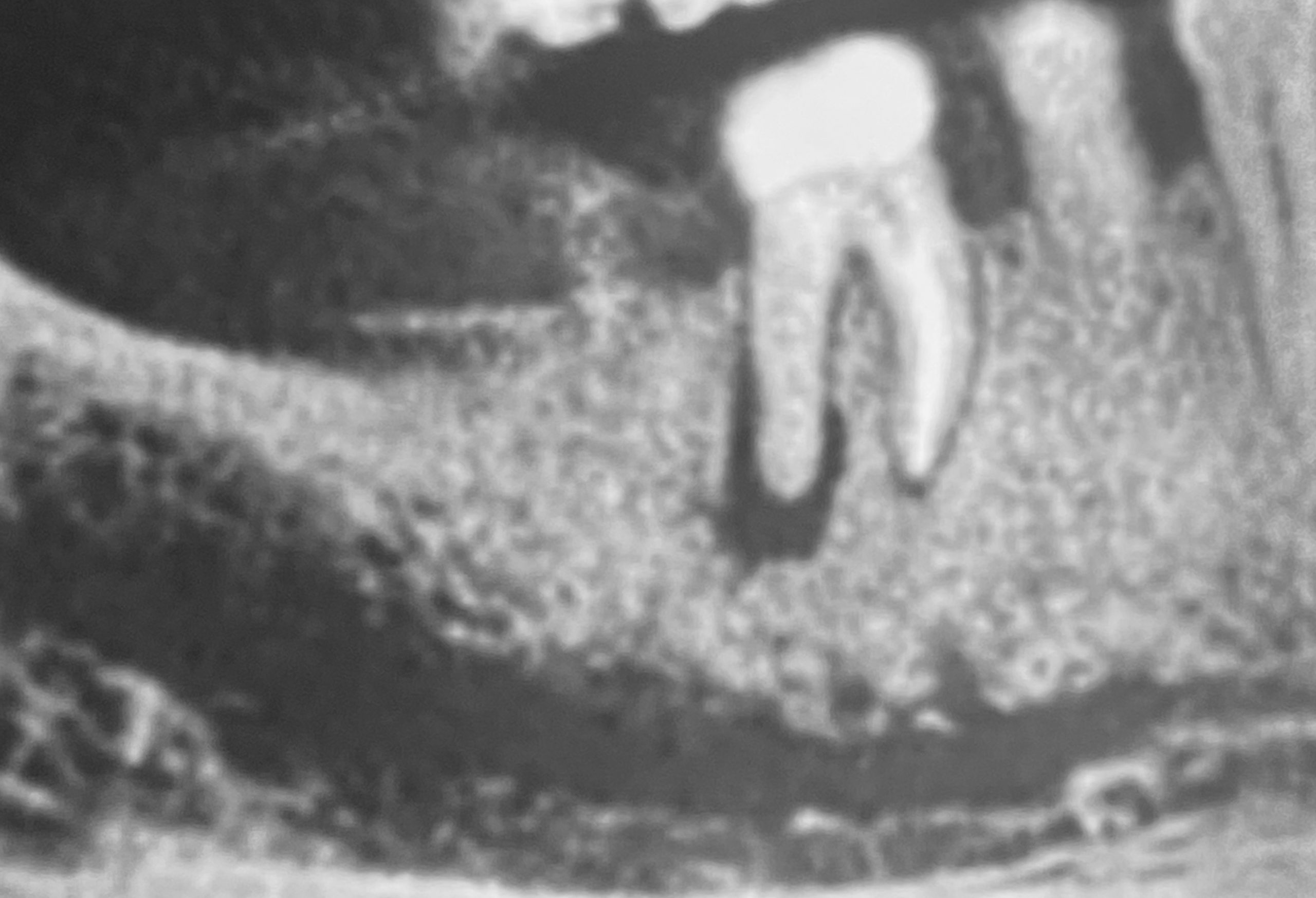
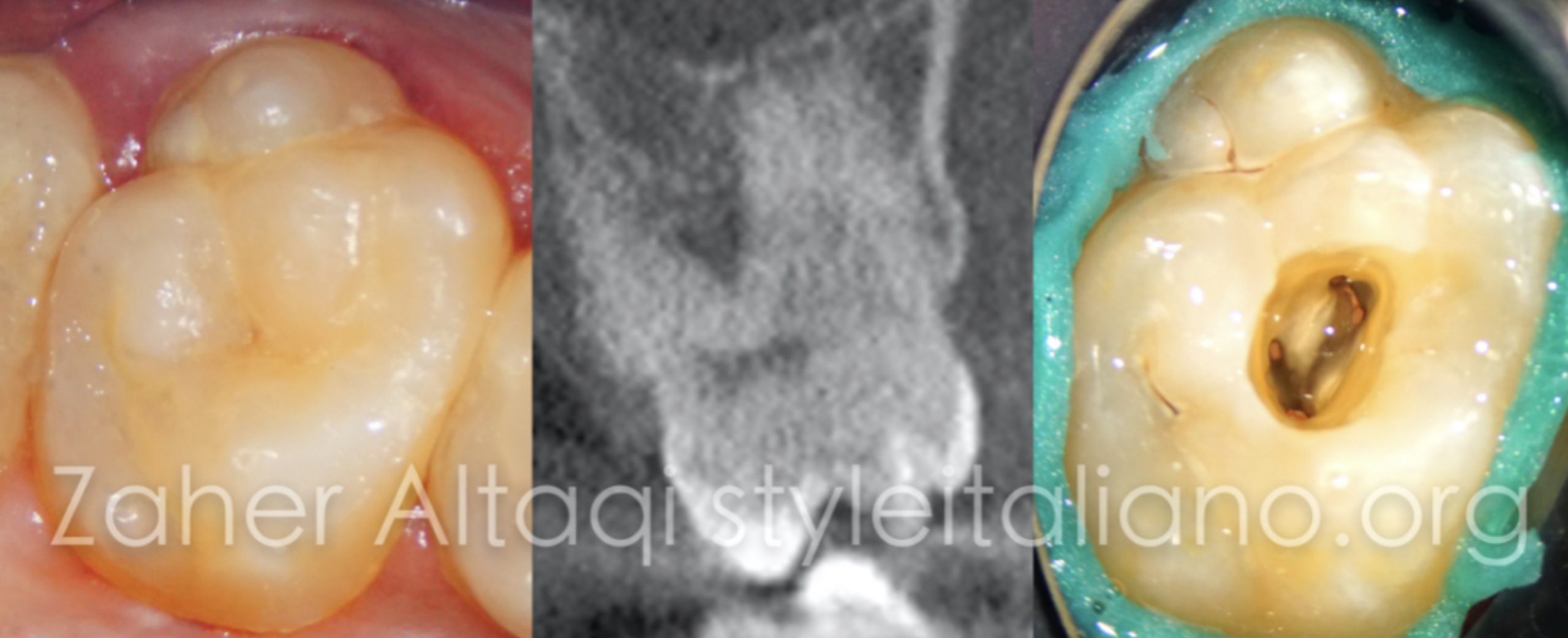
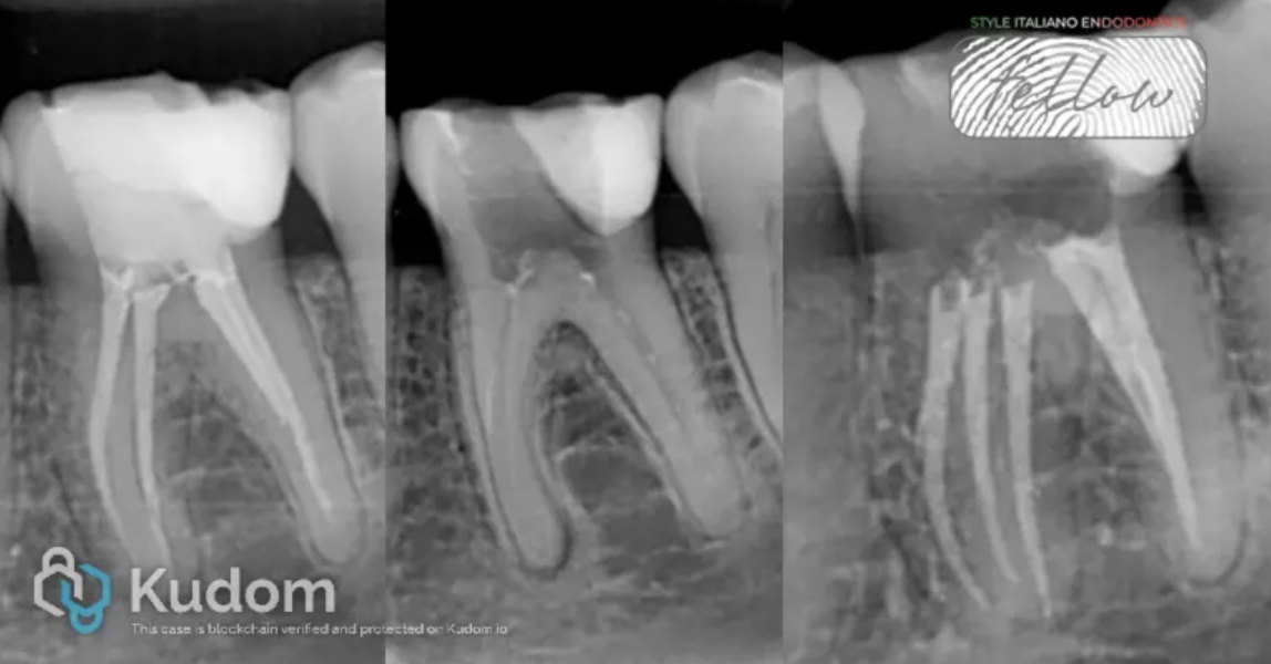 Detection and Management of a Missed Middle Mesial Canal: CBCT and Operating Microscope-Guided Retreatment. A Case Report
Mandibular molar retreatment due to an missed middle mesial canal. CBCT and Operanting Microscope are essential to identify the etiology and achieve three-dimensional disinfection. A Case Report
 Treat the invisible canal: a clinical challenge
During primary treatment, even in simple cases, many root canal anatomy alterations and errors can occur such as access cavity over enlargement, perforations, blocage or file breakage. Sometimes, many iatrogenic […]
 Correcting own errors: missed canal on lower left second molar
The purpose of this clinical article is to highlight possible root canal treatment error due to a misinterpretation of root canal morphology using 2 dimensional digital X-rays (Peri Apical Xray) […]
 Micro Rescue
Endodontic Microsurgery is one of the most important ways to save teeth rather than extracting and placing implant.
Here we will talk about a tooth that failed multiple trials of endodontic treatment, endo-surgery was made and follow ups shows complete healing.
 Dental Diagnosis Dilemma
Dentistry is all about combining clinical art & science of diagnosis to improve the quality of patient’s life.
Here we will talk about a patient with a molar needed to be retreated and instead she was misdiagnosed with trigeminal neuralgia.
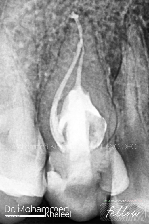 Dens invaginatus
Dens invaginatus is malformation of teeth probably resulting from an infolding of the dental papilla during tooth development. Affected teeth show a deep infolding of enamel and dentine starting from the foramen coecum or even the tip of the cusps, and which may extend deep into the root. Teeth most affected are maxillary lateral incisors and bilateral occurrence is not uncommon. The malformation shows a broad spectrum of morphologic variations and frequently results in early pulp necrosis. Root canal therapy may present severe problems because of the complex anatomy of the teeth.
 Management of a Type III Radix Entomolaris in a first lower molar
Understanding the anatomical variations of the root canal system is a must to achieve the main goals of our treatment. The radix entomolaris is the most common variation in mandibular […]
 CBCT guided treatment planning and management of instrument fracture in the mesial root of a second mandibular molar
Instrument fracture can be a very frustrating complication in Endodontics. Many factors determine whether a separated instrument should be removed or not from the root canal system. The aim of […]
 A recipe for disaster: no pre-op x-ray and wrong access cavity
Endodontic success often depends on canal debridement, disinfection, and canal obturation. Access to the canal is one key to debridement, in fact an appropriately designed access cavity assures unobstructed straight-line […]
 Management of Missed Canal Part 1: Lower First Molar
It is reported that almost 40% of retreatment with chronic apical periodontitis or symptomatic apical periodontitis are due to missed canals. Karabucak & all reported in 2016 an overall incidence […]
 Unusual anatomy in upper second molar
The hard tissue repository of the human dental pulp takes on numerous configurations and shapes. A thorough knowledge of tooth morphology, careful interpretation of angled radiographs, proper access preparation and […]