CBCT guided treatment planning and management of instrument fracture in the mesial root of a second mandibular molar
10/03/2021
Konstantinos Kalogeropoulos
Warning: Undefined variable $post in /home/styleendo/htdocs/styleitaliano-endodontics.org/wp-content/plugins/oxygen/component-framework/components/classes/code-block.class.php(133) : eval()'d code on line 2
Warning: Attempt to read property "ID" on null in /home/styleendo/htdocs/styleitaliano-endodontics.org/wp-content/plugins/oxygen/component-framework/components/classes/code-block.class.php(133) : eval()'d code on line 2
Instrument fracture can be a very frustrating complication in Endodontics. Many factors determine whether a separated instrument should be removed or not from the root canal system. The aim of this case report is to highlight the usefulness of LFOV CBCT in the treatment planning of a second lower molar with a fragment in the mesial root with merging canals by determining the exact location of the fragment. Moreover, it is a great tool for patient communication because prognosis of teeth with a instrument fragment is not affected if cleaning and shaping have not been compromised whereas unnecessary dentine removal leads to weakening of the tooth and increases the possibility for root fracture.
Endodontic instruments can, rarely, fracture inside the root canal system. This undesirable complication happens more in posterior teeth and may compromise the subsequent cleaning and shaping and final obturation. Dealing with the separated instrument is dependant on many factors. Some of them are the location of the fragment, the size and length, the preoperative pulp status the presence of a lesion, training and experience of the dentist, armamentarium and the risk of possible complications from the removal procedure and the strategic importance of the tooth. Even though removal of the fragment seems to be logical, all the factors mentioned before must be taken into consideration when determining the ideal treatment plan for each case. Dentine preservation should be the goal of all approaches when dealing with similar cases. Moreover, literature suggests that failure to remove the fragment does not affect the prognosis of the endodontic treatment when the fragment has been bypassed or the root canal system has been properly disinfected.
Fig. 1
Female patient age 24 was referred in order to remove a separated instrument from the mesial root of a lower second molar. The patient had been informed from the referring dentist about the complication and was very frustrated. When an instrument brakes inside a root canal, a periapical radiograph must be taken in order to try to identify the location of the fragment.
According to the European Society of Endodontology (ESE) position statement, limited field of view (LFOV) Cone Beam Computed Tomography (CBCT) should also be considered as a diagnostic tool in cases where treatment complications have occurred. CBCT) provides even more information and can be helpful in treatment planning of such cases.
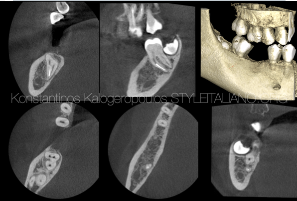
Fig. 2
Studying the LFOV CBCT clearly demonstrates the confluent mesial root canal system and the possibility to reach to the apical part via the mesial lingual canal and probably would not impede the cleaning and shaping. Moreover, the impacted third moral does not seam to have affected in any way the second molar. Nevertheless, afterwards the patient has been referred to an oral surgeon in order to extract the impacted tooth after the endodontic treatment.
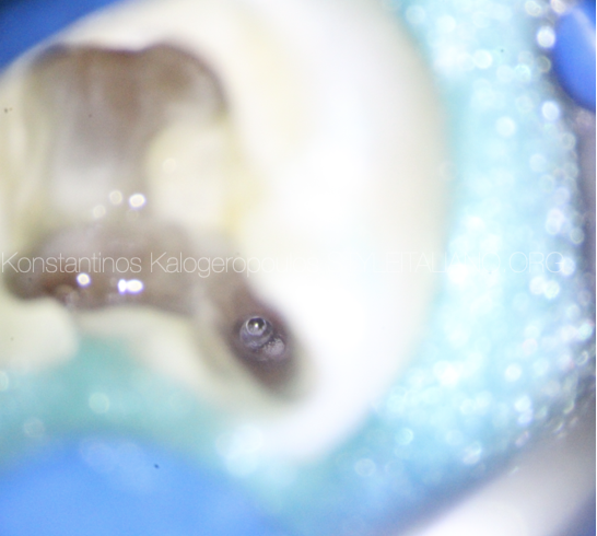
Fig. 3
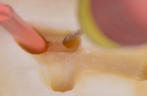
Therefore, after explaining to the patient and the referring dentist the benefits in sound dentine preservation of not trying to remove the fragment and the potential risks from the removal attempt, the treatment was initiated. Following administration of local anaesthesia and rubber dam isolation, the rest of the root canal system was cleaned and shaped. Emphasis was given to irrigation with 2,5% NaOCl via activation of the irrigant with the Endoultra device. Final irrigation was done with 17% EDTA.
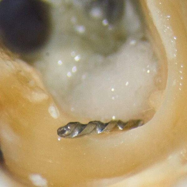
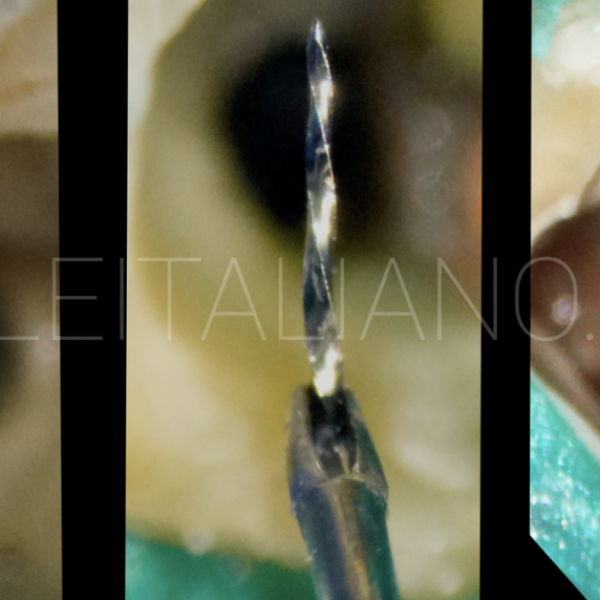
Fig. 4
View of the coronal part of the instrument under high magnification after the preparation of the canals. No tooth structure has been sacrificed in order to remove the fragment
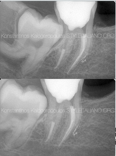
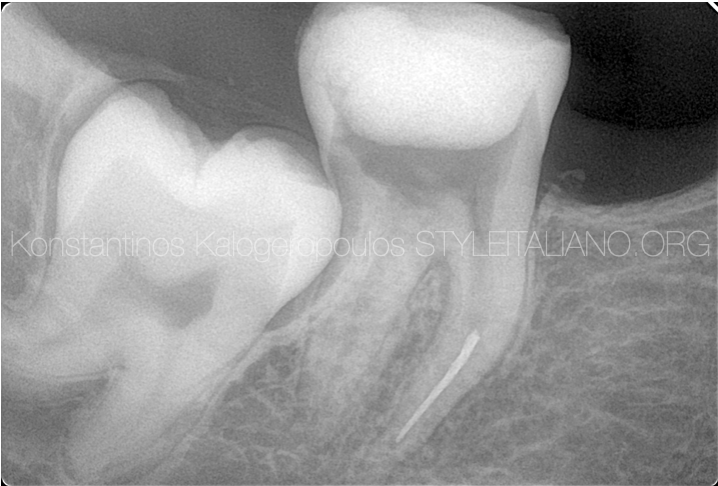
Fig. 5
Final obturation radiograph with two different angles. It is evident that the mesial root canal system has been obturated beyond the apical limit of the fragment. The whole procedure was carried out in a single session and the technique used was warm vertical compaction of gutta-percha with resin based sealer.
Conclusions
- CBCT imaging can be very helpful when treating teeth with instrument fracture.
- Determining the exact location of the fragment influences the decision-making process, especially when canals merge
- Prognosis of teeth with a instrument fragment is not affected if cleaning and shaping have not been compromised.
- Every effort must be made to prevent unnecessary dentine removal that leads to weakening of the tooth and increases the possibility for root fracture.
Bibliography
Panitvisai P, Parunnit P, Sathorn C, Messer HH (2010) Impact of a retained instrument on treatment outcome: a systematic review and meta-analysis. Journal of Endodontics 36, 775–80.
Tzanetakis GN, Kontakiotis EG, Maurikou DV, Marzelou MP (2008) Prevalence and management of instrument fracture in the postgraduate endodontic program at the Dental School of Athens: a five- year retrospective clinical study. Journal of Endodontics 34, 675-8.
Spili P, Parashos P, Messer HH (2005). The impact of instrument fracture on outcome of endodontic treatment. Journal of Endodontics 31, 845-50.
Shen Y, Peng P, Cheung GS (2004) Factors associated with the removal of fractured NiTi instruments from root canal systems. Oral Surg Oral Med Oral Pathol Oral Radiol Endod 98, 605–10.
Souter NJ, Messer HH (2005) Complications associated with fractured file removal using an ultrasonic technique. Journal of Endodontics 31: 450–2.
Patel S, Brown J, Semper M, Abella F, Mannocci F (2019) European Society of Endodontology position statement: Use of cone beam computed tomography in Endodontics: European Society of Endodontology (ESE) developed by. International Endodontic Journal 52, 1675-8
Ramos Brito AC, Verner FS, Junqueira RB et al. (2017) Detection of Fractured Endodontic Instruments in Root Canals: Comparison between Different Digital Radiography Systems and Cone Beam Computed Tomography Journal of Endodontics 544-9.
Rodríguez G, Abella F, Duràn-Sindreu F et al. (2017a) Influence of cone-beam computed tomography in clinical decision-making among specialists. Journal of Endodontics 43, 194–9.
Rodríguez G, Patel S, Durán-Sindreu F, Roig M, Abella F (2017b) Influence of Cone-beam Computed Tomography on Endodontic Retreatment Strategies among General Dental Practitioners and Endodontists. Journal of Endodontics 43, 1433-7.
Rosen E, Venezia NB, Azizi H et al. (2016) A Comparison of Cone-beam Computed Tomography with Periapical Radiography of Separated Instruments Retained in the Apical third of root-canal filled teeth. Journal of Endodontics 1035-9.
Mota de Almeida FJ, Knutsson K, Flygare L (2015) The impact of cone beam computed tomography on the choice of endodontic diagnosis. International Endododontic Journal 48, 564–72.