What a surprise!
24/05/2020
Riccardo Tonini
Warning: Undefined variable $post in /home/styleendo/htdocs/styleitaliano-endodontics.org/wp-content/plugins/oxygen/component-framework/components/classes/code-block.class.php(133) : eval()'d code on line 2
Warning: Attempt to read property "ID" on null in /home/styleendo/htdocs/styleitaliano-endodontics.org/wp-content/plugins/oxygen/component-framework/components/classes/code-block.class.php(133) : eval()'d code on line 2
When I started treating the tooth number 3.7, I understood from the beginning that the case was peculiar. During the emergency session a radix paramolaris was not detected and a SX rotary file created immediately an accidental stripping during orifice opening.
The management of an unconventional anatomy and the repair of a iatrogenic error will be described in this article.
#radixentomolaris
#radixparamolaris
#apicalplug
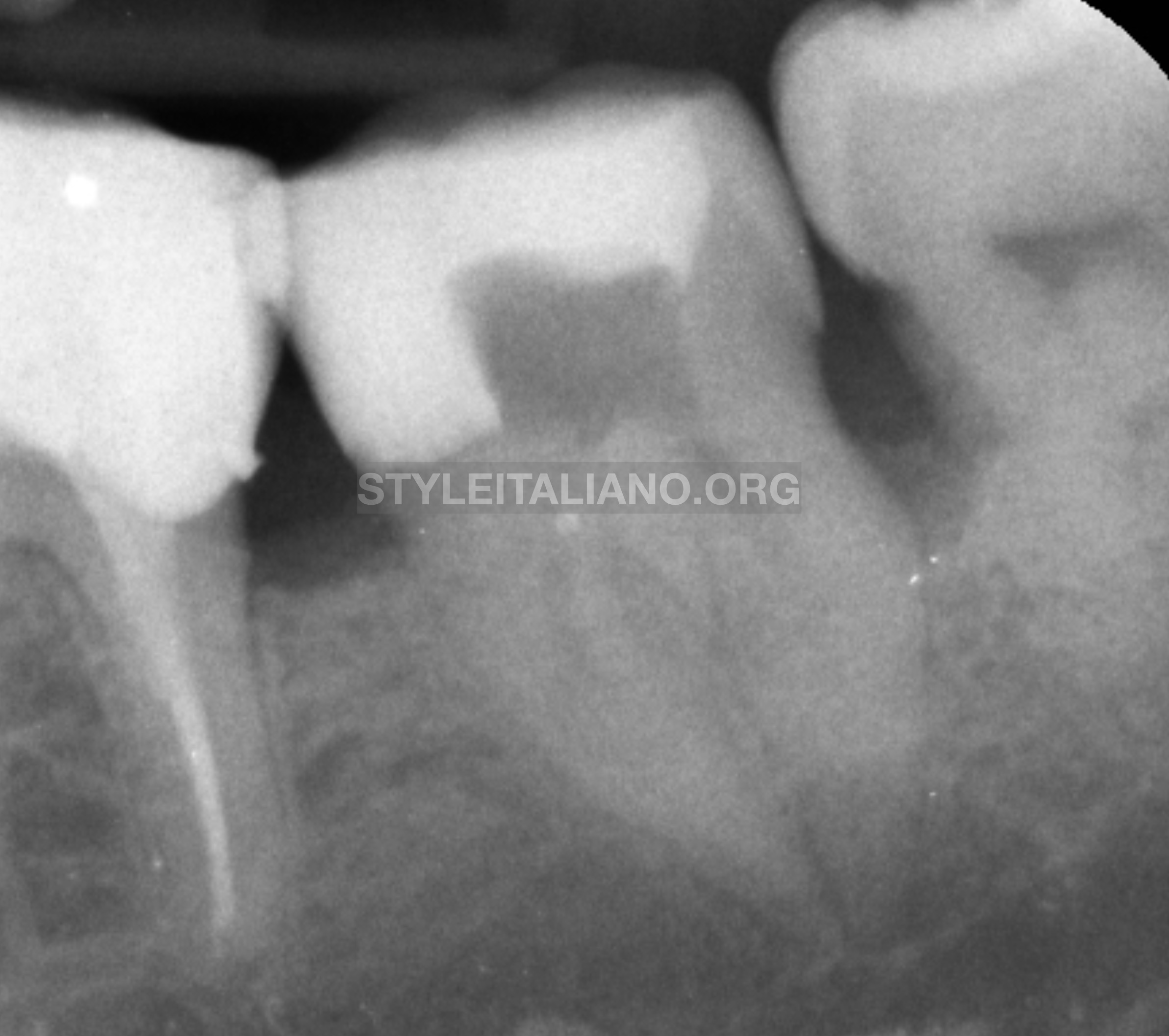
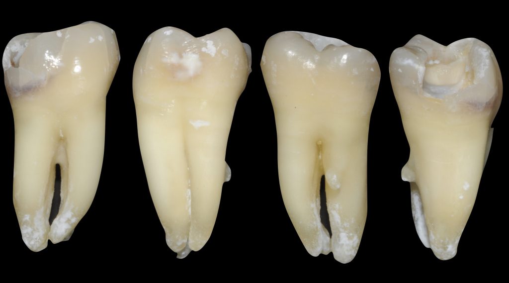
Fig. 1
Mandibular molars can have an additional root located lingually (the radix entomolaris) or buccally (the radix paramolaris). The size of the RP can be, like in this case, as a shorter root with a canal shorter than the root itself.
Unfortunately, the detection of this anatomy has not been done pre-operatively and during the first emergency appointment, thus the root has been shaped with an SX causing a stripping. In order to identify the problem, the apex locator was really useful, because the working length of RP was 13mm, shorter than the 21 mm of ML. Moreover, positioning the k file with a lingual orientation, the apex locator immediately reported OVER, the typical sign of a perforation.
For this reason a CBCT has been immediately performed, allowing to detect the RP and the stripping.
In the next visit the treatment plan was:
- Distal canal and ML shaped and filled with warm GP technique
- RP root filled completely with MTA, with apical plug technique
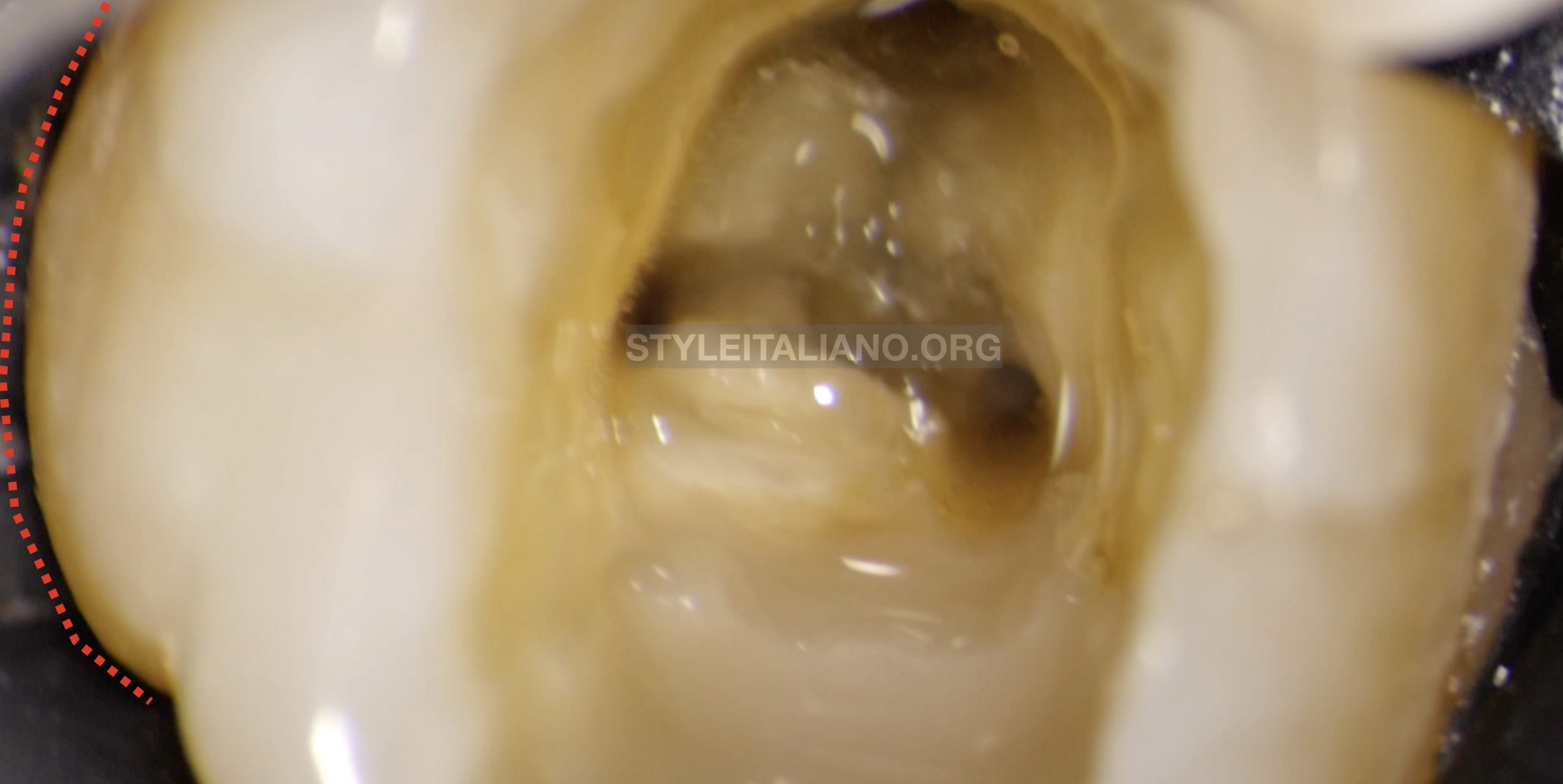
Fig. 2
Buccal aspect of the crown, typical sign of different anatomy
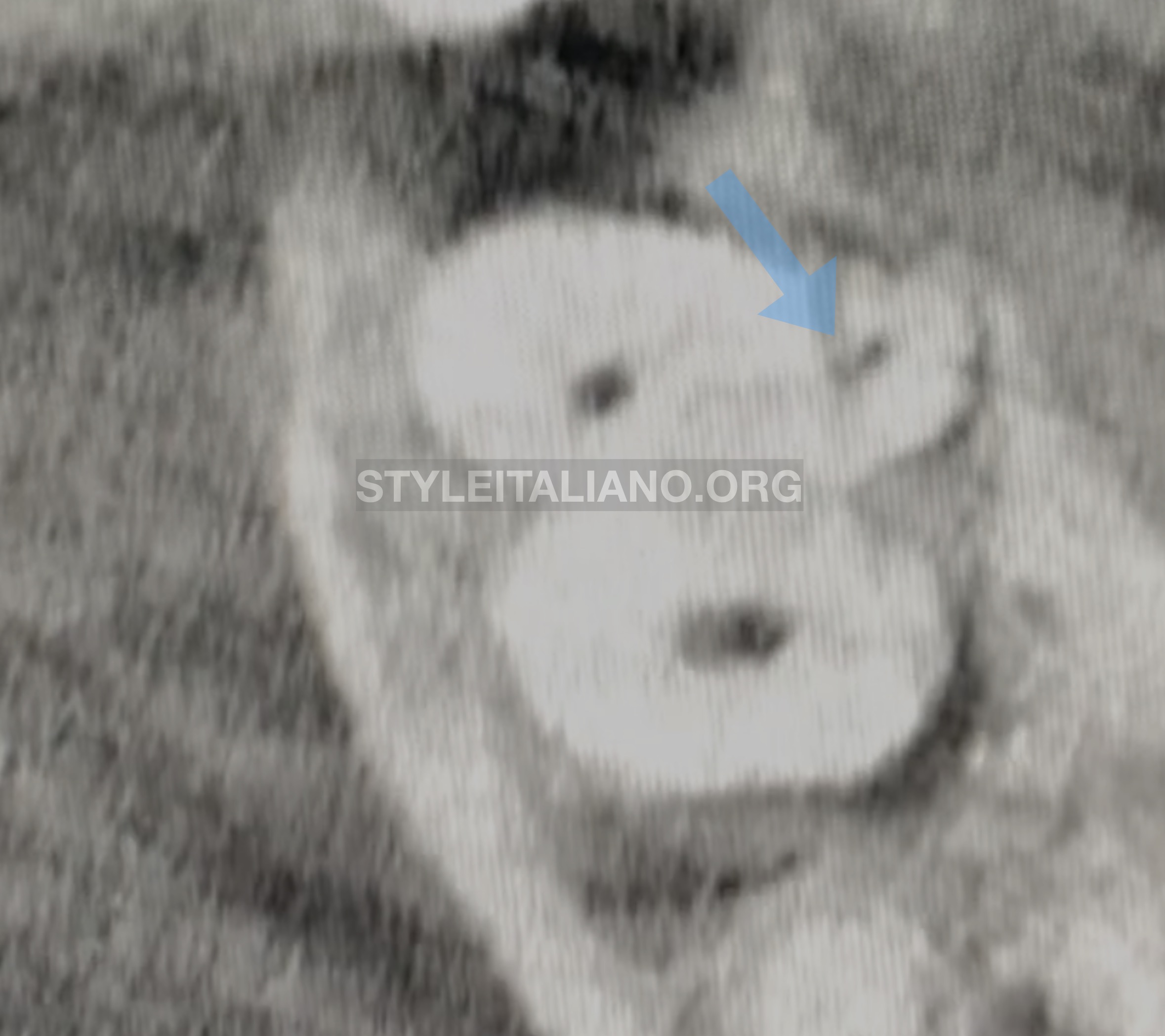
Fig. 3
Buccal aspect of the crown, typical sign of different anatomy
CBCT: Axial view.
RP navigation: it can be observed that anatomical root is longer than the canal.
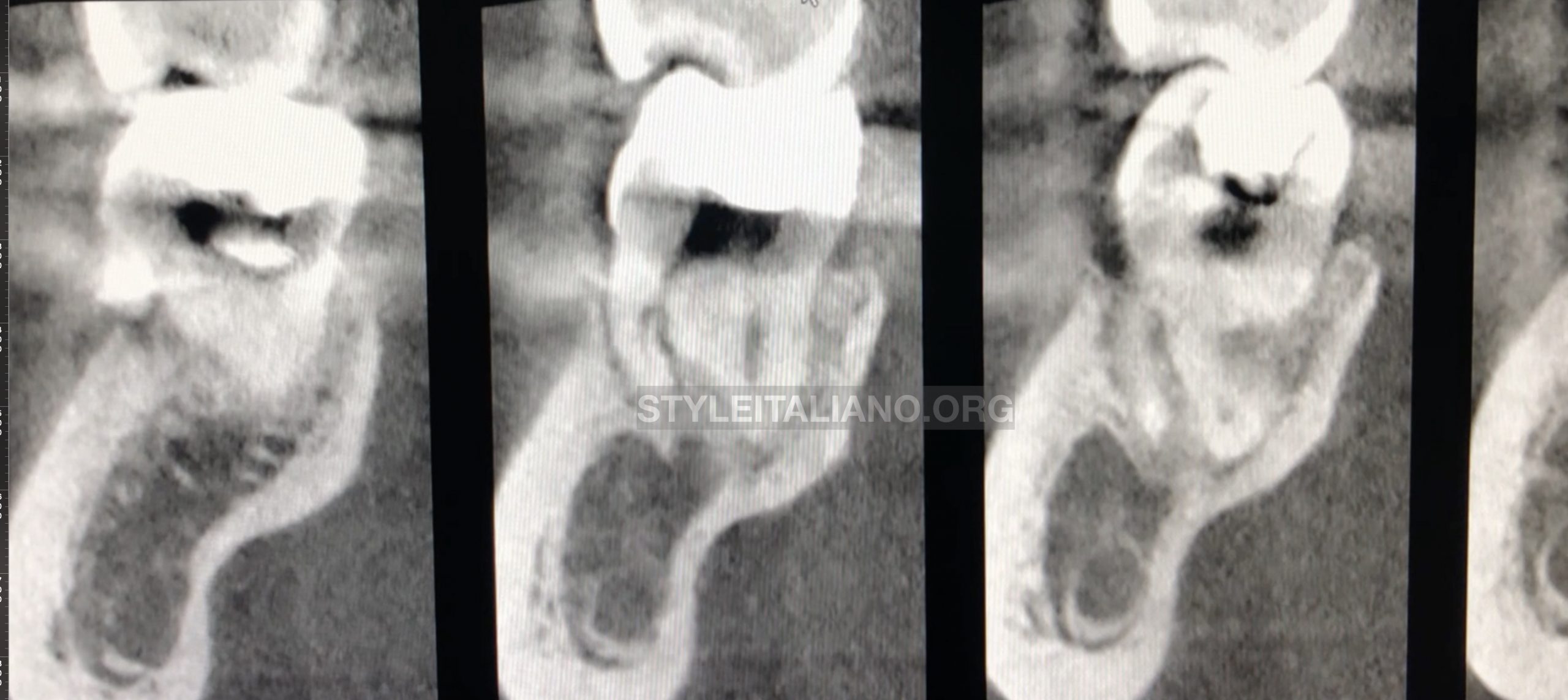
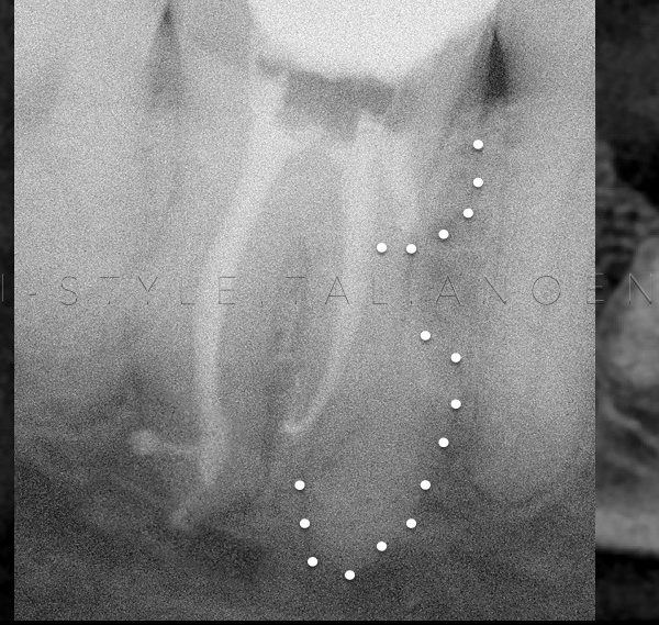
Fig. 4
CBCT: sagittal view
In the second image appears the coronal stripping and the anatomy of the root is better visible
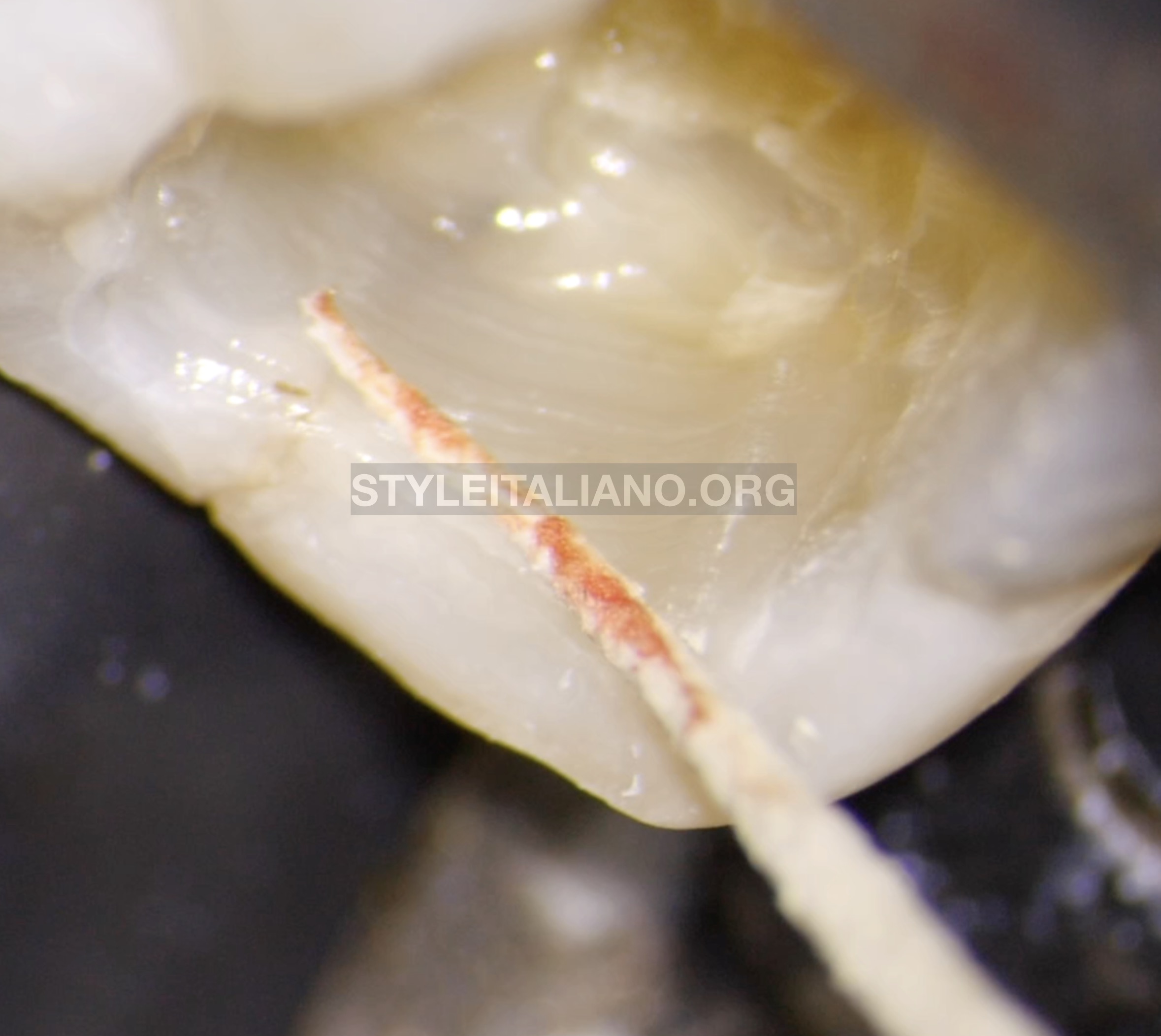
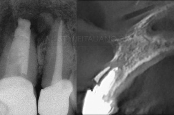
Fig. 5
Stripping detected and confirmed by a Paper point
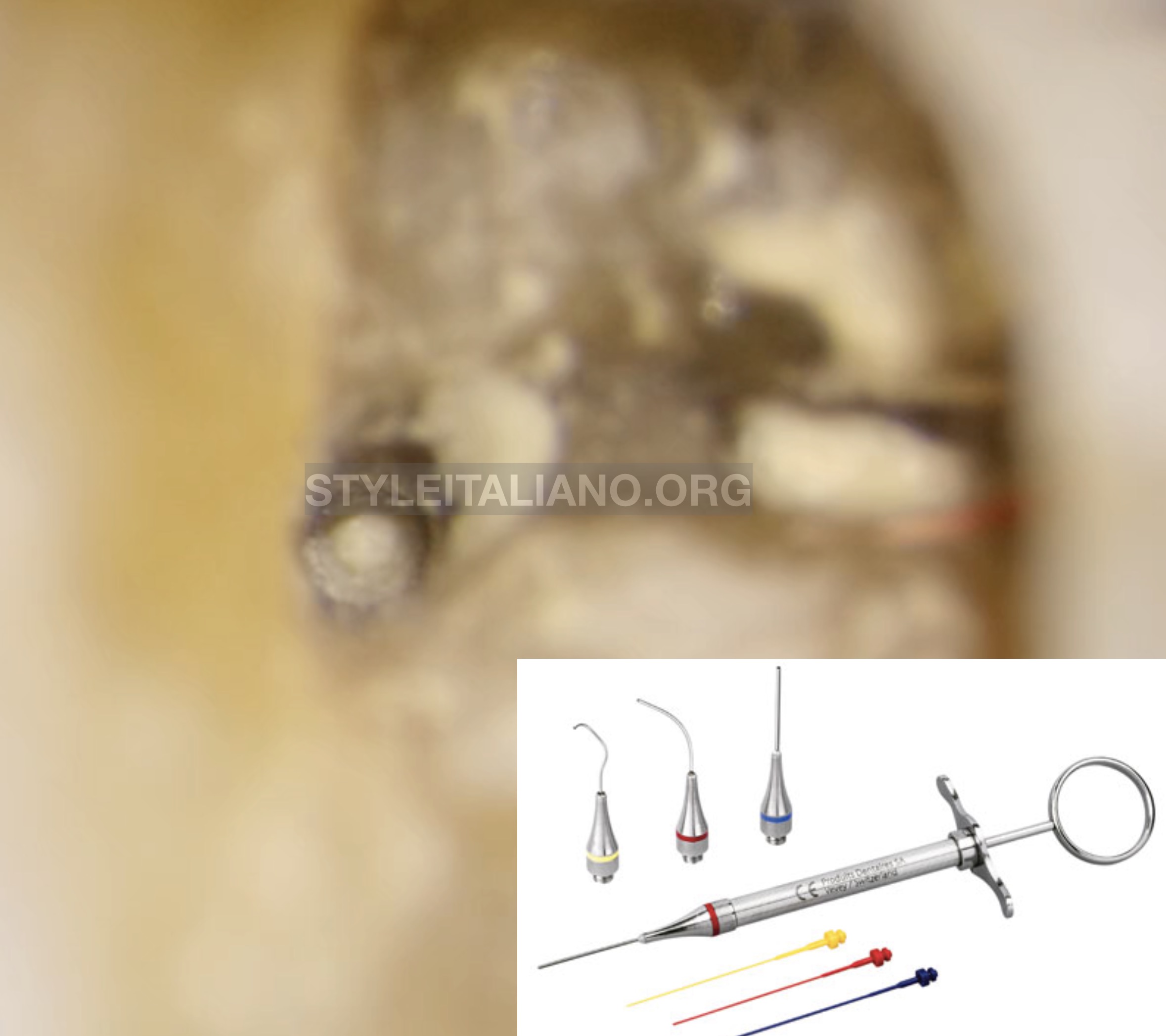
Fig. 6
Apical plug technique performed with Map System
The apical plug has been performed using:
- PD MTA
- Map system
- Plastic plugger (Thermafil)

Fig. 7
The root has been filled completely with PD MTA White. A temporary filling with a cotton pellet moistened with water.
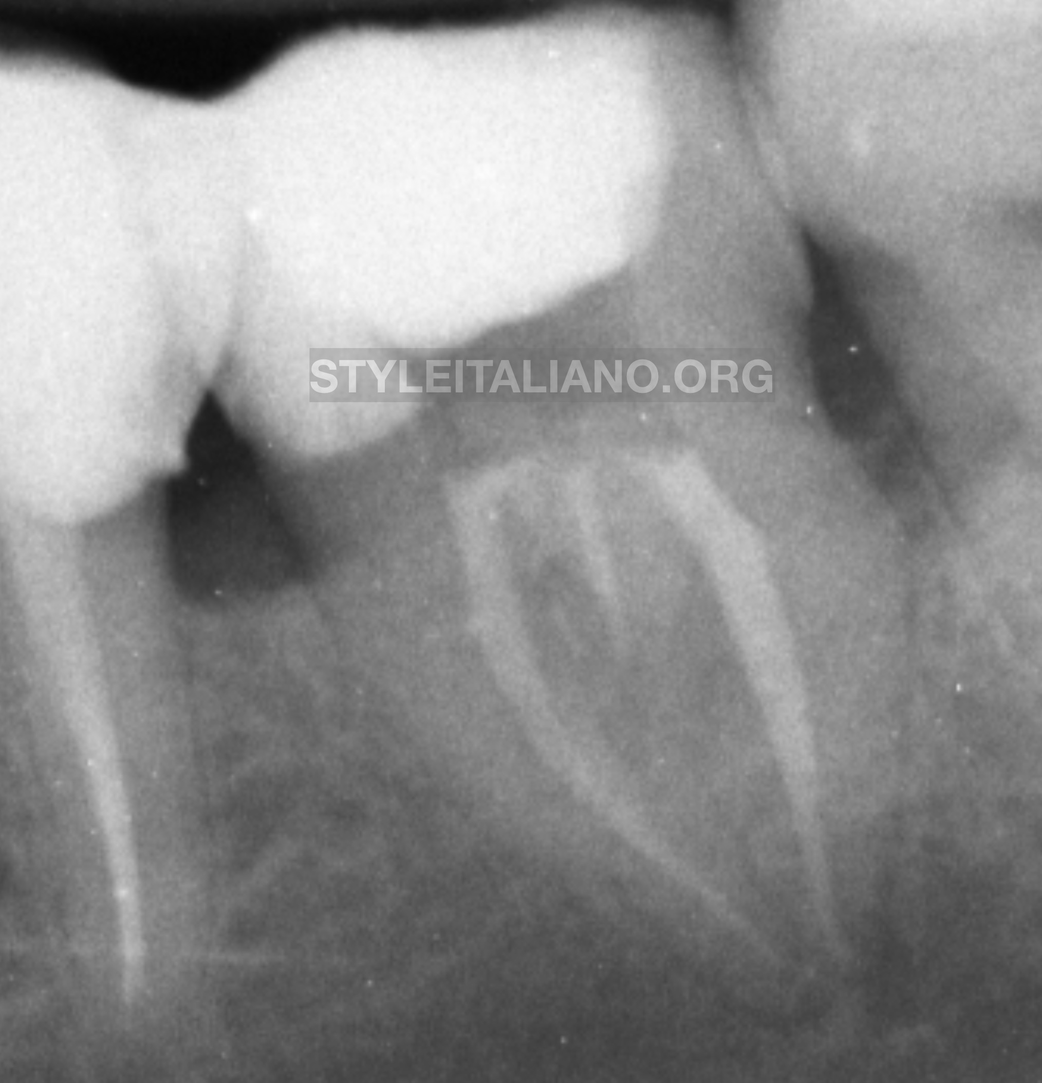
Fig. 8
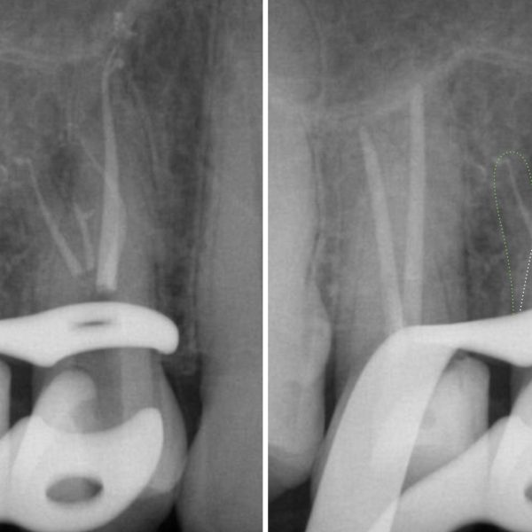
Post op X ray
MTA placed in RP root
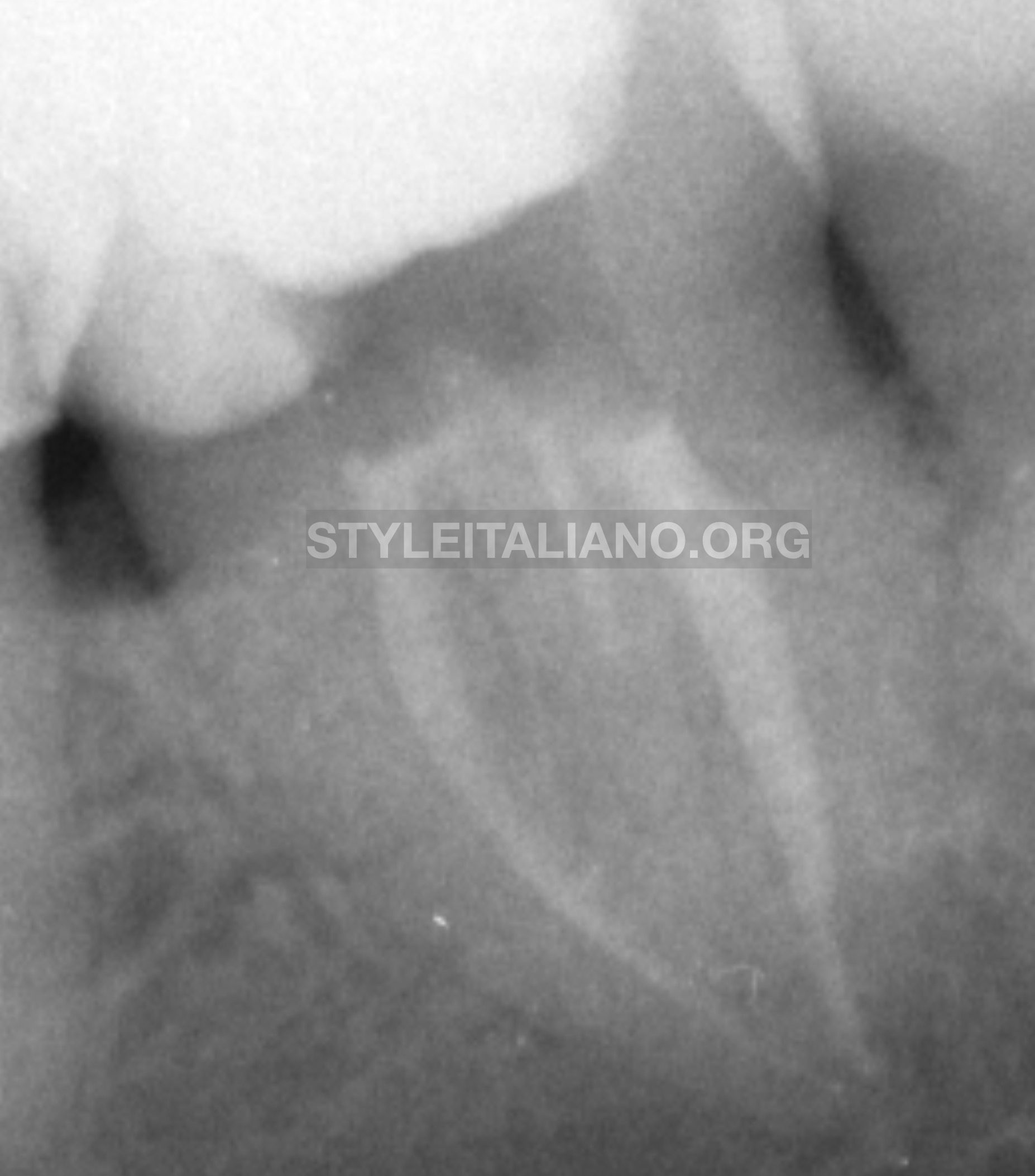
Fig. 9
A Post op with a different angulation
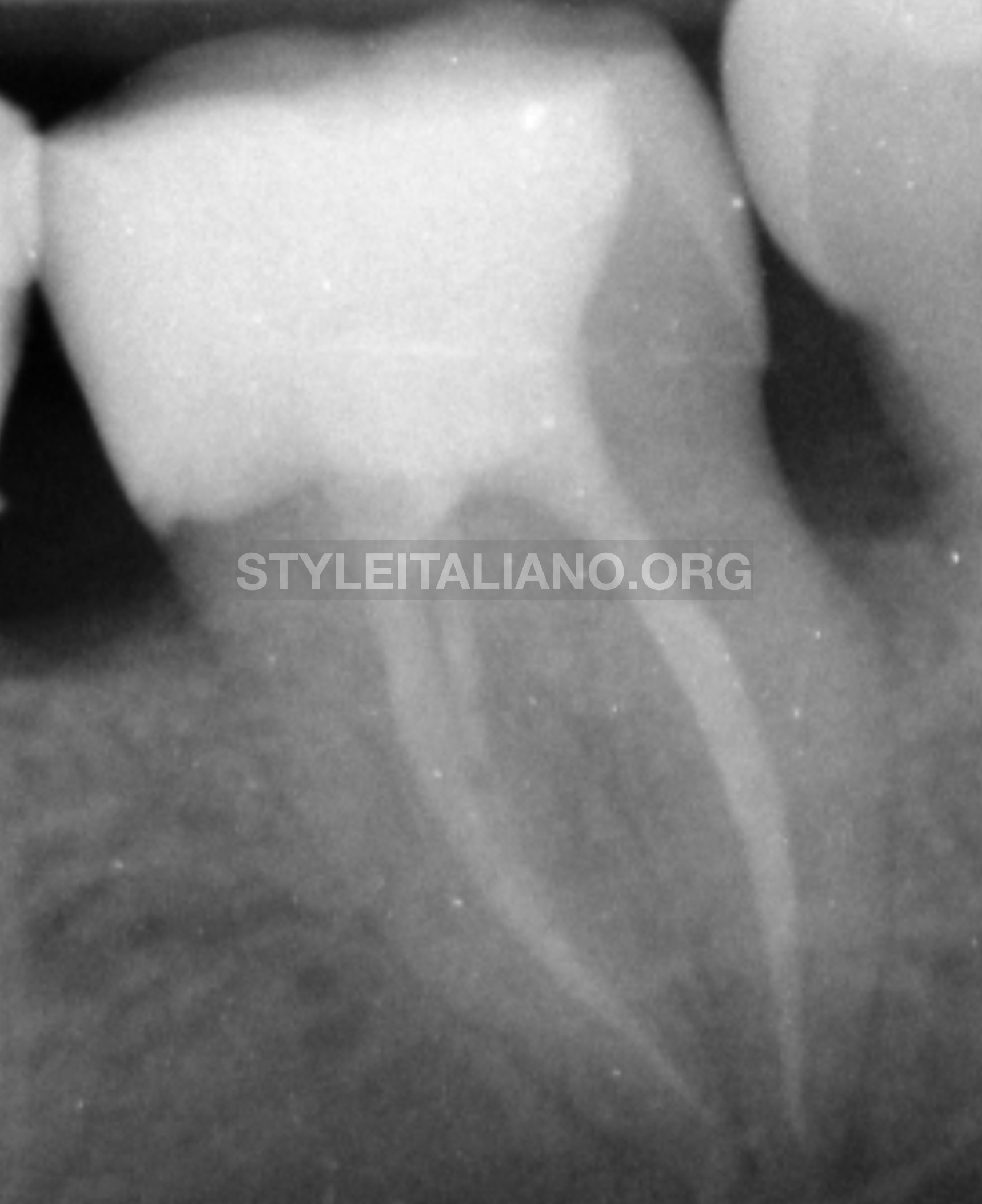
Fig. 10
Build up, waiting for an onlay
Conclusions
This clinical case demonstrated us how unpredictable anatomy can be and how important is a conservative mechanical glide path. If we have any doubt about anatomy, it is better to stop the clinical session and perform a CBCT in order to avoid iatrogenic errors. The other aspect that must be considered is that the MTA apical plug is an easy clinical procedure and it should be applied more in our daily practice .
Bibliography
Carabelli G. Systematisches Handbuch der Zahnheilkunde, 2nd ed. Vienna: Brau- muller und Seidel, 1844:114.
Carlsen O, Alexandersen V. Radix entomolaris: identification and morphology. Scan J Dent Res 1990;98:363–73.
Carlsen O, Alexandersen V. Radix paramolaris in permanent mandibular molars: identification and morphology. Scan J Dent Res 1991;99:189 –95.