Curvy Wisdom Management
20/11/2023
Fellow
Warning: Undefined variable $post in /home/styleendo/htdocs/styleitaliano-endodontics.org/wp-content/plugins/oxygen/component-framework/components/classes/code-block.class.php(133) : eval()'d code on line 2
Warning: Attempt to read property "ID" on null in /home/styleendo/htdocs/styleitaliano-endodontics.org/wp-content/plugins/oxygen/component-framework/components/classes/code-block.class.php(133) : eval()'d code on line 2
Third molars, or, more popularly, wisdom teeth usually erupt in most patients after the age of 17. Abnormal eruption patterns make them more susceptible to dental decay, as well as gingival and periodontal diseases.
If there is insufficient anatomic space to accommodate normal eruption, extraction of third molars is the preferred option, to prevent acute and chronic inflammation or infection, resorption phenomenon, carious lesions (including those of adjacent teeth), cysts, and displacement or destruction of adjacent hard tissue structures.
Despite third molar extraction being a common dental procedure, retaining every functional component of the dental arch should be among the principle goals of contemporary dental practice; when fully erupted, third molars may work as a natural component of occlusion. They can become abutments for partial fixed or removable prosthesis, or constitute the donor tooth for an autotransplantation procedure.
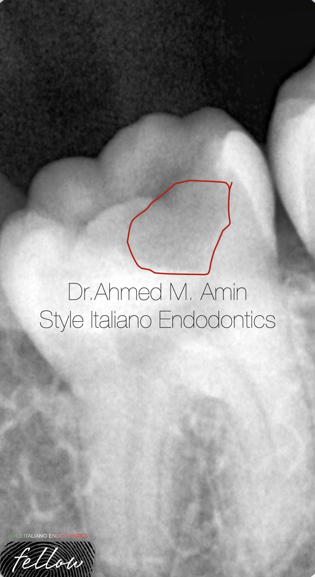
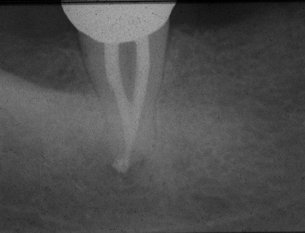
Fig. 1
Pre-op. Radiograph showing Mesial caries extended to mesial pulp horn leading to Irreversible Pulpitis “ also the patient was a Dentist “
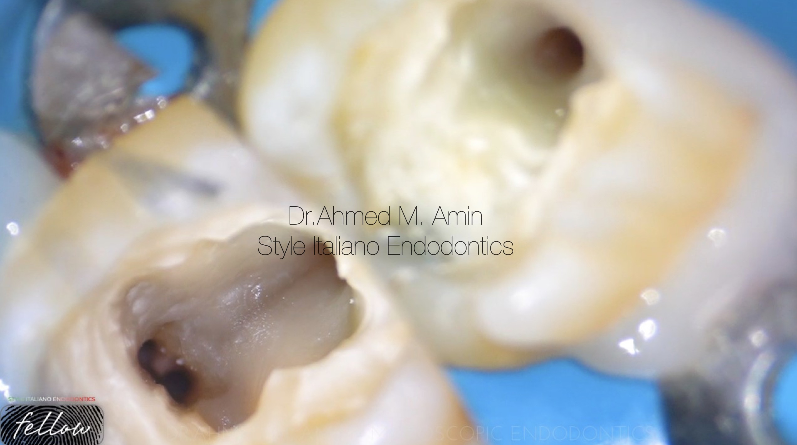
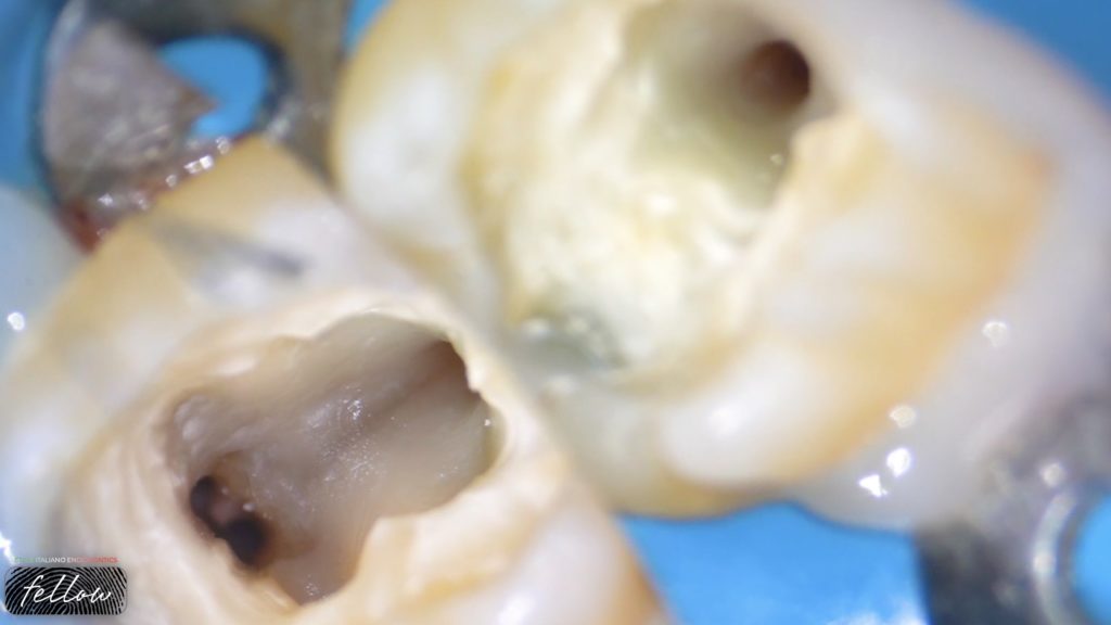
Fig. 2
Access Cavity
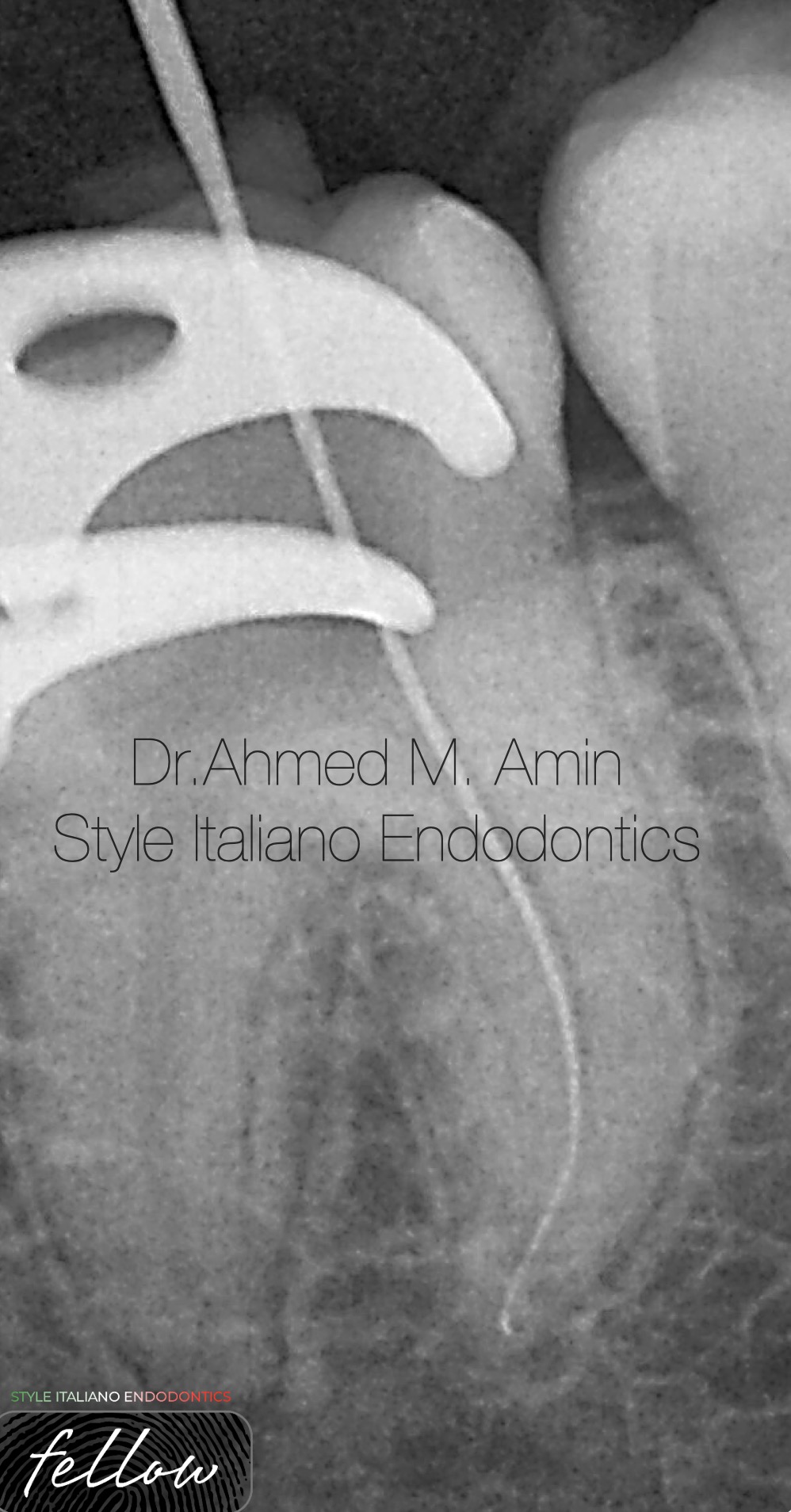
Fig. 3
Working length determination
“ the difficuilty here was the curvature & tightness of the mesials so , um used d-finder files in sequence 8,10,12 till 15 in order to get patency “
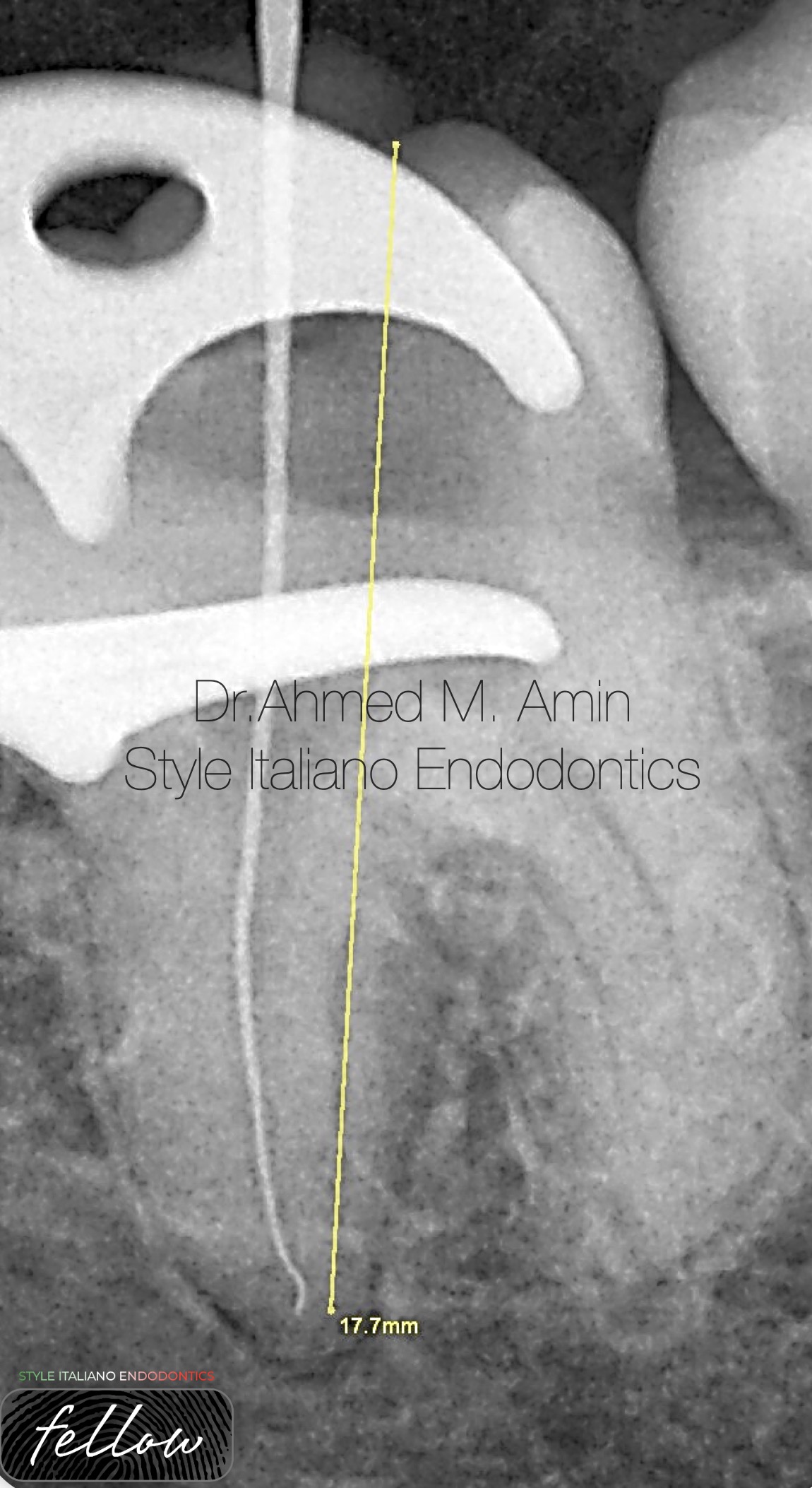
Fig. 4
Working length determination for distal
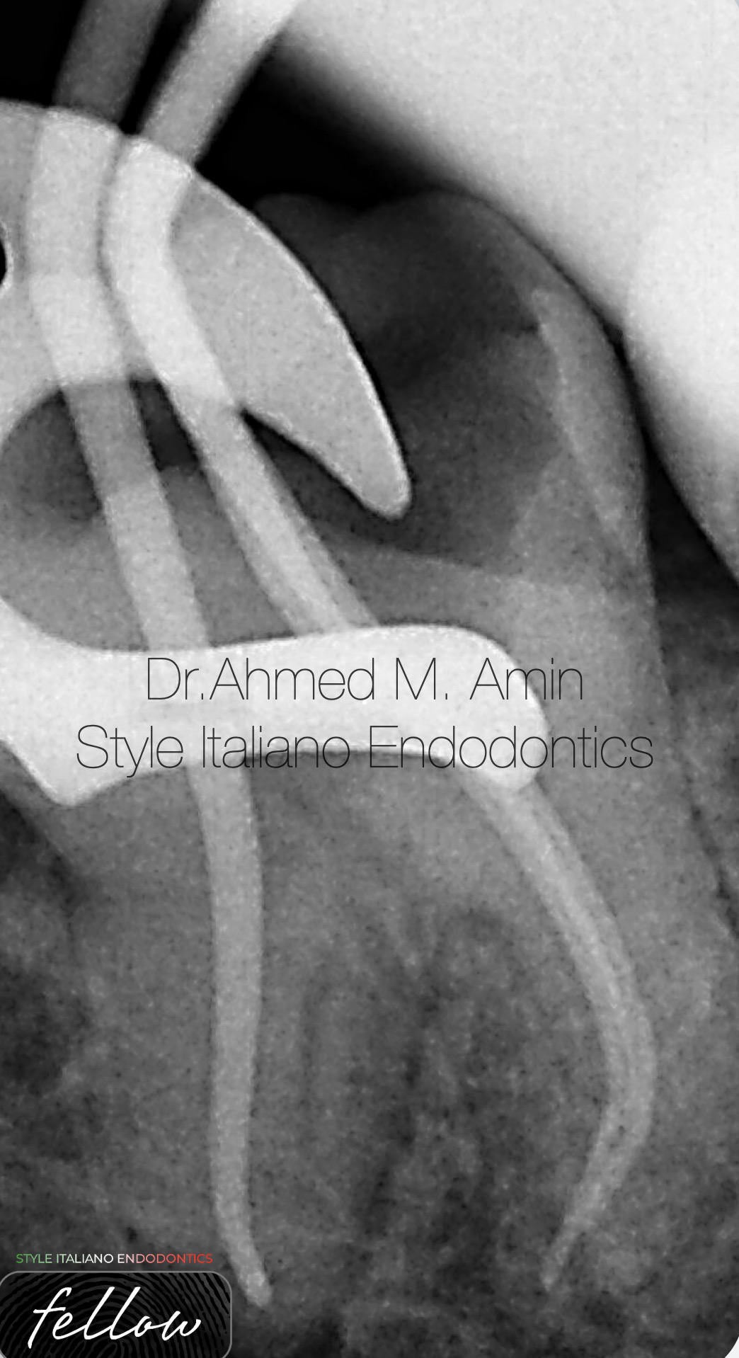
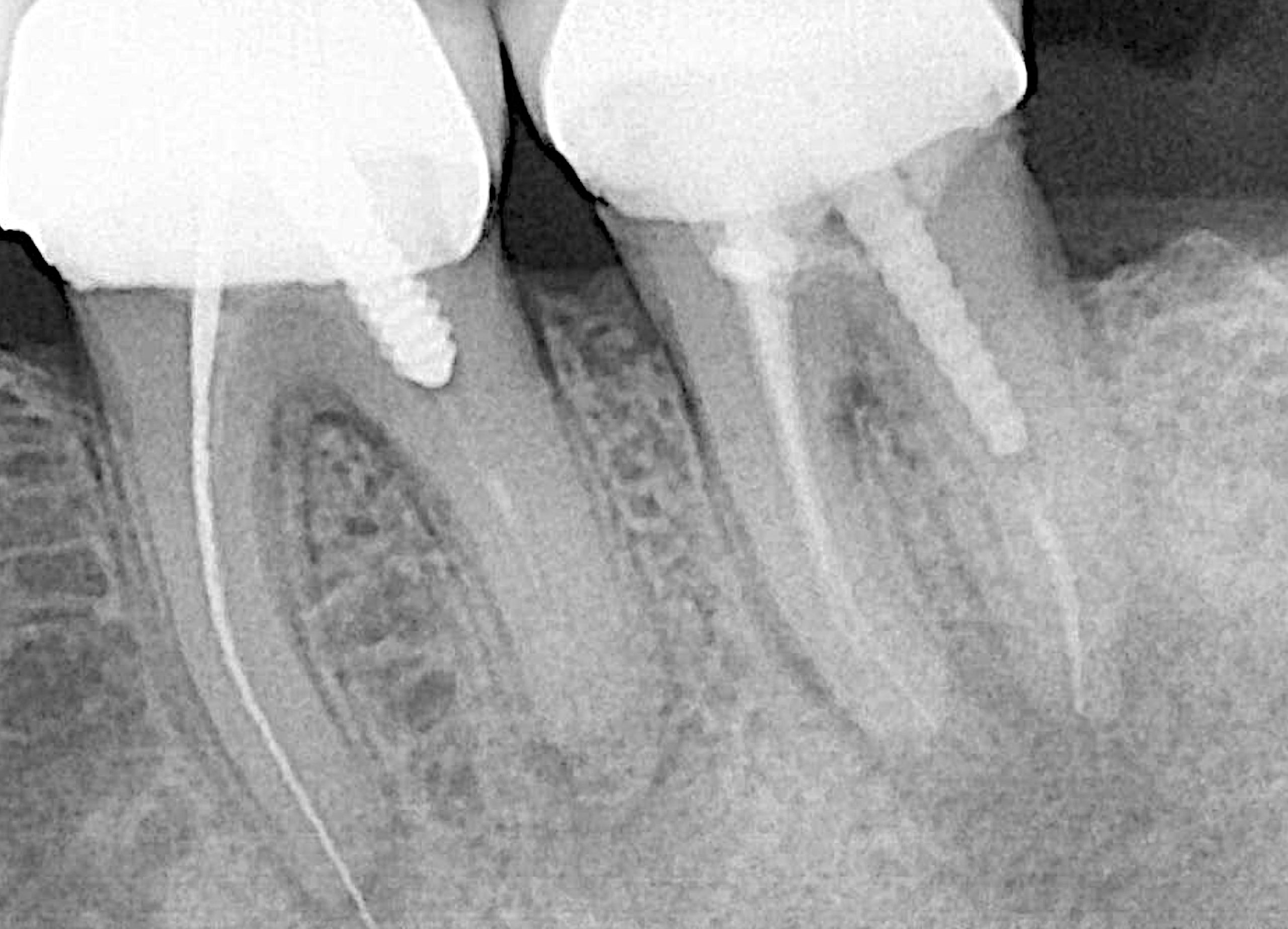
Fig. 5
Cone Fitting
“ now u can notice the severe curvature of mesials regardless of limited mouth opening of the patient during preparation “
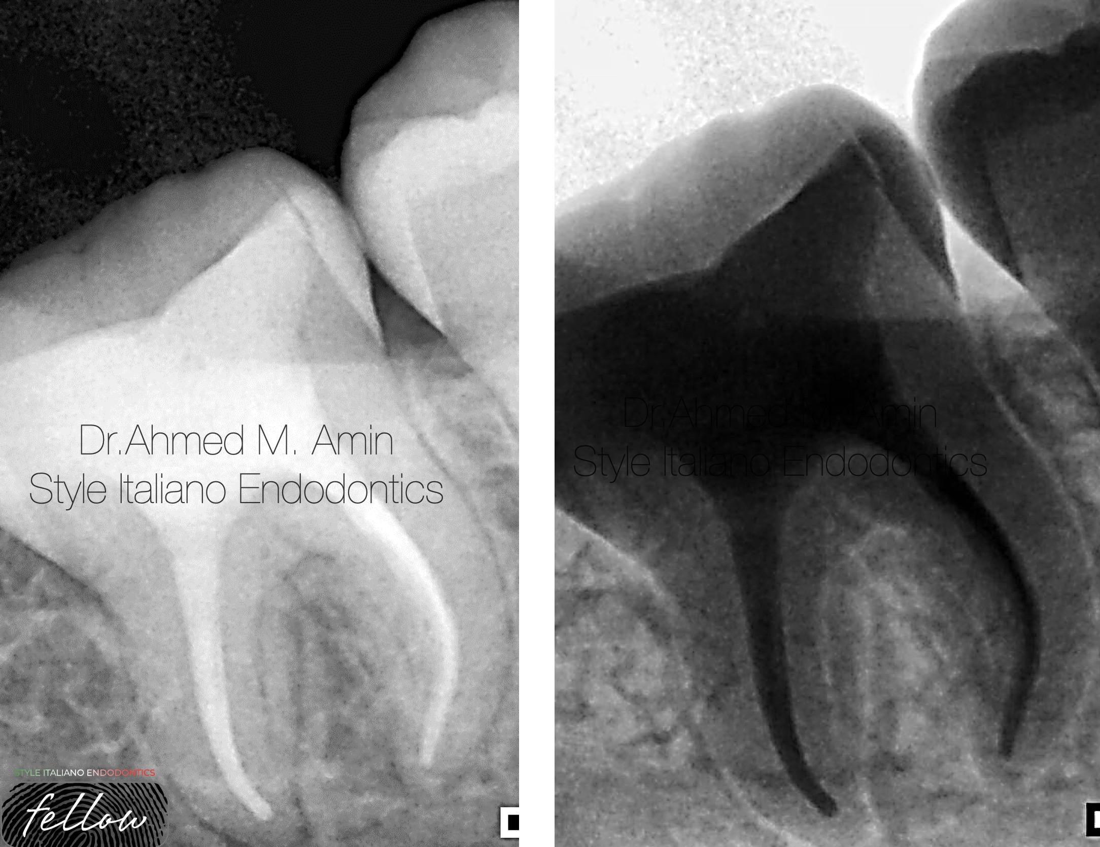
Fig. 6
Final Obturation “ Radiograph “
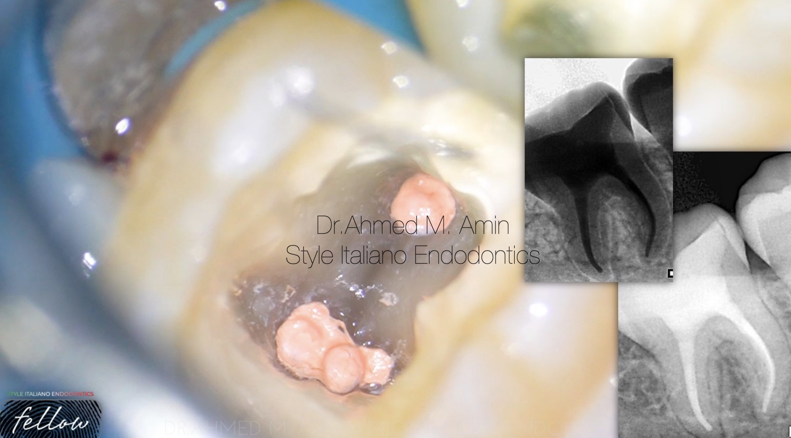
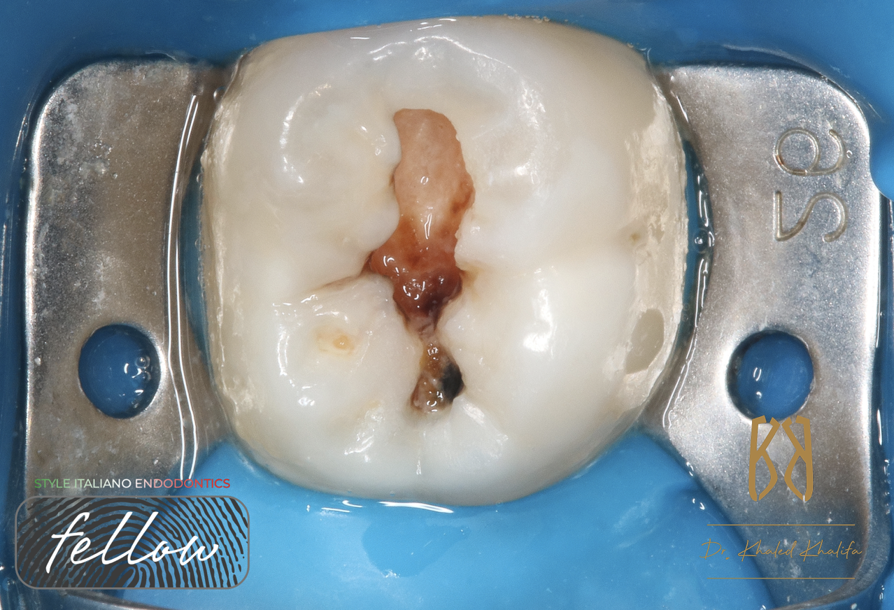
Fig. 7
Final Obturation “ Clinical “
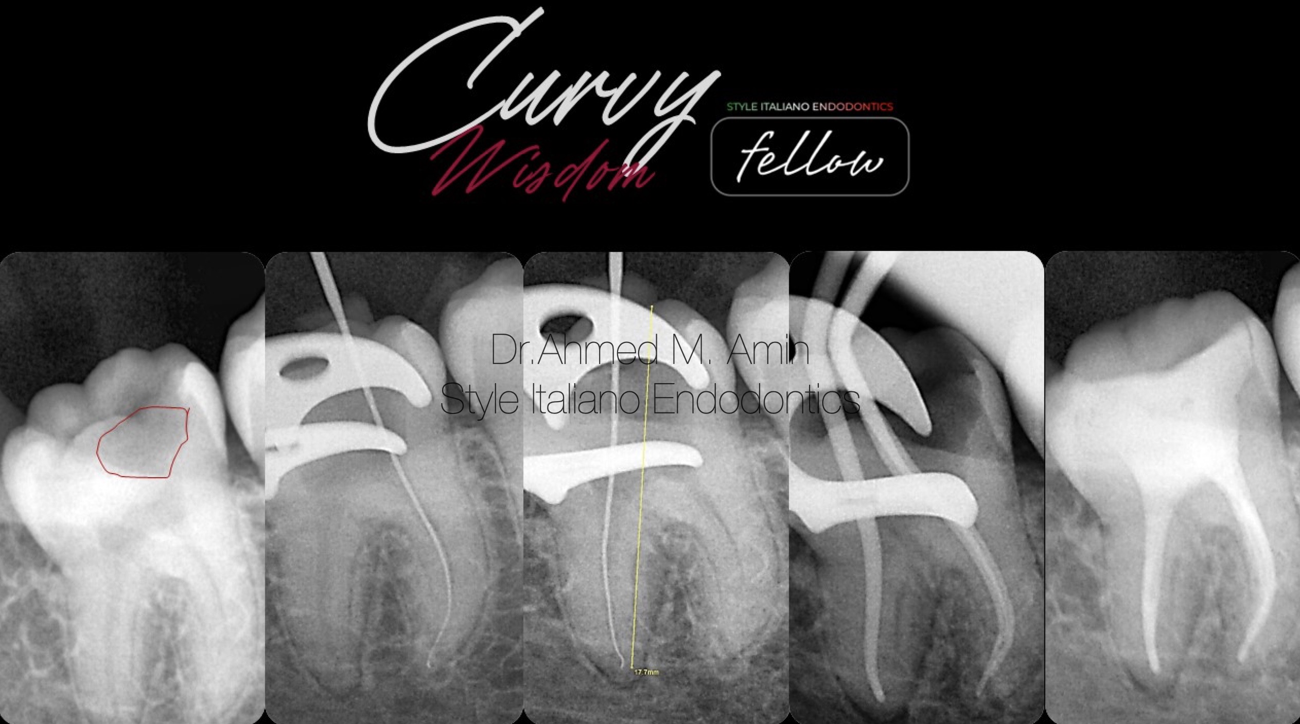
Fig. 8
Case Workflow
A short film showing the obturation Process
 : eval()'d code</b> on line <b>19</b><br />
)
Fig. 9
Conclusions
Endodontic treatment of third molars with varying root-canal morphology and difficult access can be challenging. Nevertheless, modern techniques and devices make third molar endodontics predictable
The root canal morphology of third molars shows an increased likelihood for aberrations, such as dilacerations, C-shaped canals and unpredictable morphological features, that should be identified accurately before commencing endodontic treatment. Intelligent evaluation of clinical and radiographic data makes possible to understand the unpredictable anatomy of these teeth. Magnification and coaxial illumination allow the identification of canal orifices, along with the "Laws" postulated by Krasner and Rankow.
Bibliography
Ahmed HM. Management of third molar teeth from an endodontic perspective. Eur J Gen Dent 2012;1:148-60
Sidow SJ, West LA, Liewehr FR, Loushine RJ. Root canal morphology of human maxillary and mandibular third molars. J Endod. 2000; 26(11):675-8.
Plotino G. A mandibular third molar with three mesial roots: A case report. J Endod 2008;34:224-6.
Grover AK, Grover SK . Mandibular third molar-endodontic perspective. Endodontology. 1992 Jun.; 4(1): 31-6
Krasner P, Rankow HJ. Anatomy of the Pulp-Chamber Floor. J Endod 2004; 30: 516.
Krithikadatta J, Kottoor J, Karumaran CS, Rajan G. Mandibular first molar having an unusual mesial root canal morphology with contradictory cone-beam computed tomography findings: a case report. J Endod. 2010;36(10):1712-6.