Premolars with 4 canals is not a daily case
10/12/2023
Fellow
Warning: Undefined variable $post in /home/styleendo/htdocs/styleitaliano-endodontics.org/wp-content/plugins/oxygen/component-framework/components/classes/code-block.class.php(133) : eval()'d code on line 2
Warning: Attempt to read property "ID" on null in /home/styleendo/htdocs/styleitaliano-endodontics.org/wp-content/plugins/oxygen/component-framework/components/classes/code-block.class.php(133) : eval()'d code on line 2
The purpose of root canal treatment is to clear pathogenic microbes and infected pulp in the root canal, prevent it from producing toxic products, and protect the periapical tissue. The presence of root canal variation increases the difficulty of this treatment. It has been reported that 42% of retreatment cases are due to missing canals. Therefore, there is great clinical significance for dentists to master the morphological characteristics of root canals. Branch root canals are common morphological variations in root canal systems, whose morphology can present either as one or more small lateral branches from the main root canal or as two equally large bifurcated root tips on the apical segment.
The anatomical variations in the root canal system of the mandibular premolar are known. Mandibular second premolars typically have one root canal with a reported prevalence of 98.8%. However, it is quite common for the mandibular premolars II to have two canals; with an occurrence range of 1.2% to 29%. Amongst those with three canals, the prevalence varies from 0.4% to 0.5%, and four/five canals have been reported only in case reports.
Therefore, present case shows premolars with 4 canals to provide a reference for similar cases.
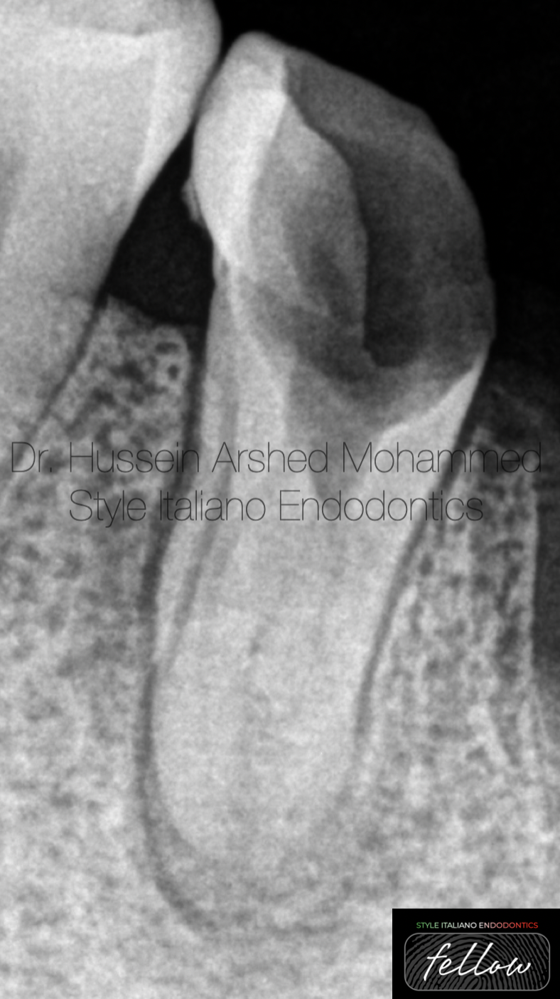
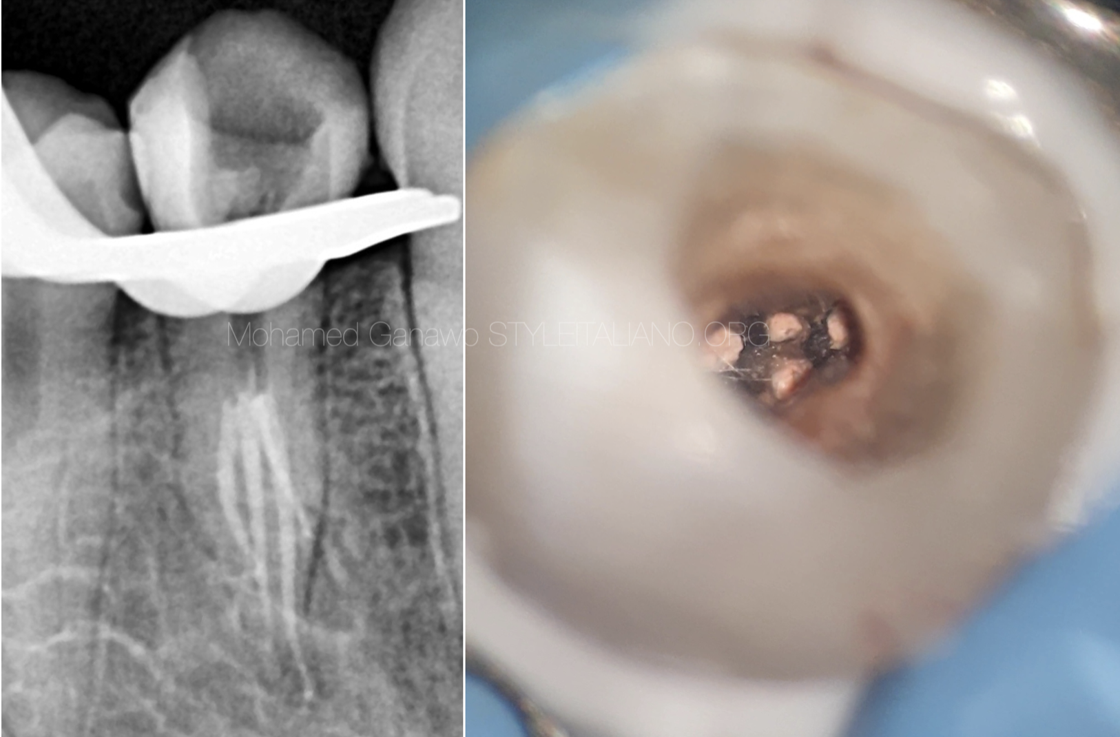
Fig. 1
Pre-op X-Ray showed lower second premolar with destructive crown with fast-breack phenomenon.
In this type of configuration there is expectation of extra anatomy
Diagnostic radiograph revealed there is periodical radiolucent area
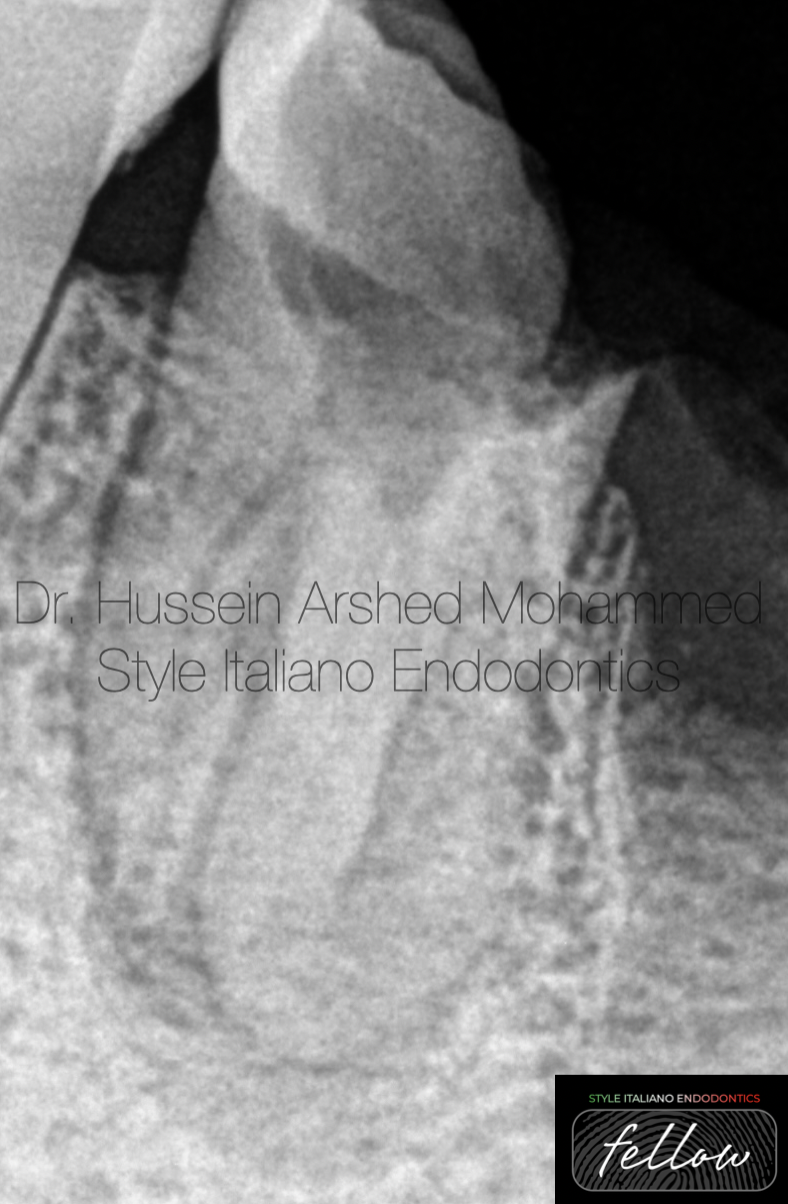
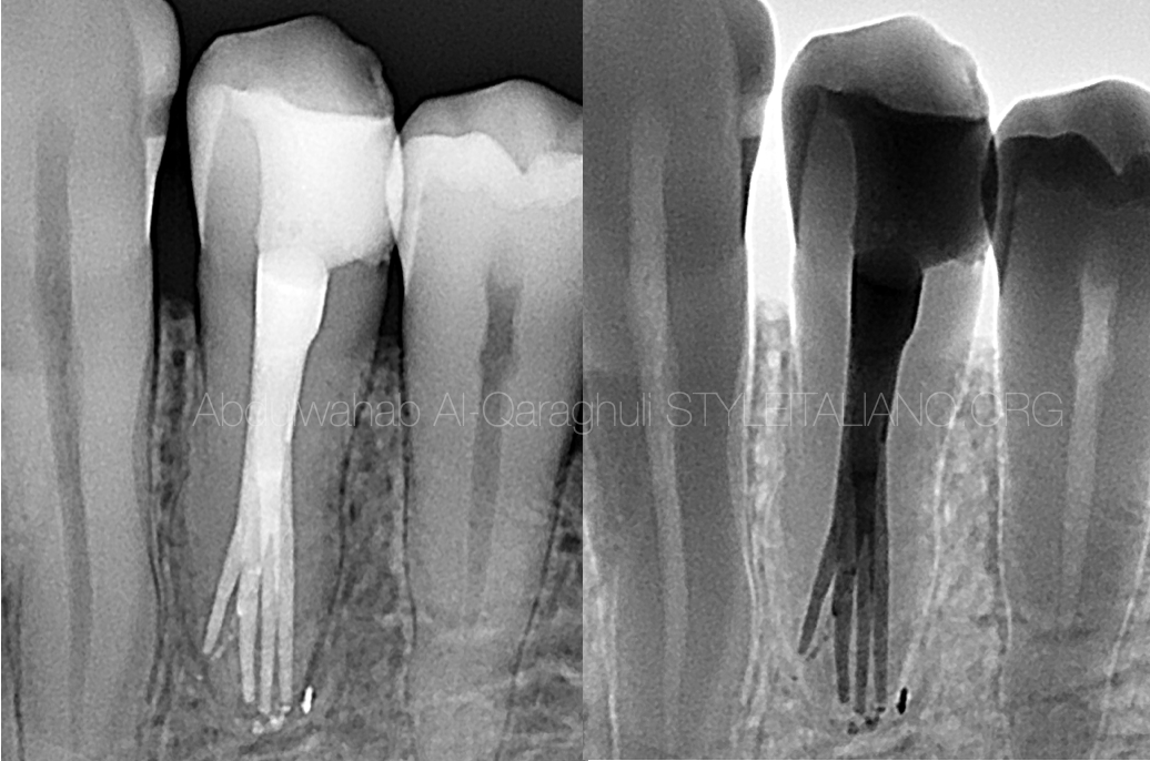
Fig. 2
After distal shift X-Ray showed there is two roots (buccal and lingual).
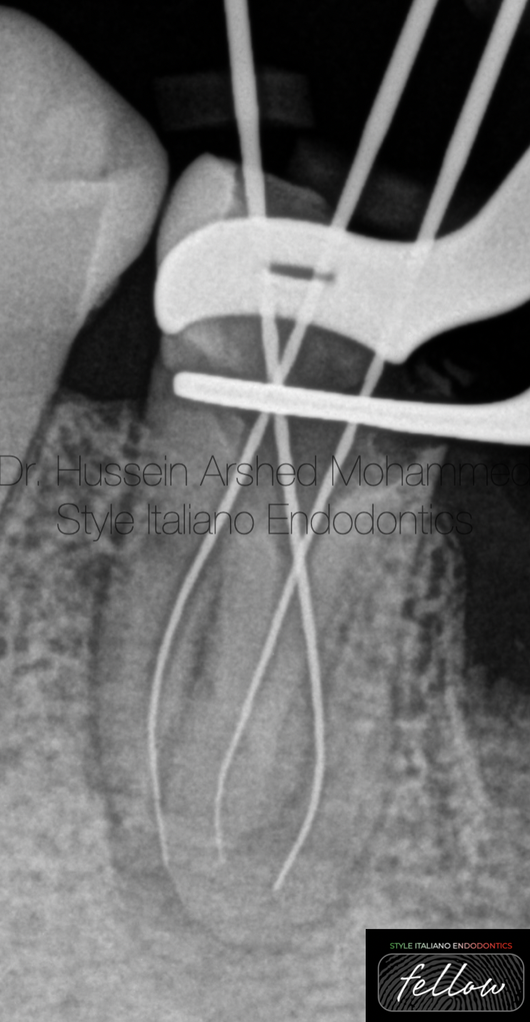
Fig. 3
Negotiation of the canals.
There is one lingual and two buccal orifices.
The buccal canals were very tinny and patent.
Working length was determined by apex locator and confirmed by x-ray for all the canals.
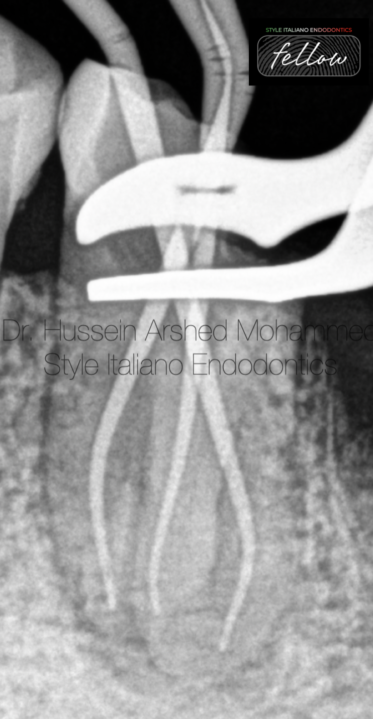
Fig. 4
Shaping till the full working length 30/04 for all the canals and master apical cone confirmed.
The shaping protocol :-
- coronal flaring with orifice opener file
- D finder file # 10.
- Rotary file 15/03
- Rotary file 17/04
- Rotary file 25//04
- Rotary file 30/04
With copious amount of NaOCl 5.25% between each file, EDTA 1cc for all the canals with sonic activation for 1 minutes to remove the smear layer and followed again with NaOCl to disinfect again
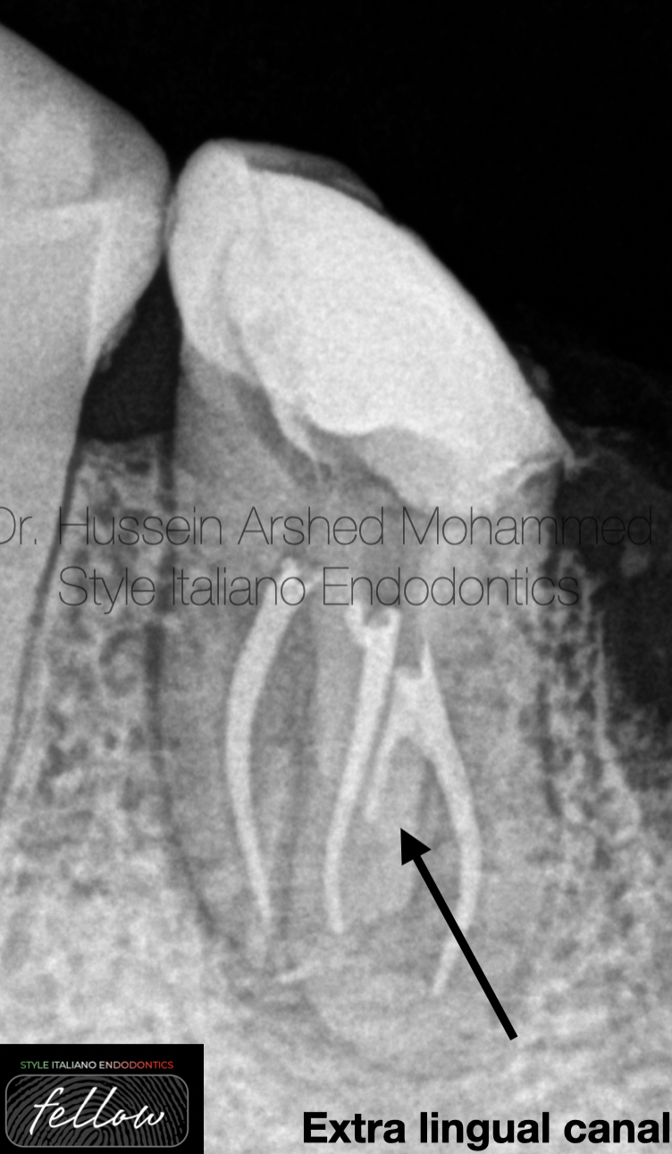
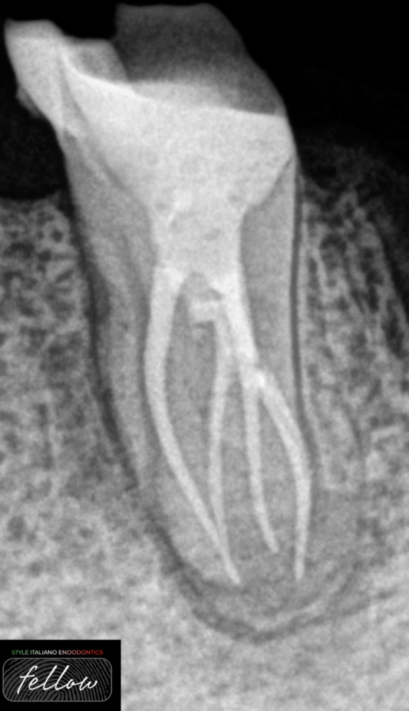
Fig. 5
Obturation of the canals by single cone with neosealer bioceramic sealer from Avalon Biomed. After cutting the guetta-percha with obturation pen with the confirmation of the final x rays deep apical split of the lingual canal by the flowability of the bioceramic sealer, this indicate there is extra mesiolingual canal.

Fig. 6
Removal of the temporary filling material, coronal flaring of the lingual canal with pesso reamer size 2 then negotiation of the mesiolingual canal with D finder file #10, instrumentation till 30/04 with irrigation as previously described.
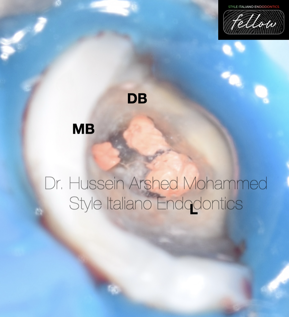
Fig. 7
Sealing of the orfices with fast fill softened guetta-percha
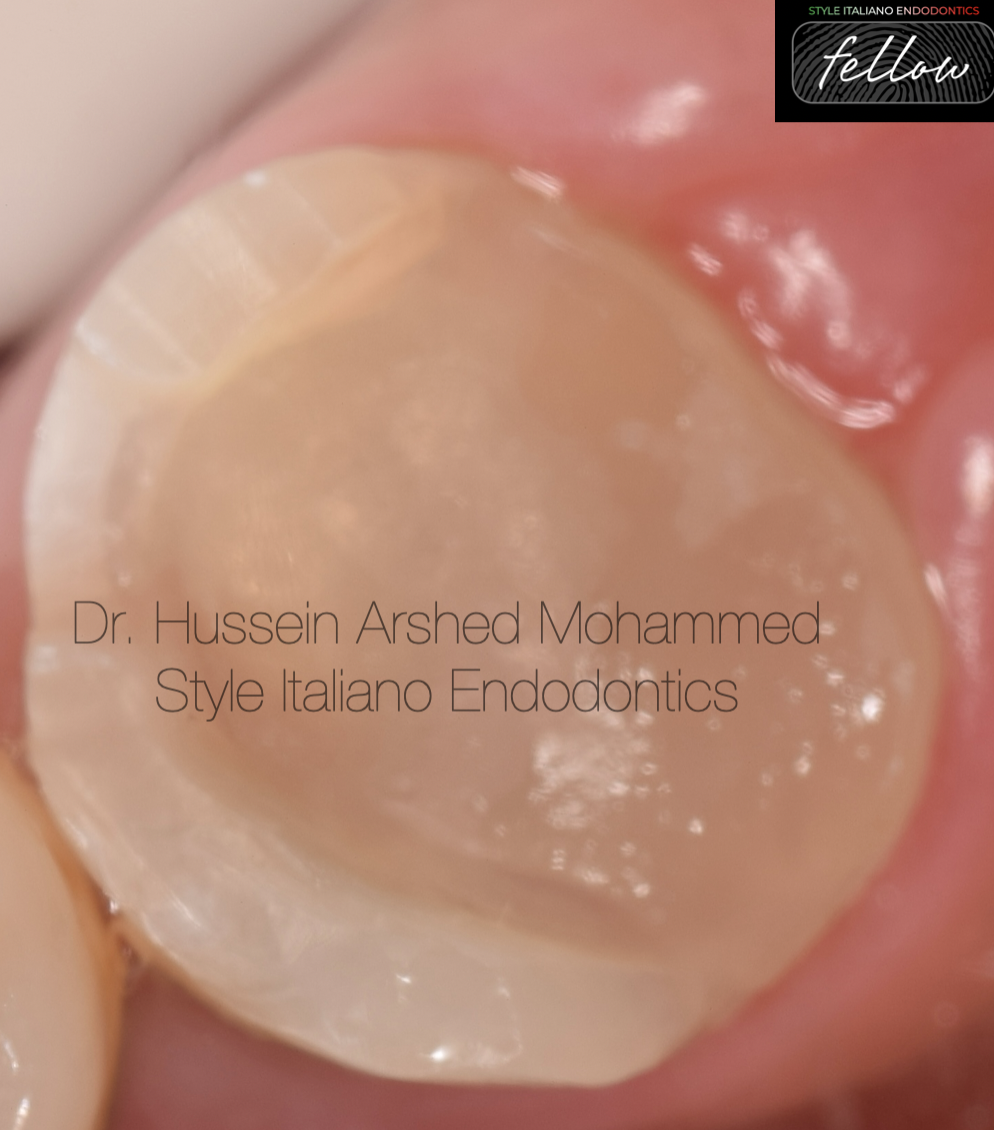
Fig. 8
Reduction of the unsupported enamel, marginal elevation of the distal and lingual gingival seats with flowable composite, replacement of the dentin with Ever-x fiber re-inforced composite to make biobase for the indirect restoration.
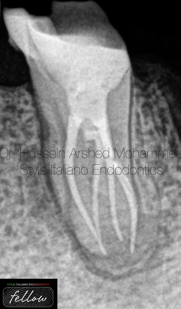
Fig. 9
Mesial shift post-operative X-ray.
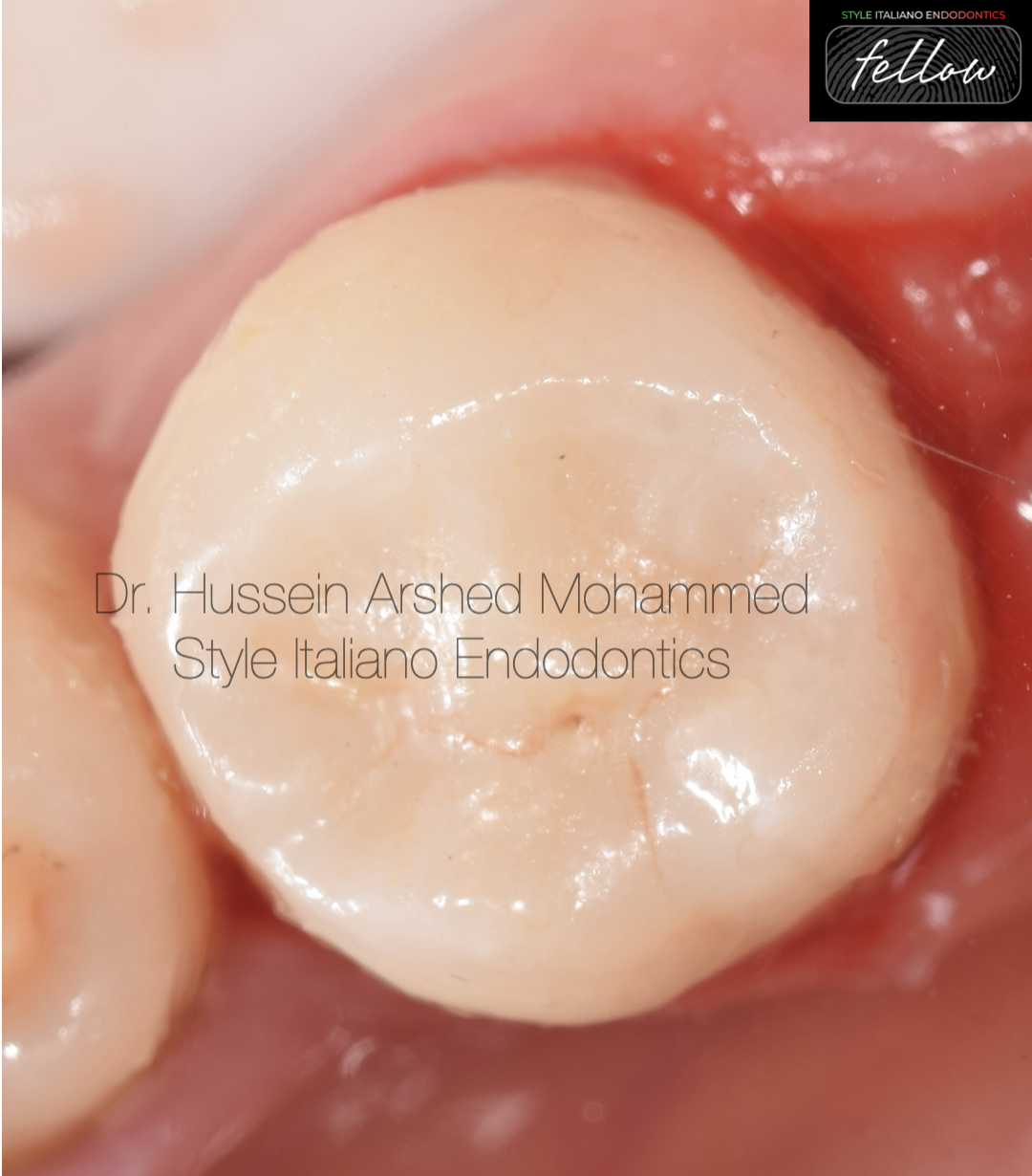
Fig. 10
Cementation of the indirect overlay with resin cement
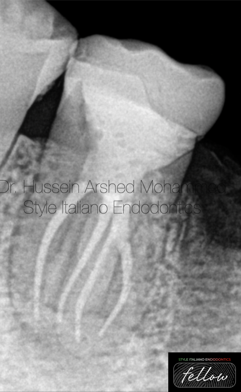
Fig. 11
Distal shift post-operative X-ray with final restoration.
Fig. 12
About the author:
Dr. Hussein Arshed graduated in 2012 from Mustansyria university in Iraq.
- in 2018, he got high diploma in Endodontics from Mustansyria university
- In 2023, he got master degree in Endodontics from Mustansyria university
- He works in Rivan dental clinic in Baghdad
- He has been offering lectures, training courses, webinars for dentists.
- He is international lecturer and gives many lectures locally and internationally.
- He is a fellow member of Style Italiano Endodontics.
- He is key opinion leader for Avalon Biomed company in Iraq.
Conclusions
This case report presents the successful endodontic treatment of a mandibular second premolar with Four Canals and provides a reference for other similar cases. Before starting root canal treatment it is essential to always consider variations in pulp anatomy and morphology. using magnification tools, to magnify the internal anatomy of a root canal increases the probability of finding additional canals. Sometimes the fowability of the sealer can also aid to detect there is apical splitting of the canal.
Bibliography
- Patel S, Dawood A, Ford TP, et al. The potential applications of cone beam computed tomography in the management of endodontic problems. Int Endod J. 2007;40(10):818–30
2. Zhang R, Wang H, Tian YY, et al. Use of cone-beam computed tomography to evaluate root and canal morphology of mandibular molars in Chinese individuals. Int Endod J. 2011;44(11):990–9.
3. Siqueira JF Jr. Aetiology of root canal treatment failure: why well-treated teeth can fail. Int Endod J. 2001;34(1):1–10.
Cohen S, Hargreaves KM. Pathways of the Pulp. 9th ed: St Louis: Elsevier Mosby; 2006. p. 216-7
4. Vertucci FJ. Root canal anatomy of the human permanent teeth. Oral Surg Oral Med Oral Pathol. 1984;58(5):589–99
Yoshioka T, Villegas JC, Kobayashi C, et al. Radiographic evaluation of root canal multiplicity in mandibular first premolars. J Endod. 2004;30(2):73–4
5. Yang L, Han J, Wang Q, Wang Z, Yu X, Du Y. Variations of root and canal morphology of mandibular second molars in Chinese individuals: a cone-beam computed tomography study. BMC oral health. 2022;22(1):1–12.