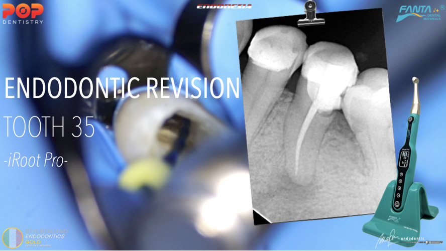
ENDODONTIC REVISION ON DEEP SPLIT PREMOLAR: A Case Report
29/11/2023
Marino Sutedjo
Warning: Undefined variable $post in /home/styleendo/htdocs/styleitaliano-endodontics.org/wp-content/plugins/oxygen/component-framework/components/classes/code-block.class.php(133) : eval()'d code on line 2
Warning: Attempt to read property "ID" on null in /home/styleendo/htdocs/styleitaliano-endodontics.org/wp-content/plugins/oxygen/component-framework/components/classes/code-block.class.php(133) : eval()'d code on line 2
Patient came to my clinic with main symptoms of spontaneous pain on his lower left. Tooth was not be able to use for eating.
From clinical examination, tooth 35 was very tender to percussion. Palpation on buccal side of tooth 35 was also very tender. There was a class II (disto occlusal) composite restoration on this tooth.
Radiographic examination was taken to evaluate the main cause of the problem (image 1)
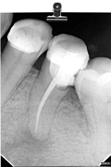
Fig. 1
From the PA radiograph, we can immediately see:
1.Endodontically treated tooth with poor marginal restoration on distal area that can lead to leakage
2. Periapical lesion
3. 1 canal has been treated, but the position of the canal it doesn’t look right. It’s not located centrally from the apex of the root
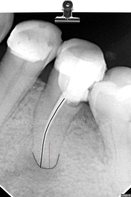
Fig. 2
Black Line:
Showed the projection of the treated canal and root apex. It doesn’t located centrally from the apex
Red Line:
Possible missed canal
After seeing the PA radiograph and missed canal suspected, cbct examination was then performed to confirmed this.
Cbct scan in axial view showed confirmation of missed canal at the lingual. The root canal started 1 canal at the orifice level and than at the apical 1/3, the root canal split into two canals.
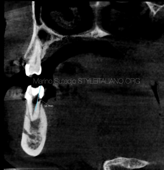
Fig. 3
From the sagital view, we can identified the opening of missed lingual canal was at 13mm from the lingual cusp.
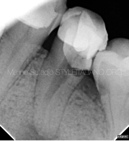
Fig. 4
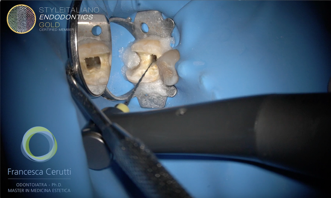
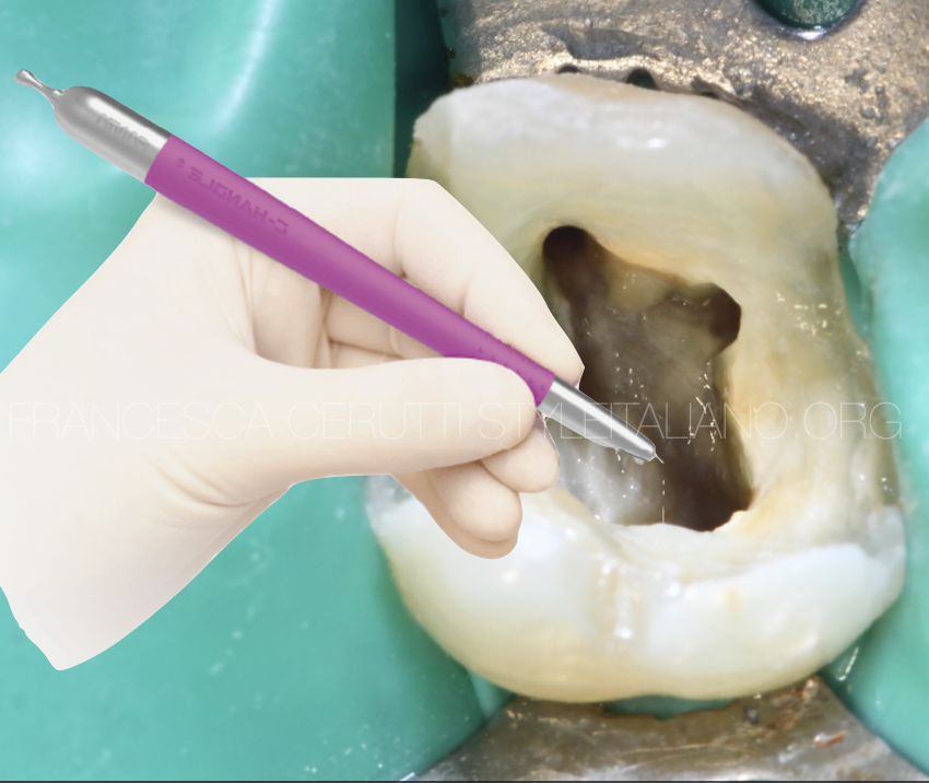
After removal of root canal filling material, the split level was seen. Access refining to create radicular access need to be made to find the entrance of the orifice of lingual canal
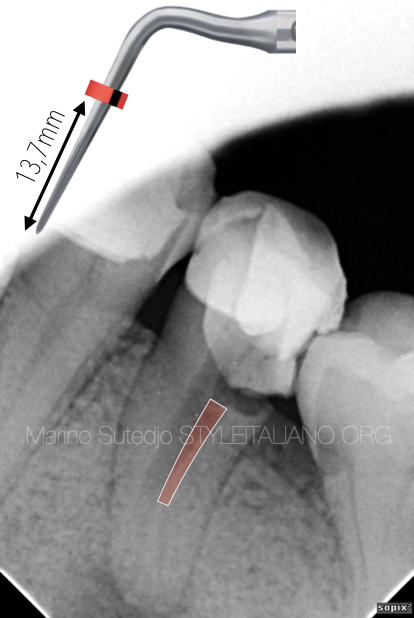
Fig. 5
Access refining area
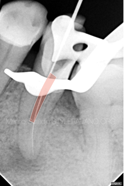
Fig. 6
After access refining, lingual canal found
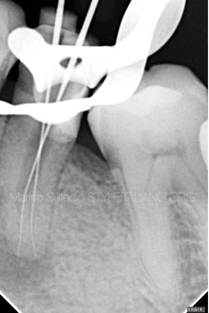
Fig. 7
Radiograph confirmation of 2 canals
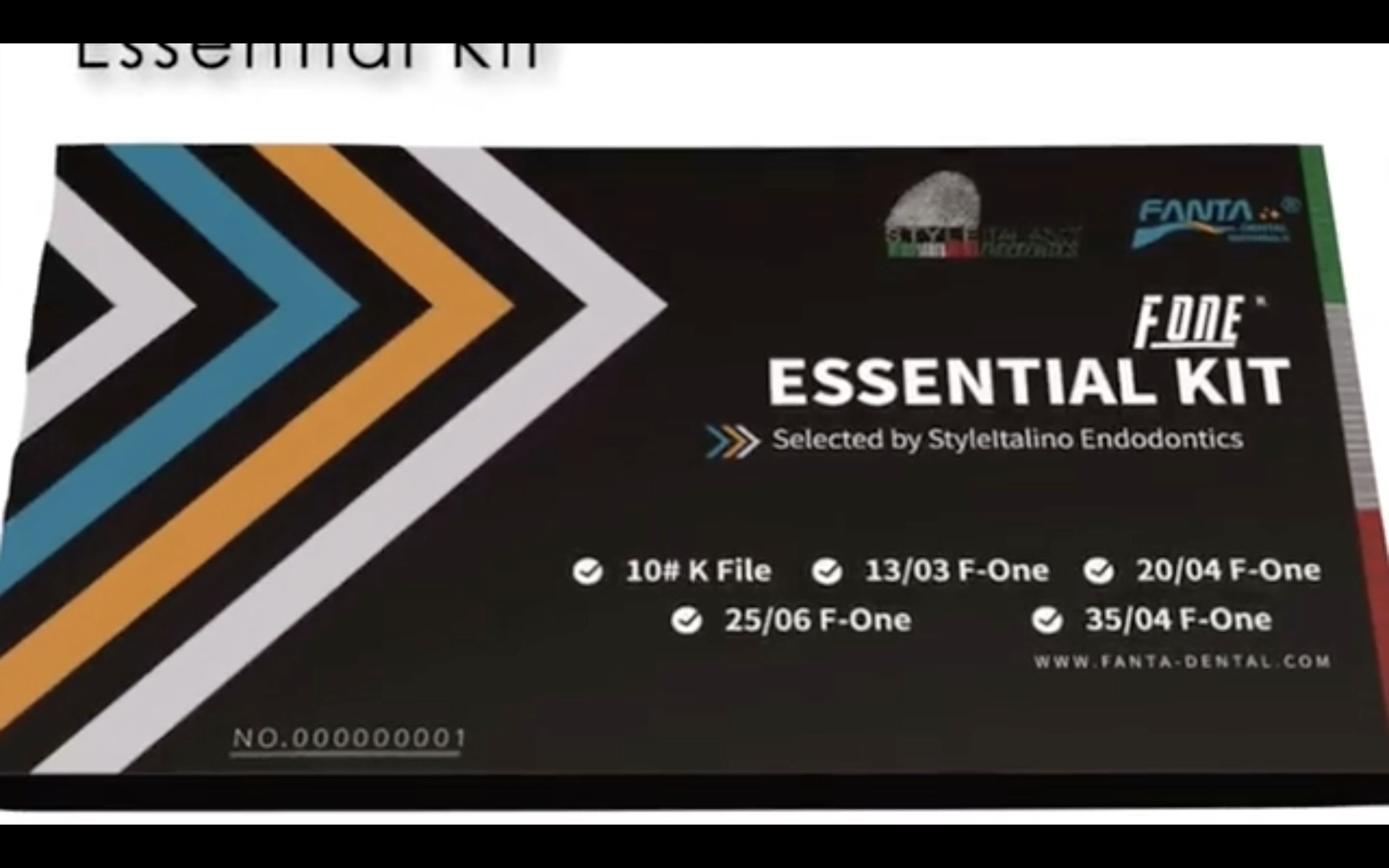
Due to difficult entrance of the canal, a precurved martensitic file used for better and easier insertion of instruments. In this video AF Blue S One Rotary File was prebend and used to shape the canal.
In this case, AF Blue S One Rotary file was used with new endodontic motor from FANTA, that has several advantages that are very useful in endodontic treatment. Some important features can be seen in this video
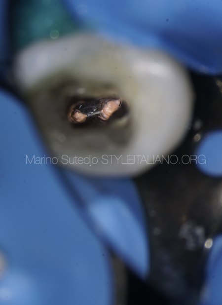
Fig. 8
After obturation of 2 canals with BC sealer and cold molding technique up until the split level
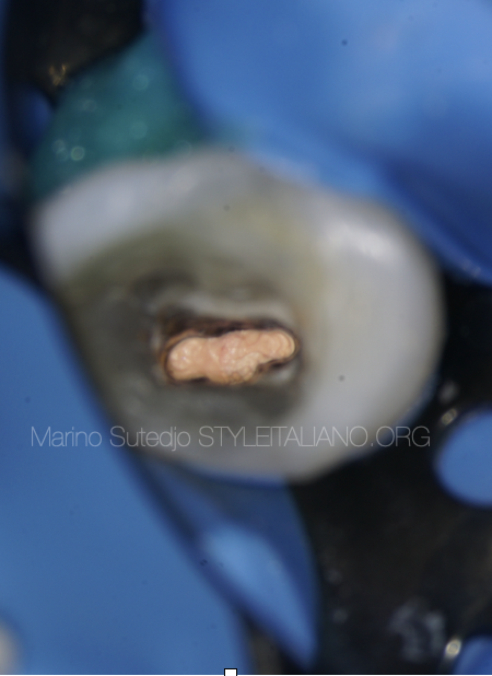
Fig. 9
Injectable heated gutta percha than used to fill remaining space of the root canal
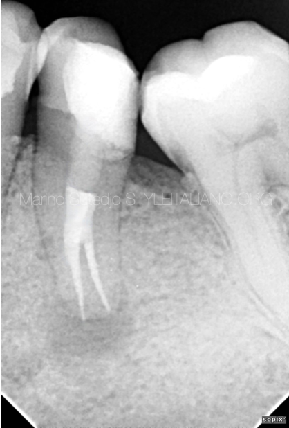
Fig. 10
Post op radiograph (1) :
2 canals were obdurated, marginal restoration was revised and tooth scheduled for indirect restoration
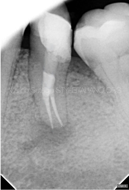
Fig. 11
Post op radiograph (2)
Conclusions
Endodontic retreatment is not just retreating, but also revised every aspect that need to be revised. Choosing the right method of treatment, the right instruments and the right equipment is necessary to produce adequate treatment.
And once again root canal anatomy is always a challenge and it’s constant, therefore we will always be a student for endodontic anatomy