Endodontic surgery of the lower incisor: how important is it to verify the presence of a lingual canal?
06/11/2023
Fellow
Warning: Undefined variable $post in /home/styleendo/htdocs/styleitaliano-endodontics.org/wp-content/plugins/oxygen/component-framework/components/classes/code-block.class.php(133) : eval()'d code on line 2
Warning: Attempt to read property "ID" on null in /home/styleendo/htdocs/styleitaliano-endodontics.org/wp-content/plugins/oxygen/component-framework/components/classes/code-block.class.php(133) : eval()'d code on line 2
A patient came to my attention due to pain in the lower anterior area and the presence of a fistula. She reported having undergone endodontic surgery years ago. The X-ray revealed the presence of a radiolucent area compatible with apical periodontitis and a radiopaque area that may correspond to the endodontic surgery mentioned by the patient.
Thus, a surgical re-intervention was necessary to improve the current situation.
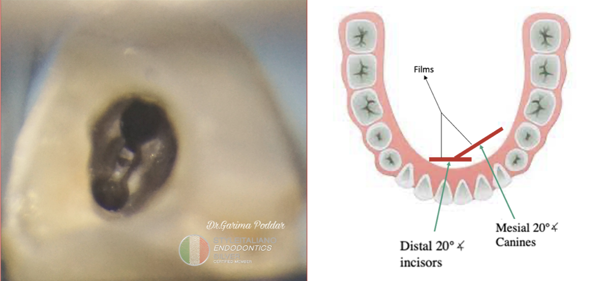
It is crucial to have a thorough understanding of radicular anatomy and the prevalence of any anatomical variations before doing a case of surgical or non-surgical failure.
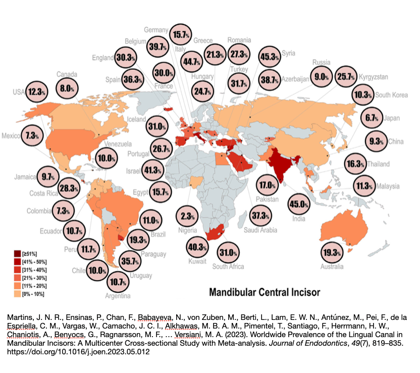
Fig. 1
The worldwide prevalence of the lingual canal in mandibular central incisors was 21.9%, ranging from 2.3% in Nigeria to 45.3% in Syria. For lateral incisors, the global prevalence was 26.0%, ranging from 2.3% in Nigeria to 55.0% in India. The overall difference in prevalence between the two types of teeth was not statistically significant.
The most common root canal configuration was the presence of two confluent canals merging into a single foramen, with central incisors at 20.5% and lateral incisors at 24.1%. Multiple root canals or double-root anatomy were rare findings.
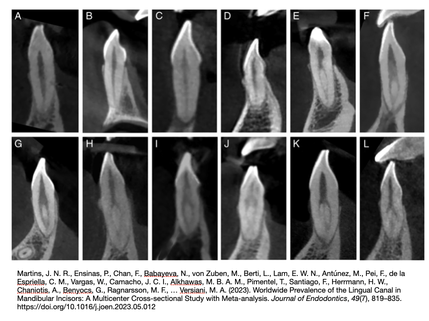
Fig. 2
Representative images of various root canal anatomic configurations found in different countries. The configurations include a single canal noted in Belgium (A ), Argentina (B ), Syria (C ), and Malaysia (D ); two independent canals with single exit observed in England (E ), Turkey (F ), South Africa (G ), France (H ), and Peru (I ); two independent canals with multiple exit recorded in Portugal (J ); more than two canals observed in Brazil (K ); and a 2-rooted incisor found in Spain (L ).
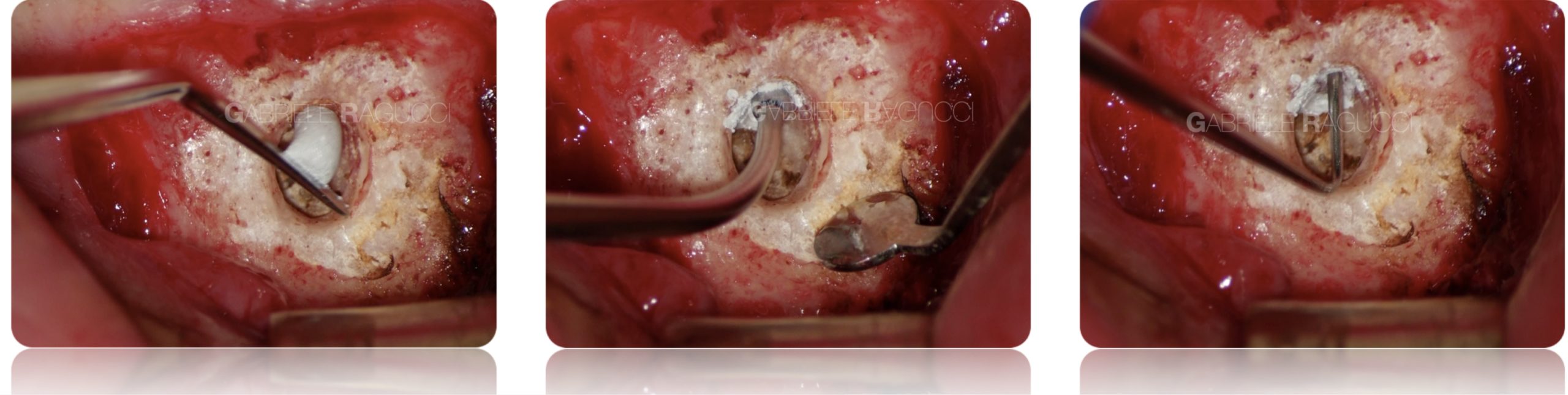
The importance of proper apical resection and the use of adequate magnification allows for the identification of any accessory lingual canals and their correct management.
Finally, to prevent visible scars, proper management of soft tissues is required, along with the use of appropriate sutures (in this case, non-absorbable 6-0 sutures).
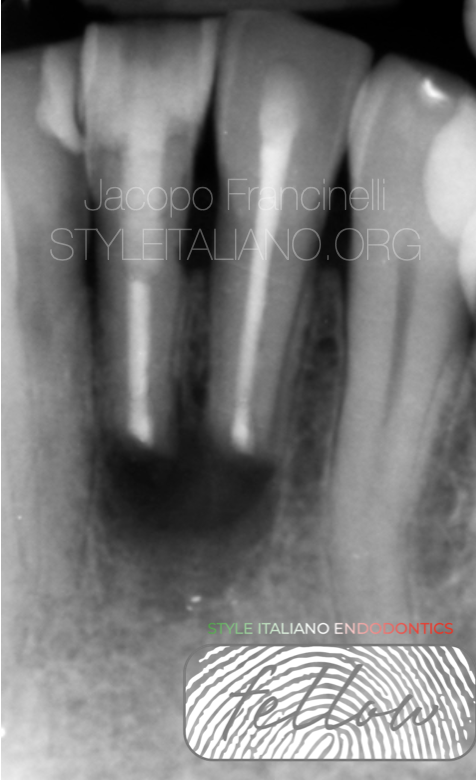
Fig. 3
The final X-ray shows the correct apical resection and the proper obturation of the two canals and the isthmus that connects them.
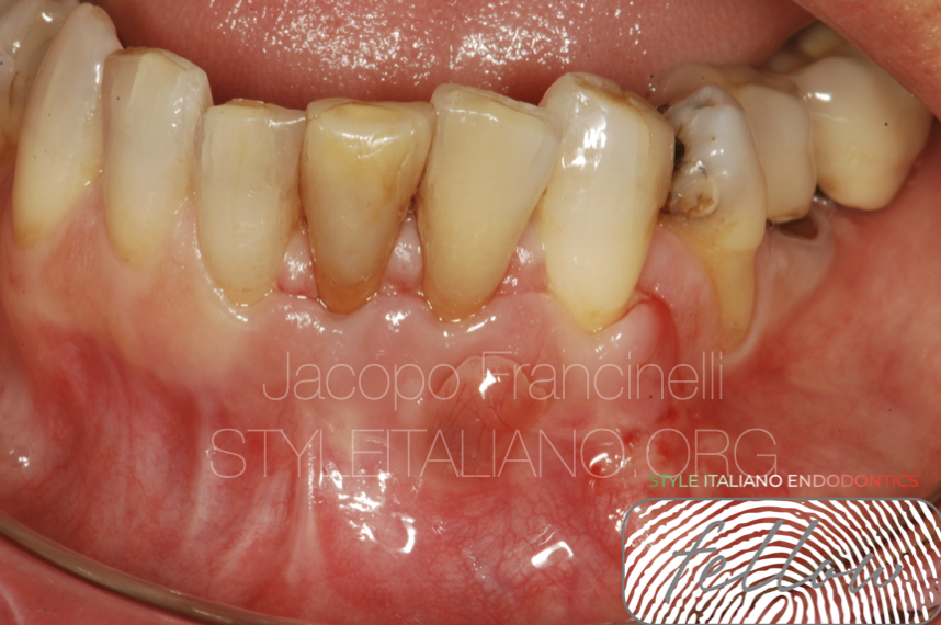
Fig. 4
Sutures removed after 72 hours.
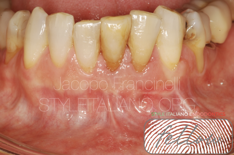
Fig. 5
Healing of the tissues 20 days later.

Fig. 6
About the author:
Jacopo Francinelli
- Degree with honors in Brescia in 2018
- Numerous private courses in the field of endodontics
- 2020 2nd level post-graduate Master in Clinical and Microsurgical Endodontics at the University of Brescia with full marks and honors
- from 2019 he is clinical tutor in the 3rd year of Dentistry alongside Prof. Salgarello
- from 2021 he is tutor for the aforementioned university master
- He collaborates with the Spedali Civili of Brescia as a freelancer
- He has published articles in national and international journals
- Contract professor for the University of Brescia
- Member of the Italian Academy of Microscopic Dentistry
- Collaborate with colleagues in some private courses focused on endodontics
- He carries out his freelance activity in the province of Brescia dedicating himself mainly to micro-endodontics
Conclusions
The importance of having a proper knowledge of anatomy and the use of magnification systems allows us to manage complex cases and manage any prior failures effectively.
Bibliography
Martins, J. N. R., Ensinas, P., Chan, F., Babayeva, N., von Zuben, M., Berti, L., Lam, E. W. N., Antúnez, M., Pei, F., de la Espriella, C. M., Vargas, W., Camacho, J. C. I., Alkhawas, M. B. A. M., Pimentel, T., Santiago, F., Herrmann, H. W., Chaniotis, A., Benyocs, G., Ragnarsson, M. F., … Versiani, M. A. (2023). Worldwide Prevalence of the Lingual Canal in Mandibular Incisors: A Multicenter Cross-sectional Study with Meta-analysis. Journal of Endodontics, 49(7), 819–835. https://doi.org/10.1016/j.joen.2023.05.012
Setzer, F. C., & Kratchman, S. I. (2022). Present status and future directions: Surgical endodontics. In International Endodontic Journal (Vol. 55, Issue S4, pp. 1020–1058). John Wiley and Sons Inc. https://doi.org/10.1111/iej.13783
S. Friedman. The prognosis and expected outcome of apical surgery. Endod Topics, 11 (2005), pp. 219-262