Micro apical surgery by SIE kit
24/12/2020
Zaher Al-taqi
Warning: Undefined variable $post in /home/styleendo/htdocs/styleitaliano-endodontics.org/wp-content/plugins/oxygen/component-framework/components/classes/code-block.class.php(133) : eval()'d code on line 2
Warning: Attempt to read property "ID" on null in /home/styleendo/htdocs/styleitaliano-endodontics.org/wp-content/plugins/oxygen/component-framework/components/classes/code-block.class.php(133) : eval()'d code on line 2
Apical surgery is an option for the management of endodontically-treated tooth with persistent periapical lesions or symptom/sign. Several epidemiological studies have suggested that 33–60% of endodontically-treated teeth still presented the pictures of apical periodontitis. The possible causes may be persistent primary infection, secondary infection after endodontic therapy, vertical root fracture or cemental tears. The success rate of apical surgery was reported to range from 37% to 91%. Complete apical healing had been observed in 37%–96% of the endodontically-treated teeth after apical surgery.9 The wide range and inconsistency of the results may be attributed to the variation in treatment planning, surgical technique, methodology, and follow-up period.
Patient presented with moderate pain on percussion related to tooth 22 . Upon radiographic and clinical examination we've found an apical lesion related to this tooth with normal PDL probing . The patient has done the RCT 15 years ago with the full arch prosthetic crowns that looks fine and healthy. So we discussed with the patient the different treatment plans and we suggest the micro apical surgery to be done for more conservative approach rather than removing the crown, post, Re-RCT, new post and new crown. The procedure was done in single visit compared to the multiple visits in the non surgical approach. The patient was very happy for her smile, so she wants to keep everything as it is, this could be accomplished by the surgical approach as well.
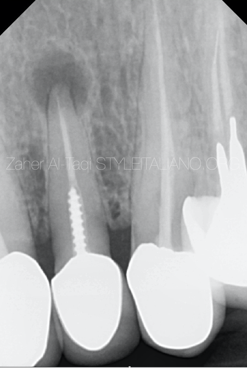
Fig. 1
Pre-op radiograph showing the apical lesion with poor RCT , post and crown .
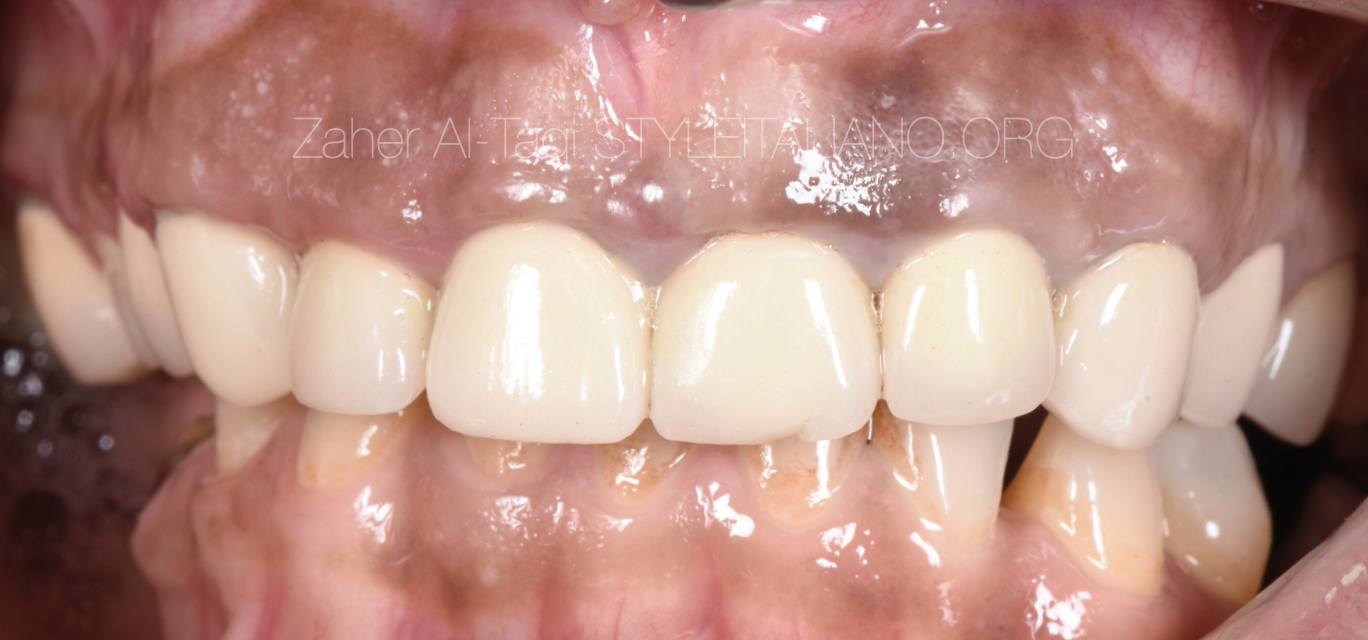
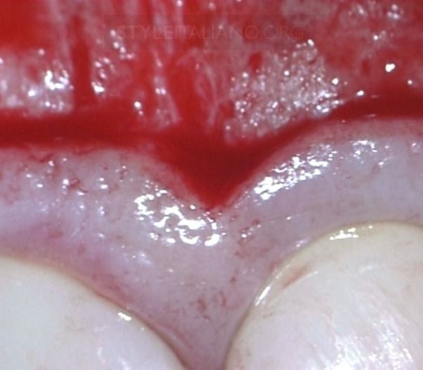
Fig. 2
Clinical image for the 15 years old crowns of the patient
Video explaining the procedure step by step :
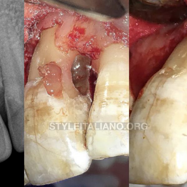
Flap
Osteotomy
Lesion curettage
Root end resection by Piezoelectric surgery
Retro prep by US tips
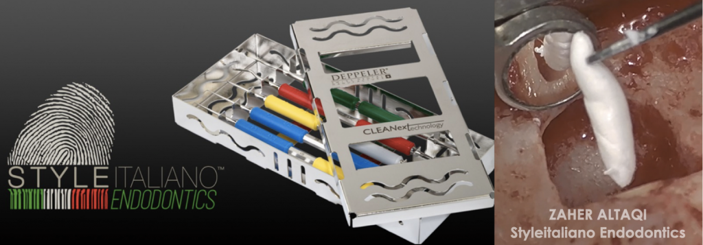
Retro Fill by bioceramic putty material and with the help of Style Italiano Endodontics micro surgical Kit .
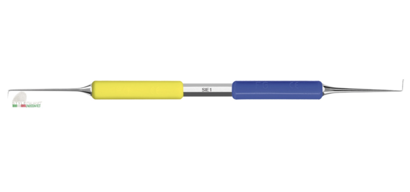
Fig. 3
PLUGGER-SPATULA SIE1
Plugger-spatula for apical microsurgery, elaborated with the team of Style Italiano Endodontics.
A real innovative instrument: from one side there is a thin and pointed explorer of 4 mm length, that is important for a correct detection of canals after apical resection. This flexible explorer gives the operator a greater sense of feedback for an easier detection of canals and isthmus.
The long plugger side has a flat tip with rounded angles and the shape is inverse tapered, in order to perform a perfect adaptation of retro filling material without removing it accidentally during instrument removal. The length is considerable (6mm) and is ideal for longer and straight canals.
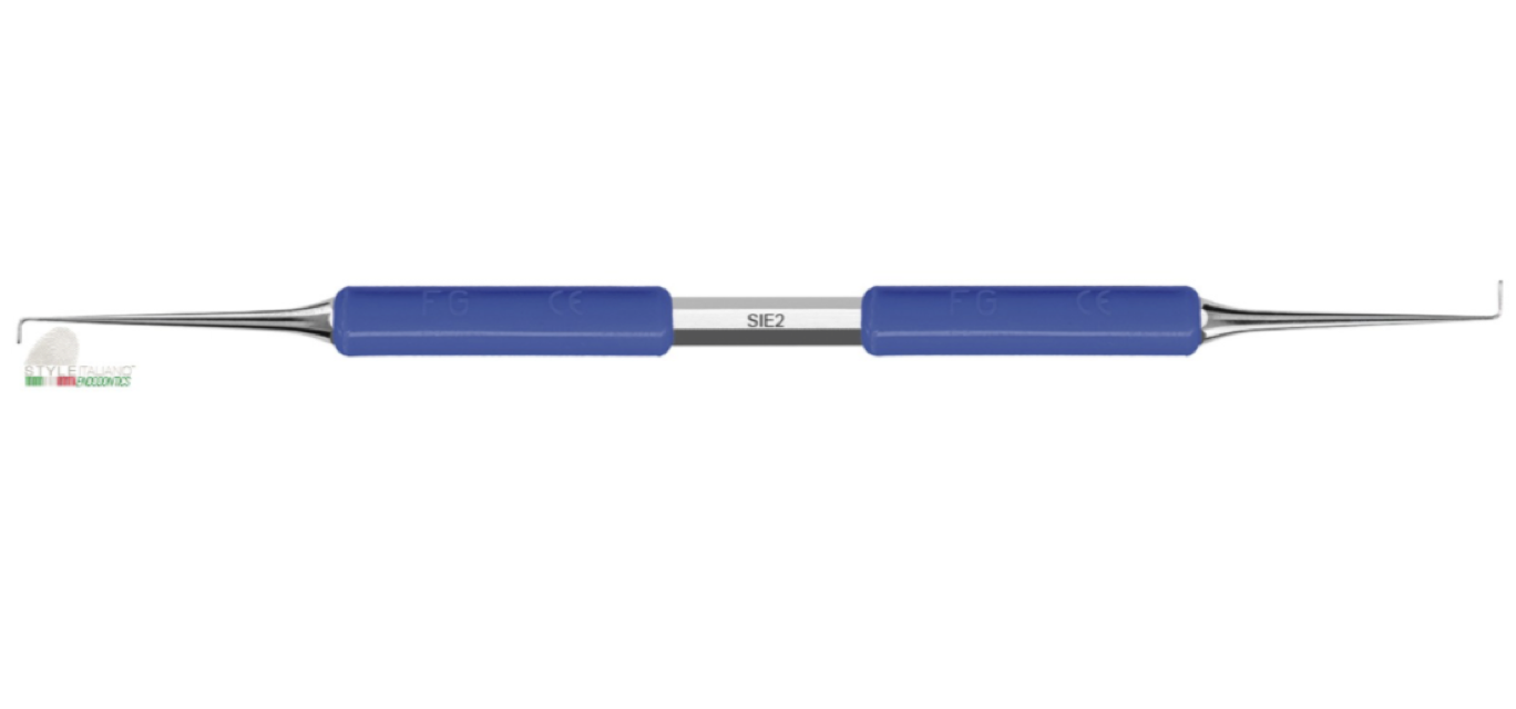
Fig. 4
PLUGGER SIE 2
A double plugger for apical microsurgery, elaborated with the team of Style Italiano Endodontics, with the same diameters, but different lengths. The pluggers have the innovative design as described before, flat tip with rounded angles and inverse taper. These two different lengths complete the 3 pluggers available in this kit. For the clinician the retro filling represents the most sensitive part of the Micro Apical Surgery
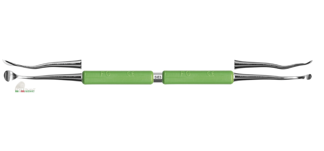
Fig. 5
PERIOSTEAL ELEVATOR SIE3 :
A double curved periosteal elevator for apical microsurgery, elaborated with the team of Style Italiano Endodontics, available in two different sizes in the same instrument, is used for a sharp dissection of periosteal tissue, without any tissue damage. The double inclination of the neck gives the instrument the perfect contact angle for a smooth undermining elevation of the flap. No additional left and right instruments are needed.
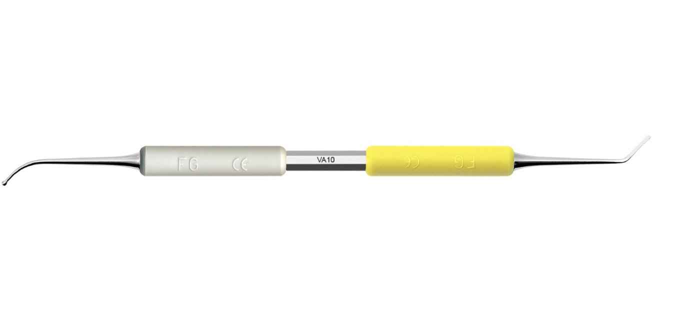
Fig. 6
BALL PLUGGER-SPATULA VA10
Ball plugger-spatula for apical microsurgery. The gold standard. From one side a thin angulated spatula for placing retro filling material and from the other side a spherical ball for first adaptation of the material itself.
Is part of the KITSIE elaborated with the team of Style Italiano Endodontics.
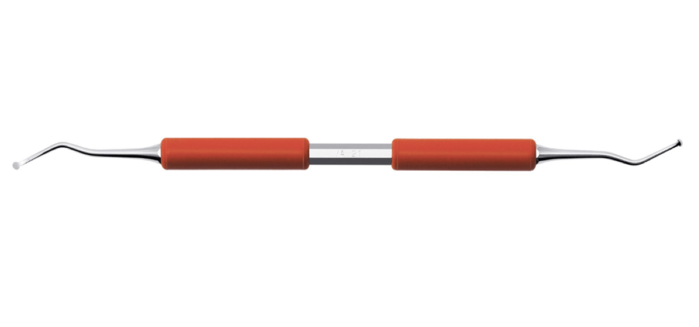
Fig. 7
SURGICAL CURETTE VA21
Surgical double curette for apical microsurgery, shaped like an excavator, but with a much more pronounced cutting edge, which helps with fast, efficient removal of the lesion.
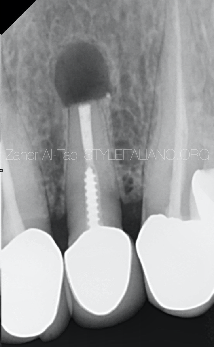
Fig. 8
Post op radiograph
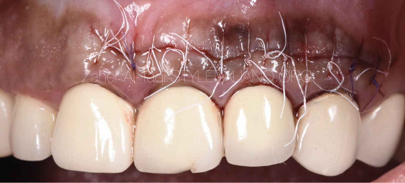
Fig. 9
Suturing
Conclusions
Micro apical surgery could be more conservative in many cases when we have heavy coronal restorations , The procedure should be done with high magnification under microscope for precise and better outcome .
Bibliography
1-H.M. Eriksen, L.L. Kirkevang, K. Petersson. Endodontic epidemiology and treatment outcome: general considerations. Endod Topics, 2 (2002), pp. 1-9
2-C. Barone, T.T. Dao, B.B. Basrani, N. Wang, S. Friedman. Treatment outcome in endodontics: the Toronto study—phases 3, 4, and 5: apical surgery. J Endod, 36 (2010), pp. 28-35
3- S. Friedman. The prognosis and expected outcome of apical surgery. Endod Topics, 11 (2005), pp. 219-262
4- T. von Arx, M. Penarrocha, S. Jensen. Prognostic factors in apical surgery with root-end filling: a meta-analysis. J Endod, 36 (2010), pp. 957-973