What signs can help us in the vertical root fracture diagnosis?

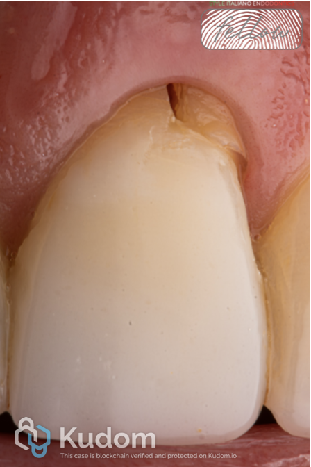 Demystifying vertical root fractures
What signs can help us in the vertical root fracture diagnosis?
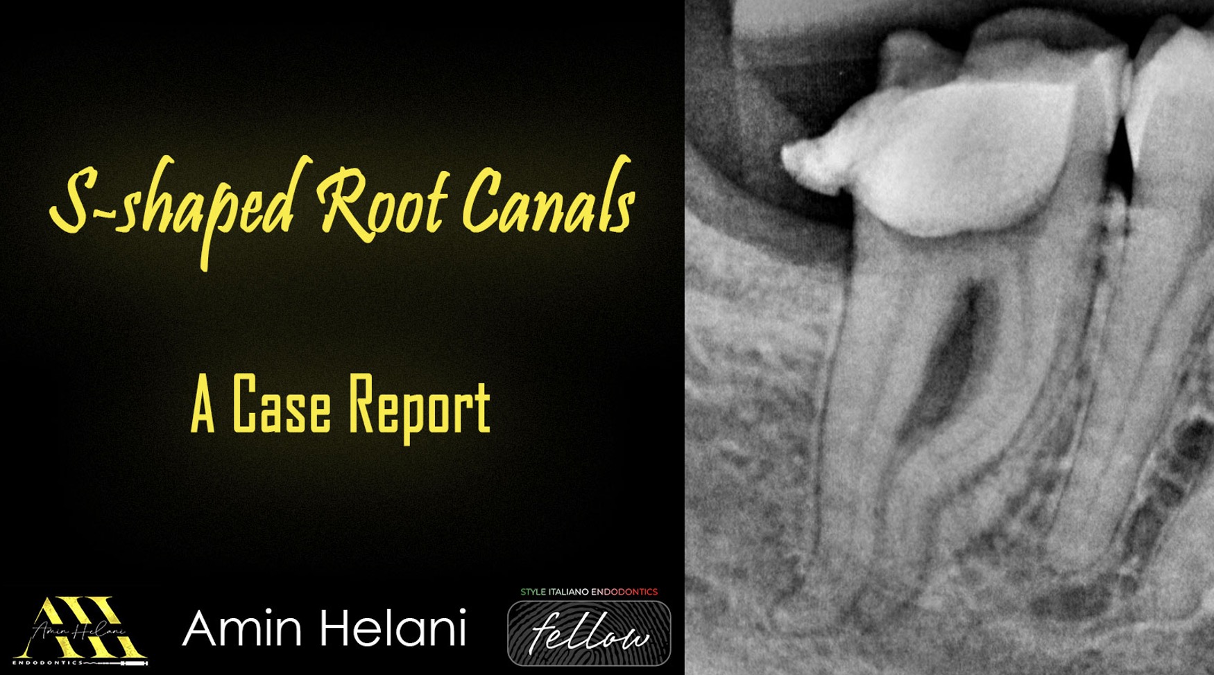 S-shaped root canals – a case report
S-shaped root canals can be one of the most challenging anatomies in endodontics. In this case, a careful treatment approach allowed us to manage a severe multiplanar curvature while preserving […]
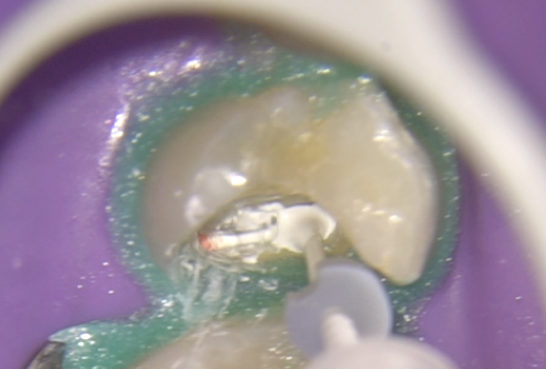 Bioceramic Sealer: “new” approach for obturation technique.
Bioceramic cements have gained increasing popularity in endodontics due to their excellent biological and sealing properties.These materials are mainly composed of calcium silicate and are characterized by their biocompatibility and […]
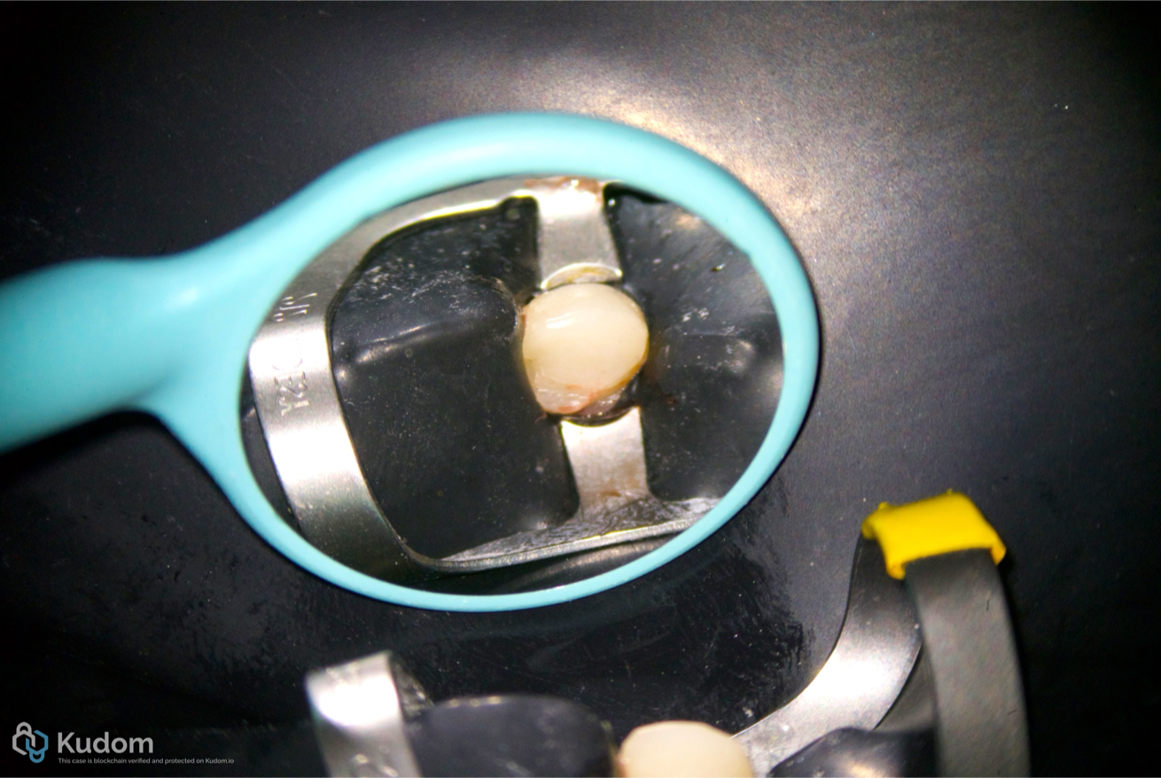 Endodontic Management of Type II Dens Invaginatus in a Maxillary Lateral Incisor Using: A Case Report
Dens invaginatus is a developmental anomaly characterized by invagination of the enamel organ into the dental papilla before calcification, resulting in highly variable internal anatomy. The complexity of the root […]
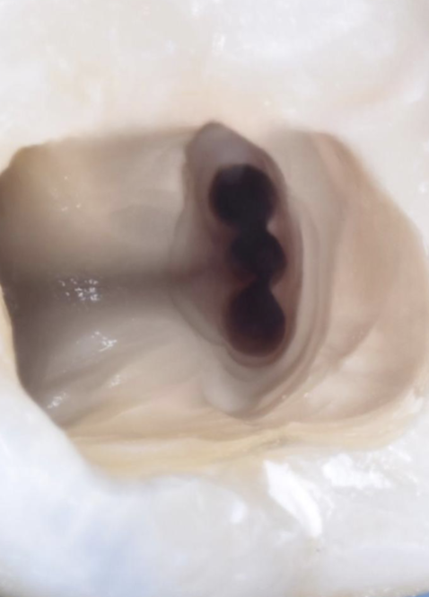 Five Canals, One Tooth: A Microscope-Assisted Endo-Resto Case
This case report presents the endodontic and restorative management of a mandibular molar with an unexpected middle distal canal. Five canals were successfully shaped, disinfected using combined sonic and ultrasonic activation, and obturated with a warm modified technique, followed by a conservative restorative workflow culminating in an adhesive overlay.
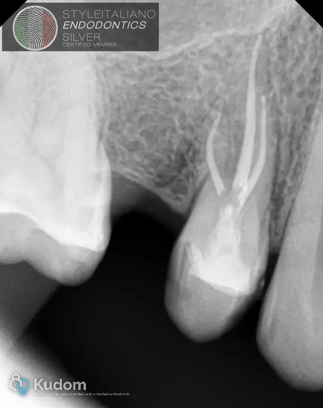 Three-rooted maxillary premolars: an uncommon anatomical variation.
Three-rooted maxillary premolars represent an uncommon but clinically important anatomical variation. The maxillary first premolar most frequently presents with two roots, while the occurrence of three separate roots is considerably […]
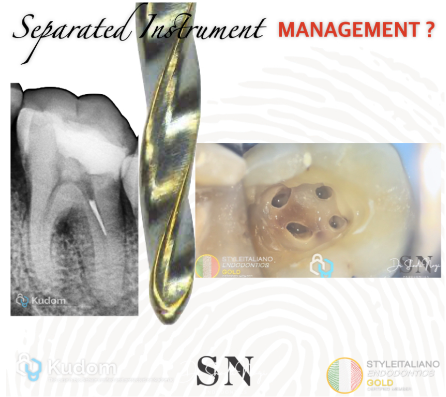 The Importance of Access Cavity Design in Endodontic Treatment
Provides Straight Line Access is mandatory.
A properly designed access cavity provides straight line access to the root canals, facilitating efficient and controlled canal shaping. This reduces unnecessary stress and torsional forces on endodontic instruments, thereby minimizing the risk of instrument separation during root canal treatment.
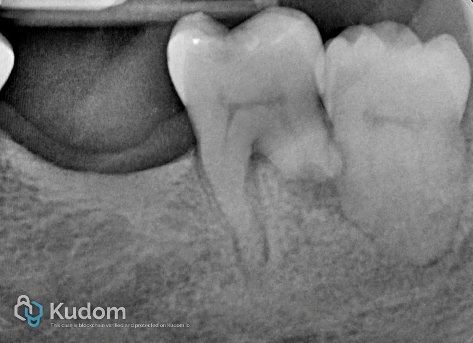 Nonsurgical Root Canal Treatment with an Apical Plug in a Patient on Active Oral Bisphosphonate Therapy with Root Resorption
Nonsurgical root canal treatment with an apical plug to avoid extraction in a patient on active oral bisphosphonate therapy and root resorption.
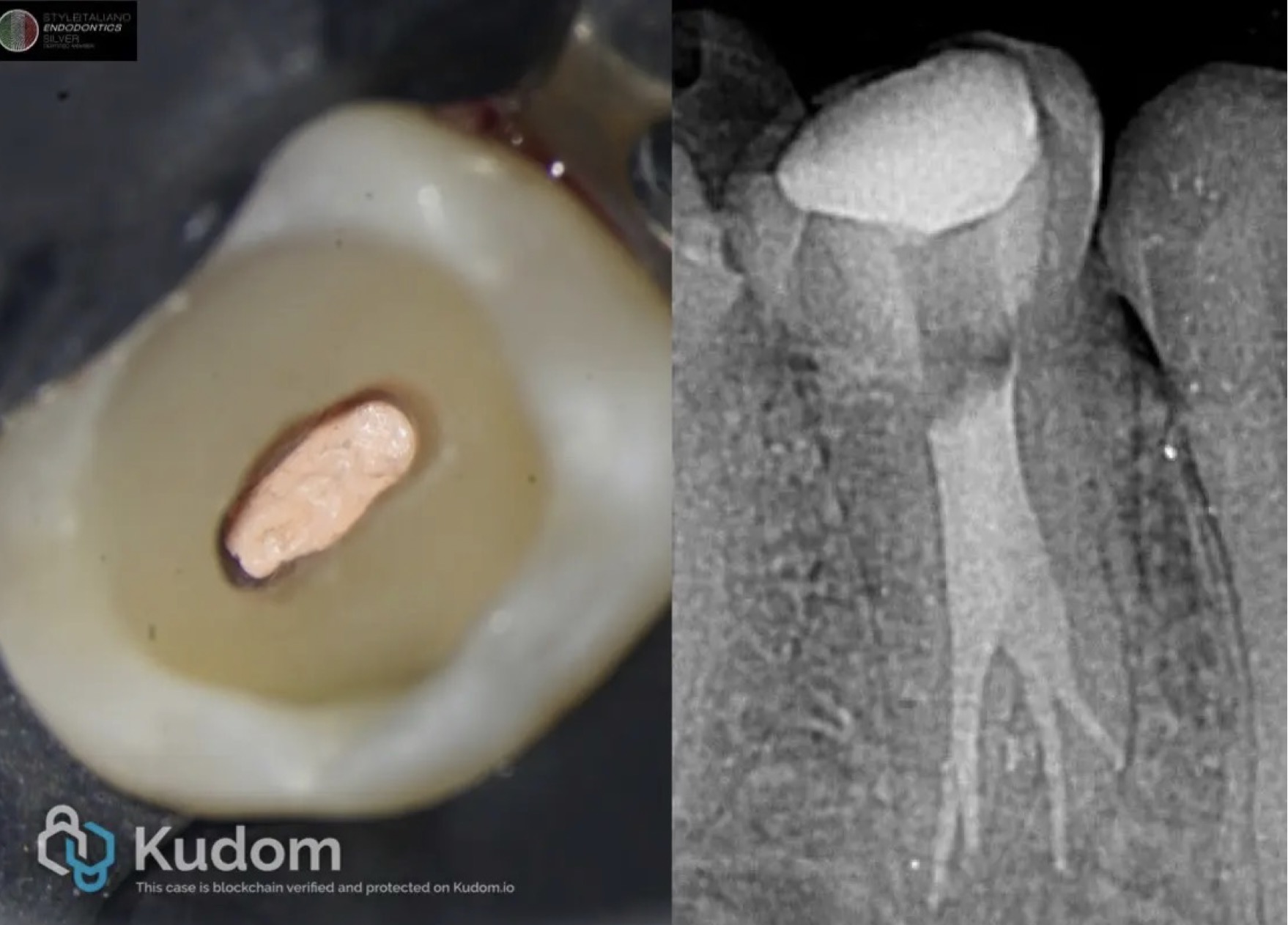 Expect the unexpected : Four canals in mandubular premolar
According to Vertucci, mandibular premolars have one single canal in approximately 74% of the cases. On occasion, mandibular premolars have two canals, in around 25.5%, but higher numbers are very […]
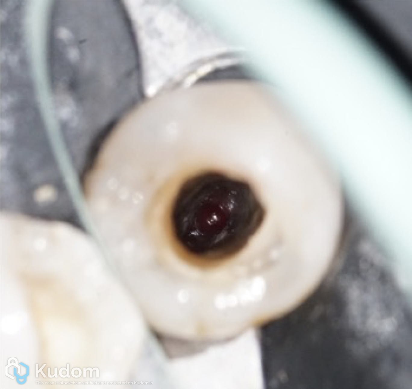 Apexification of an immature mandibular premolar: a case report
Apexification is a treatment procedure aimed at creating an apical barrier in a non-vital immature permanent tooth with incomplete root development and an open apex. The absence of apical constriction makes canal disinfection, obturation and the achievement of a predictable apical seal particularly challenging.
The purpose of this case report is to describe the non-surgical endodontic retreatment of a previously treated immature mandibular second premolar with symptomatic apical periodontitis. An MTA apical barrier was created to provide an effective apical seal, followed by root canal obturation. Follow-up demonstrated resolution of symptoms and favourable periapical healing.
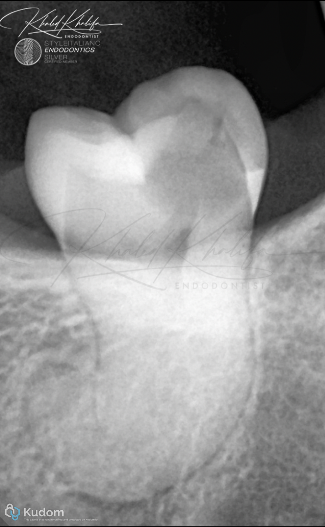 Impossible is possible with the right protocol
Impossible cases might be possible and worth your time.
But you should always approach cases like these with the right protocols to avoid making iatrogenic errors.
Dilacerated roots are one of the hardest anatomies any clinician can ever face.
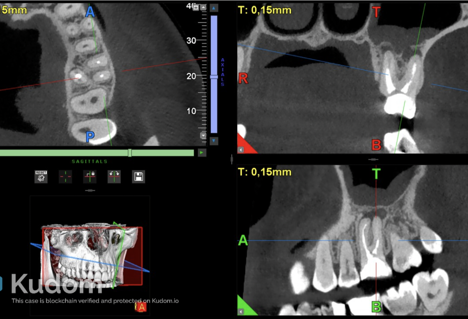 Periapical osteoperiostitis: CBCT diagnosis and healing following endodontic retreatment
CBCT revealed periapical osteoperiostitis with recurrent unilateral sinusitis, resolving after endodontic retreatmentt.