Selective Root canal retreatment
06/08/2026
Mohammed Khaleel
Warning: Undefined variable $post in /home/styleendo/htdocs/styleitaliano-endodontics.org/wp-content/plugins/oxygen/component-framework/components/classes/code-block.class.php(133) : eval()'d code on line 2
Warning: Attempt to read property "ID" on null in /home/styleendo/htdocs/styleitaliano-endodontics.org/wp-content/plugins/oxygen/component-framework/components/classes/code-block.class.php(133) : eval()'d code on line 2
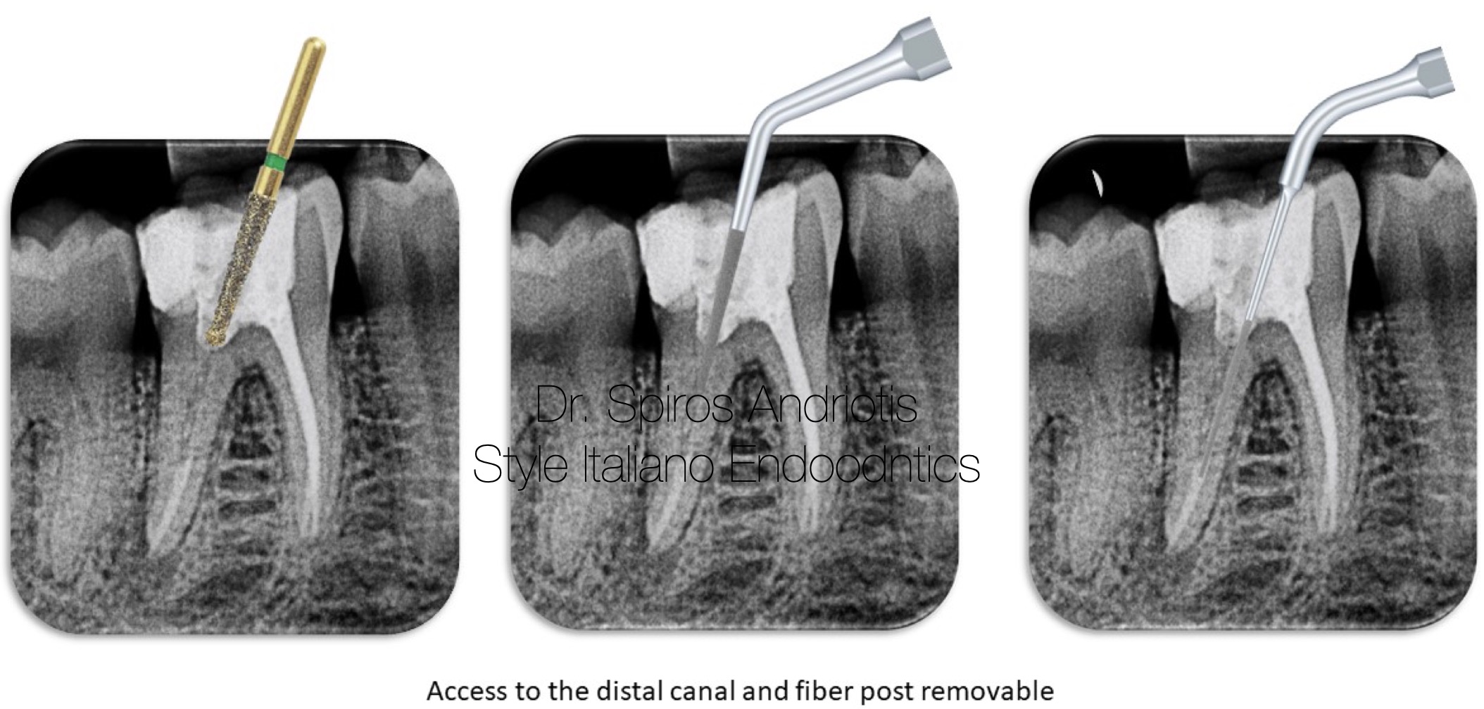
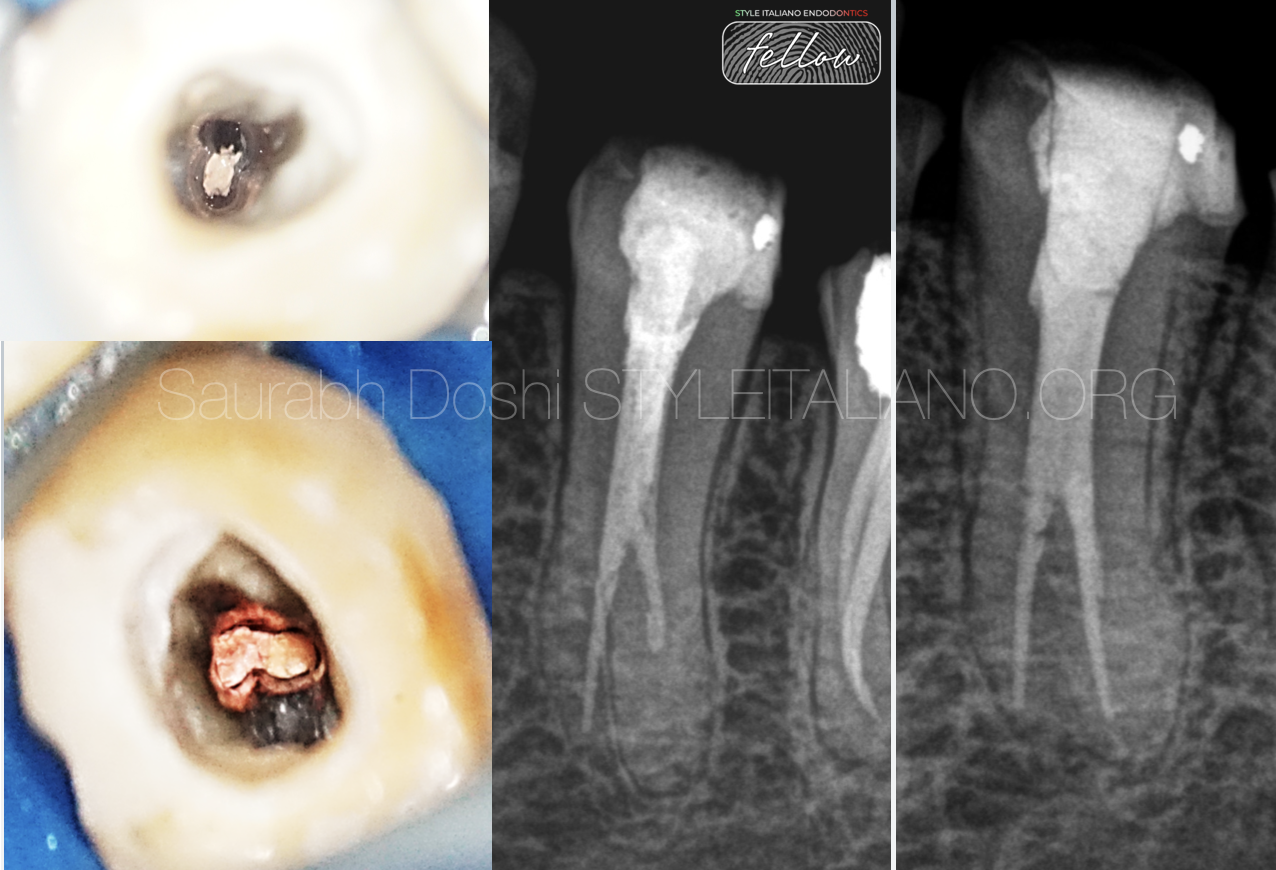
Selective root canal retreatment is a focused endodontic procedure where only the affected canal(s) of a previously treated tooth are retreated, instead of performing a complete retreatment on all canals.
When to Consider Selective Retreatment
• Radiographic evidence of periapical lesion localized to one canal.
• Persistent symptoms (pain, swelling, tenderness).
• Presence of post and core in one canal making access difficult or risky.
• CBCT imaging confirms that the rest of the canals are well obturated and disease-free
Tools That Make It Possible
• Cone Beam CT (CBCT): Essential for identifying the precise location of pathology.
• Dental Operating Microscope (DOM): Improves visualization.
• Ultrasonics: Allows conservative access refinement without removing restorations.
• Bioceramic sealers and MTA: Excellent for achieving a tight seal in selectively retreated canals.
Important Considerations
• Requires accurate diagnosis — CBCT is often a must.
• Requires good communication with the patient about benefits and potential risks.
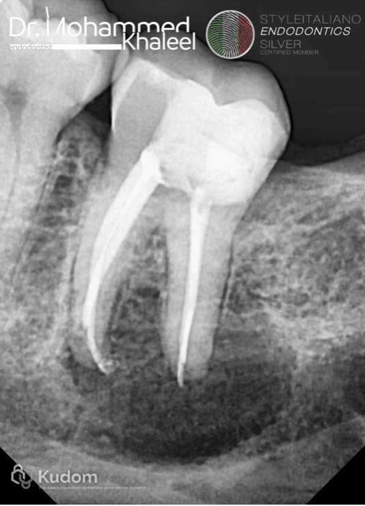
Fig. 1
A 32 years female patient was referred to my clinic, with chief complaint of pain in lower right region.
Clinical examination revealed mandibular right segment have large periapical lesion related to 2nd molar
The tooth was tender on percussion.
The radiographic examination revealed the large radiolucency involving the root apex.
I decided to take tube shift techniques…
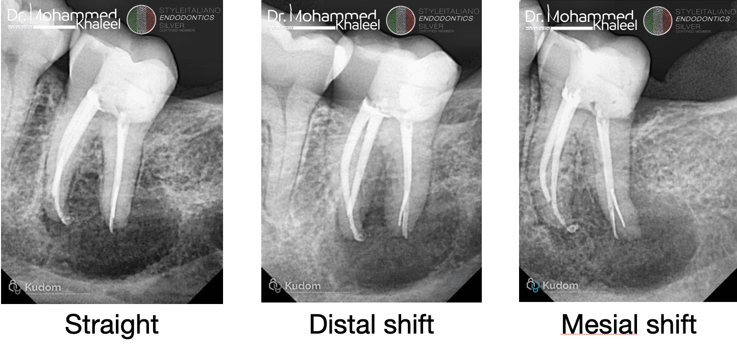
Fig. 2
In tube shift techniques showed separated instrument in distal root with perforation and extrusion of obturation material.
The mesial root have good obturation with a little bit sealer puff .
I decided to selective retreatment for distal root.
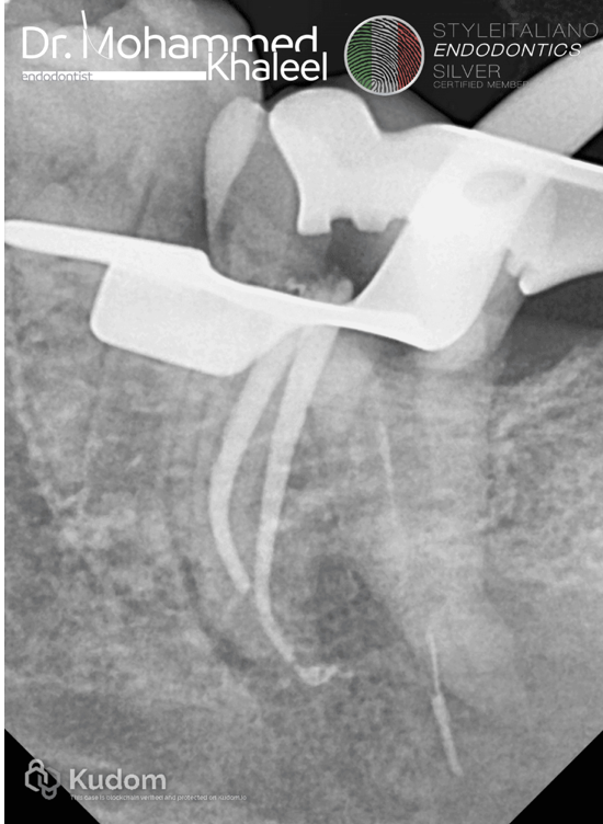
Fig. 3
Removed the gutta percha , but unfortunately remains the extruded part out of the root , and removed the separated instrument under magnification DOM by using ultrasonic tips and Btr pen for grasping.
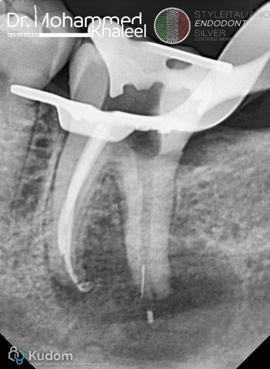
Fig. 4
Then I tried to remove the extruded G.P by XP shaper going 2 to 3 mm over at high speed 1200 rpm , so that everything inside the lesion rotating around it and coming out just like tornado.
In the first try removed some of it.
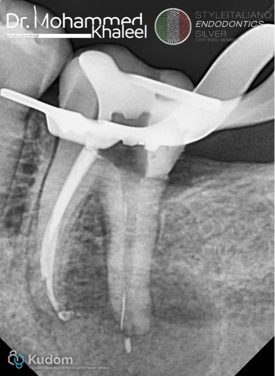
Fig. 5
2nd try by XP shaper removed more….
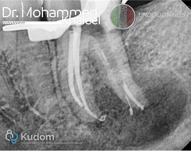
Fig. 6
Obturation till 1 mm under the perforation level by single cone with BC sealer .
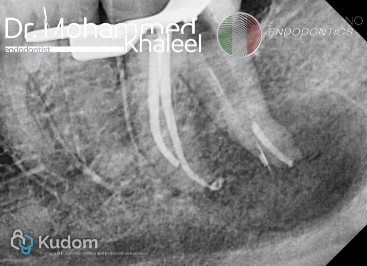
Fig. 7
Then I tried more by XP shaper to remove the extruded G.P , the remaining part came inside the canal.
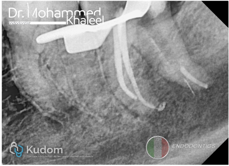
Fig. 8
Final try I can’t remove totally extruded G.P , I decided to leave it and repairing the perforation site.
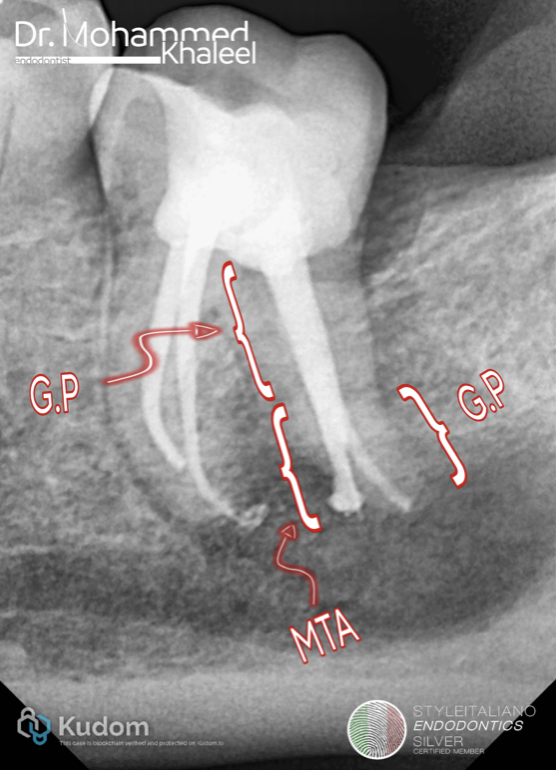
Fig. 9
Repaired the perforation site by MTA then injected G.P (sandwich technique ).
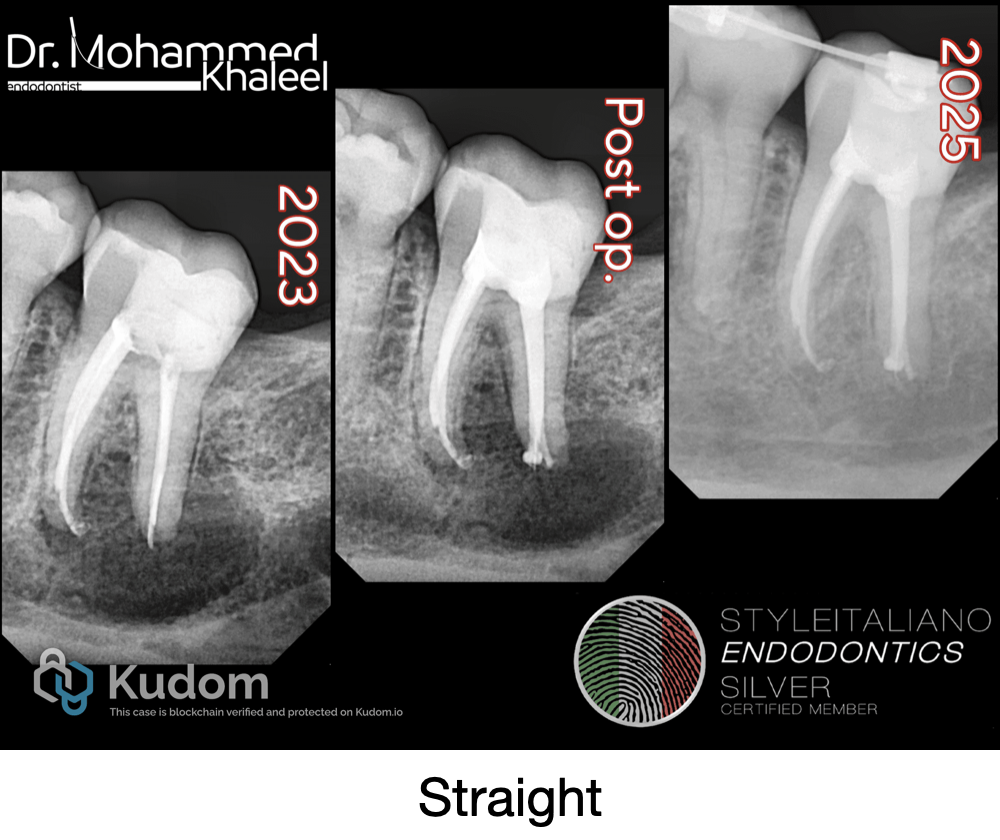
Fig. 10
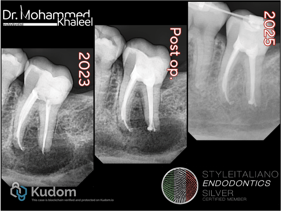
Follow up shows great healing
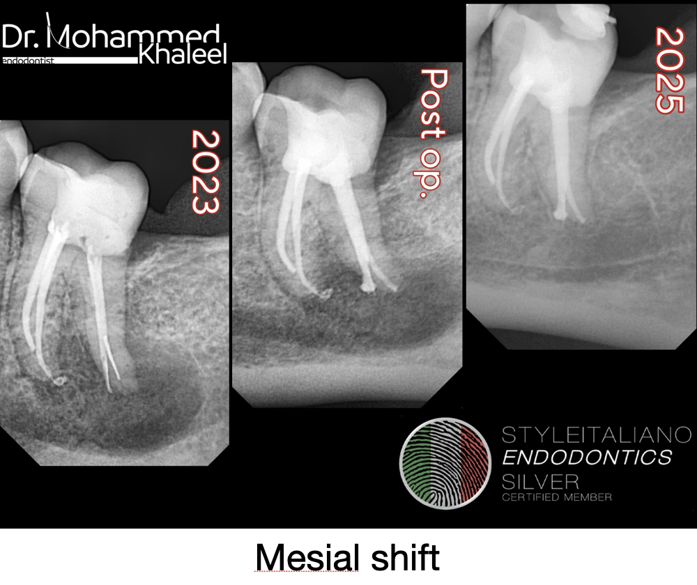
Fig. 11
Mesial shift
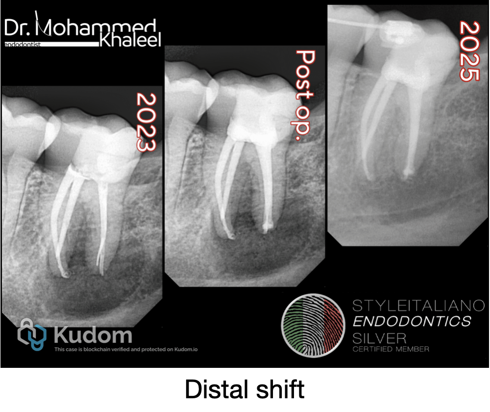
Fig. 12
distal shift
Conclusions
As endodontics becomes more evidence-driven, selective retreatment emerges as a logical evolution.
It recognizes that not all failures require full intervention and that precision, not aggression, leads to better outcomes.
With the aid of CBCT, magnification, and modern materials, selective retreatment is proving to be a safe and effective alternative in well-chosen cases.
Bibliography
- AAE . (2009) AAE consensus conference recommended diagnostic terminology. Journal of Endodontics, 35(12), 1634.
- Al‐Nuaimi, N. , Patel, S. , Austin, R.S. & Mannocci, F. (2017) A prospective study assessing the effect of coronal tooth structure loss on the outcome of root canal retreatment. International Endodontic Journal, 50(12), 1143–1157.
- Al‐Nuaimi, N. , Patel, S. , Davies, A. , Bakhsh, A. , Foschi, F. & Mannocci, F. (2018) Pooled analysis of 1‐year recall data from three root canal treatment outcome studies undertaken using cone beam computed tomography. International Endodontic Journal, 51(Suppl 3), e216–e226.
- Azim, A.A. , Griggs, J.A. & Huang, G.T. (2016) The Tennessee study: factors affecting treatment outcome and healing time following nonsurgical root canal treatment. International Endodontic Journal, 49(1), 6–16.
- Carr, G.B. , Schwartz, R.S. , Schaudinn, C. , Gorur, A. & Costerton, J.W. (2009) Ultrastructural examination of failed molar retreatment with secondary apical periodontitis: an examination of endodontic biofilms in an endodontic retreatment failure. Journal of Endodontia, 35(9), 1303–1309.