Expect the Unexpected: Anatomy Twist in Maxillary Second Premolar
20/08/2026
Andreas Louloudiadis
Warning: Undefined variable $post in /home/styleendo/htdocs/styleitaliano-endodontics.org/wp-content/plugins/oxygen/component-framework/components/classes/code-block.class.php(133) : eval()'d code on line 2
Warning: Attempt to read property "ID" on null in /home/styleendo/htdocs/styleitaliano-endodontics.org/wp-content/plugins/oxygen/component-framework/components/classes/code-block.class.php(133) : eval()'d code on line 2
Successful endodontic treatment is grounded in an in-depth understanding of root canal anatomy. While most maxillary second premolars conform to a predictable pattern—with one or two canals housed in a single root—clinicians must remain vigilant for anatomical deviations. Among the rarest of these is the three-rooted maxillary second premolar, a variation that mimics the root morphology of maxillary molars more than that of typical premolars.
Vertucci (1984) reported that roughly 75% of maxillary second premolars have a single canal, 24% have two canals, and only about 1% have three. However, these three canals rarely correspond to three separate roots. Cleghorn et al. (2007) conducted an extensive literature review and found the incidence of three-rooted maxillary second premolars to be exceptionally low—typically under 1%. Despite its rarity, failure to identify this variation can lead to missed canals, persistent infection, and treatment failure.
With the rise of cone-beam computed tomography (CBCT) and improved magnification techniques, more cases of such anatomical anomalies have come to light (Neelakantan et al., 2010).
The typical maxillary second premolar has:
- One root in 85–90% of cases
- Two roots in 9–10%
- Three roots in <1%
When three roots are present, they usually appear as:
- Two buccal roots (mesiobuccal and distobuccal)
- One palatal root
This configuration closely resembles that of a maxillary first molar, making it easy to misdiagnose unless careful evaluation is undertaken. Case studies and CBCT data suggest slightly increased prevalence in certain ethnic populations, such as Middle Eastern or East Asian groups, but the global incidence remains low.
Conventional periapical radiographs may fail to reveal the presence of additional roots due to overlapping structures. Subtle indicators include:
- A sudden disappearance or narrowing of the root canal on the radiograph
- Unusual root divergence or root outlines inconsistent with normal anatomy
Using angled radiographs (15–20° mesial or distal) may help reveal multiple roots. However, CBCT remains the gold standard for visualizing complex root canal systems and should be considered when an anomaly is suspected.
This rare anatomical twist challenges the clinician’s diagnostic and technical skills. Failure to detect and properly treat all root canals may lead to persistent infection and eventual treatment failure. Hence, clinicians must “expect the unexpected”—especially when radiographic or tactile cues suggest deviation from normal anatomy.
In this article, we explore the diagnostic clues, clinical implications, and case-based management strategies for handling the rare but significant occurrence of a three-rooted maxillary second premolar.
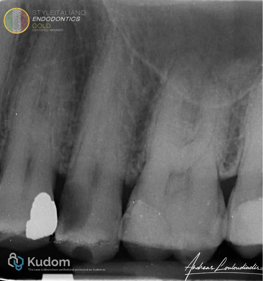
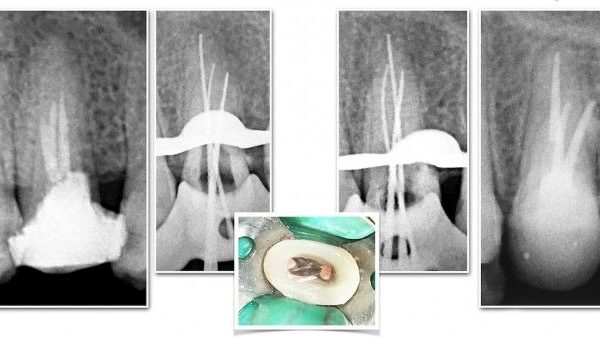
Fig. 1
A 52-year-old male presented with Symptomatic Irreversible Pulpitis and Symptomatic Apical Periodontitis in tooth #25.
Periapical radiograph revealed large decay lesion extending into pulp chamber with no periodical pathology. The radiograph also suggested unusual root canal anatomy, suggesting possibility of three canals.
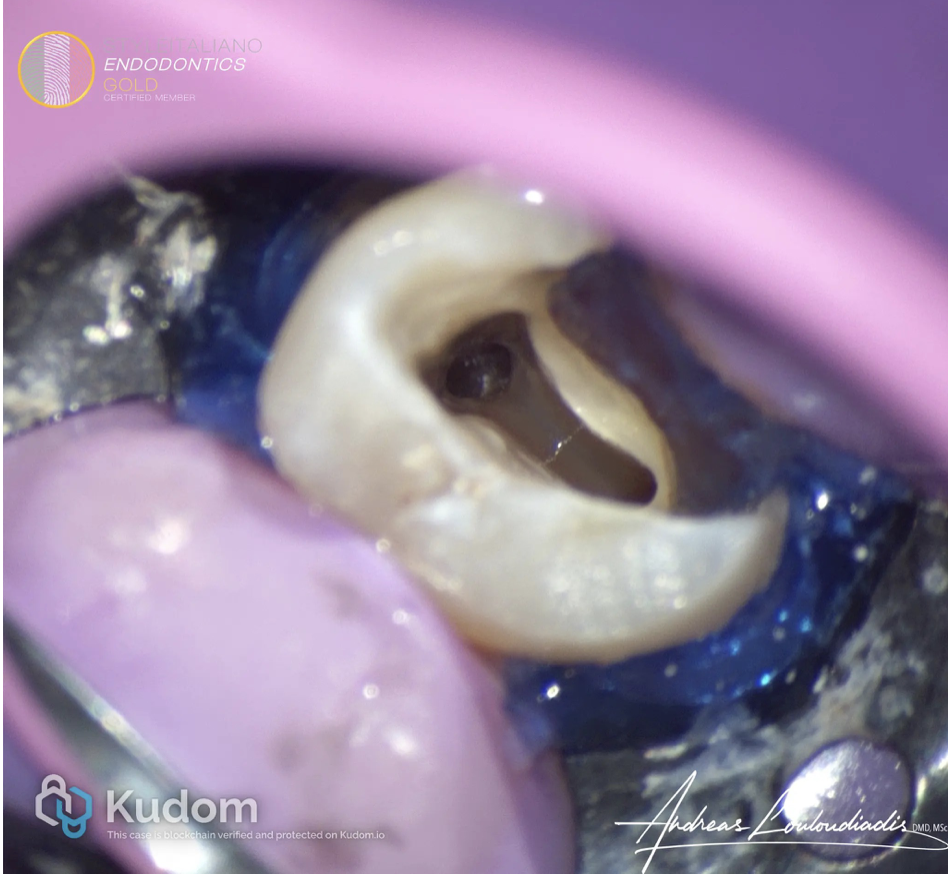
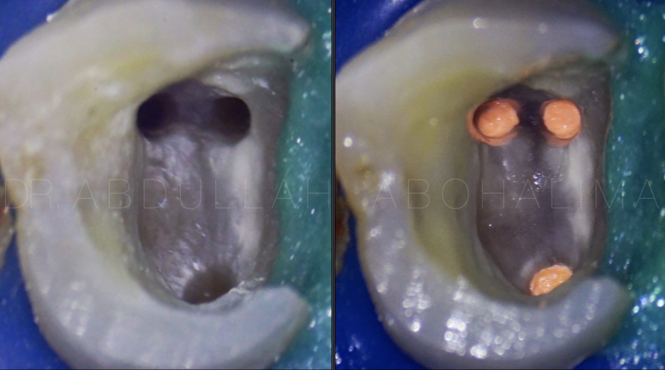
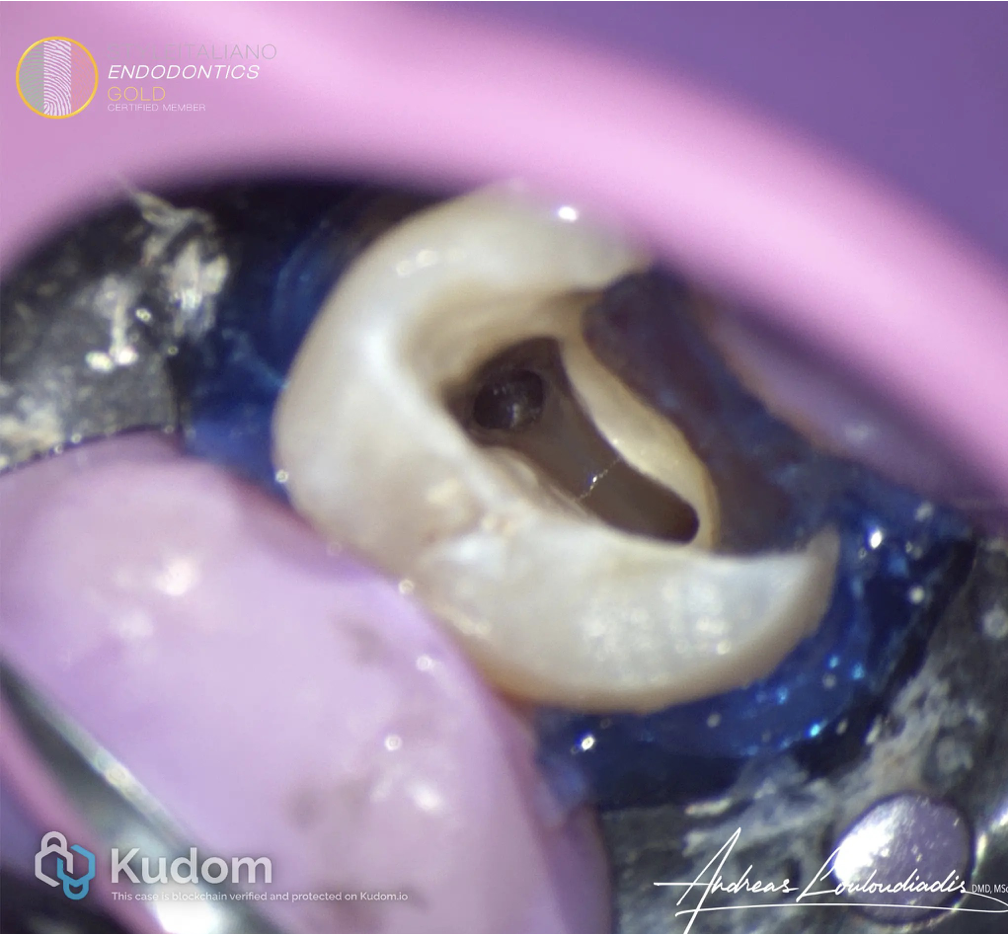
Fig. 2
Tooth was isolated, decay removed , liquid rubber dam used as temporary build up and access was made into pulp chamber, refined with USS and 3 canals were identified. The buccal canals were exhibiting a deep split at the middle 1/3.
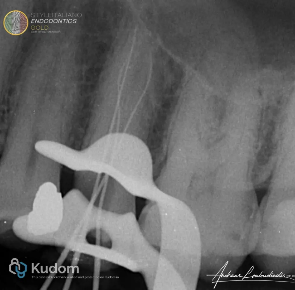
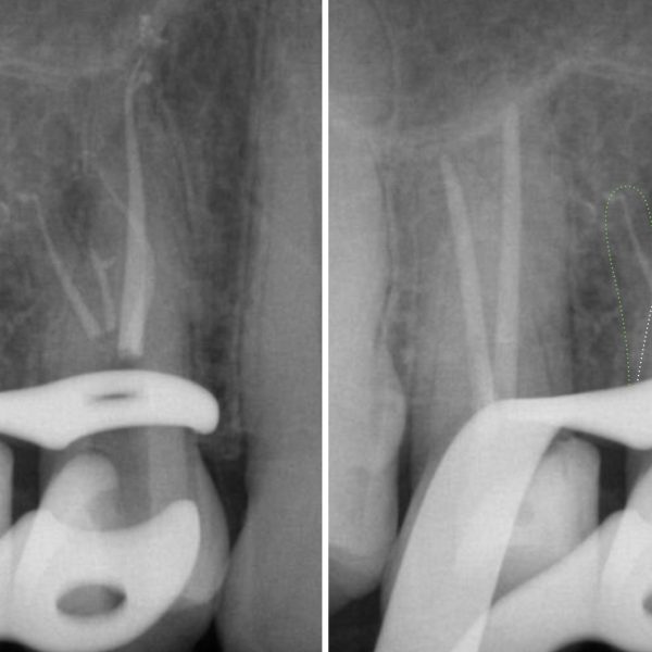
Fig. 3
WL was determined with apex locator and verified with an X-ray, where it was clearly noted the 3 separate roots, two buccal and one palatal.
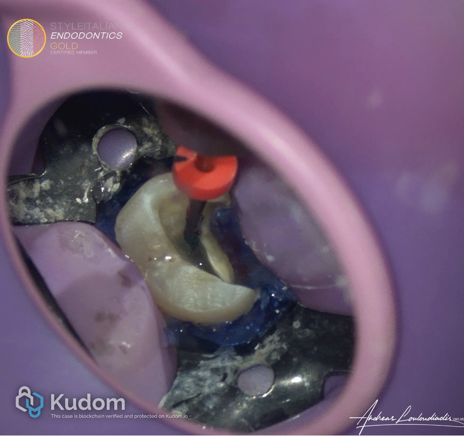
Fig. 4
Minimally invasive instrumentation was performed by crown down to ISO 25/04 on Mb and Db canals, and to ISO 25/06 on Palatal canal.
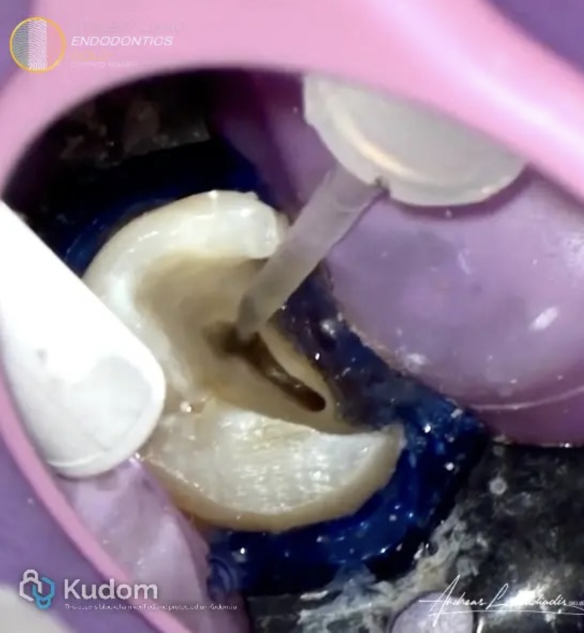
Fig. 5
Irrigation of the canals using the Iriiflex tip with 5.25% Naocl and 17% EDTA, with flush of distilled water in between.
FInal flush with distilled water.
The liquids were activated with US activation using the Ultra-X.
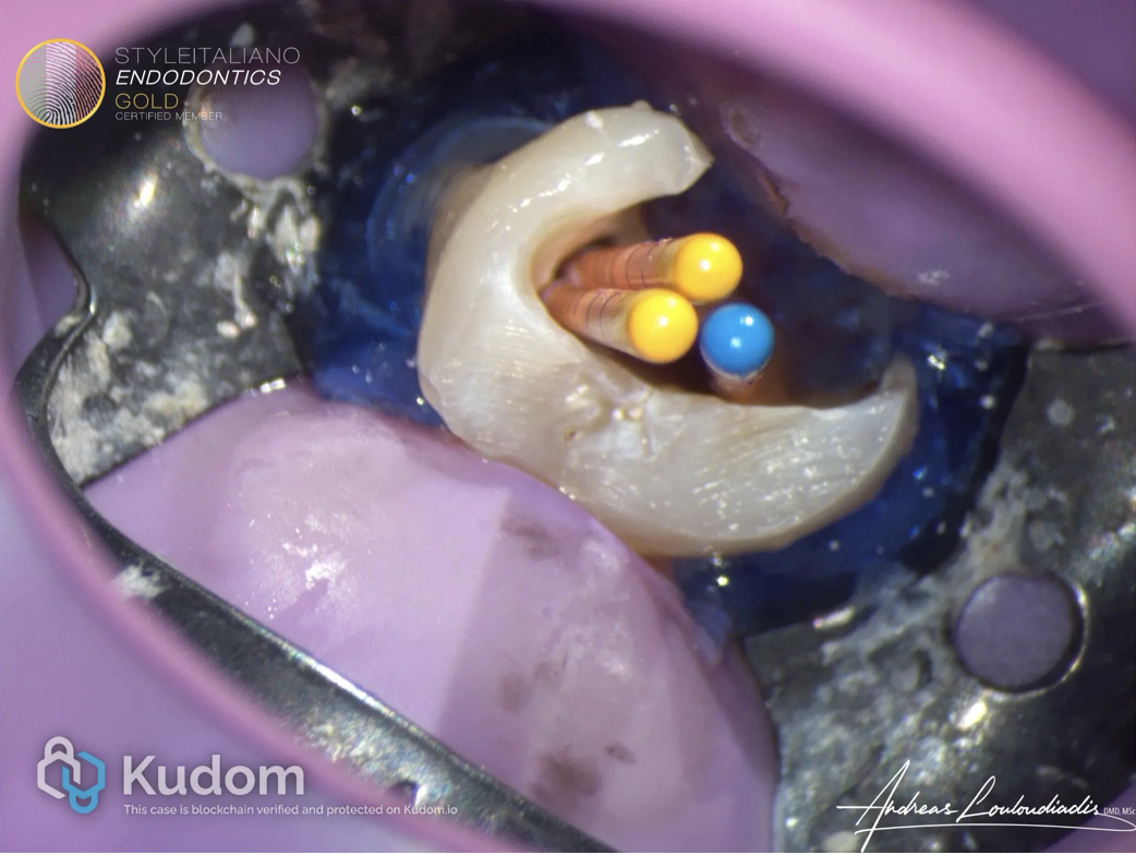
Fig. 6
Fit of GP points.
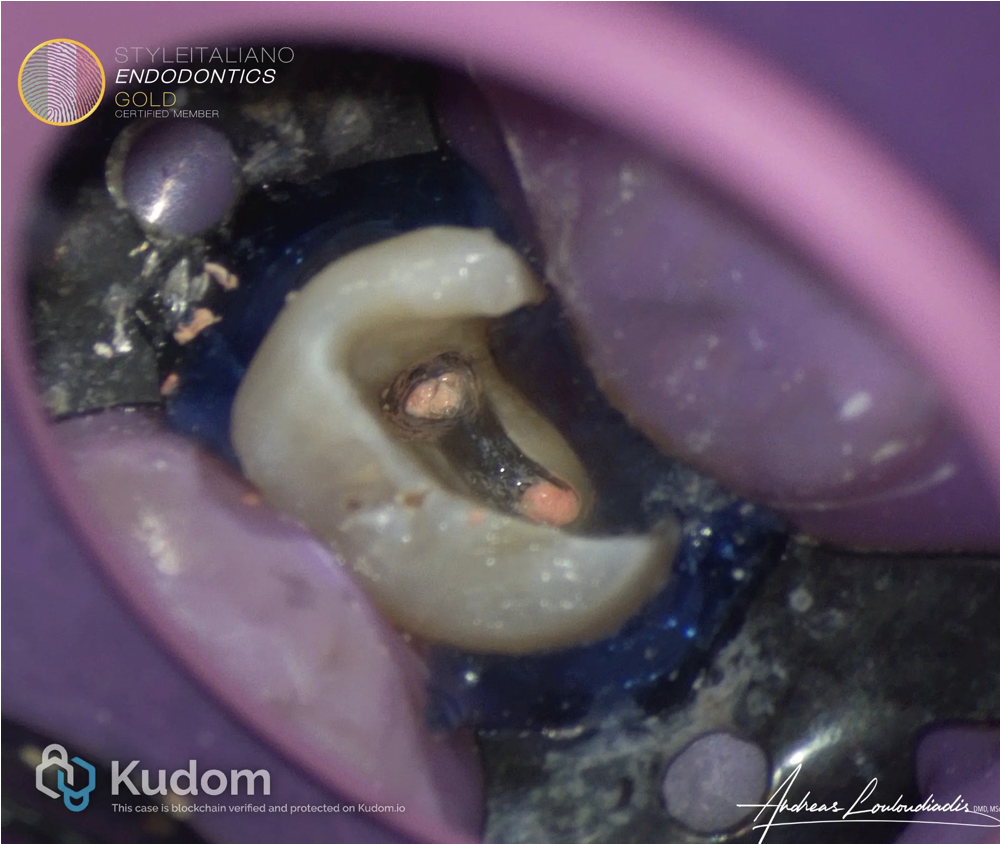
Fig. 7
Obturation performed with Bio-C BC Sealer and Cold Hydraulic Condensation.
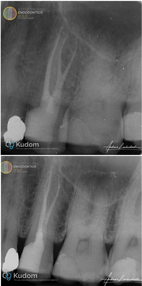
Fig. 8
Post - Operative radiographs with different angles.
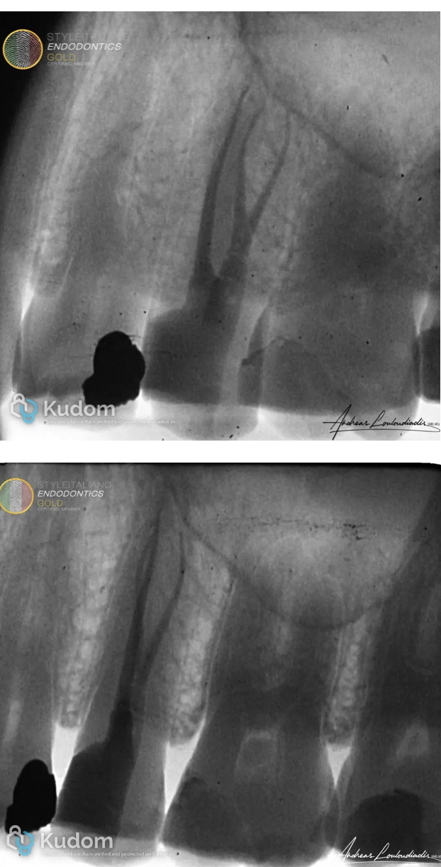
Fig. 9
Post - Operative radiographs positive images
Video of procedure
Conclusions
Three-rooted maxillary second premolars may be rare, but their clinical significance is substantial. Missed canals are a common cause of post-treatment disease. Dentists and endodontists must maintain a high index of suspicion, particularly when anatomical anomalies are suspected on radiographs or during access. Advanced imaging and magnification tools, combined with an understanding of root canal anatomy variations, are essential for delivering optimal care and achieving long-term success in endodontic therapy.
Bibliography
1. Vertucci FJ. Root canal anatomy of the human permanent teeth. Oral Surg Oral Med Oral Pathol. 1984.
2. Pécora JD, et al. Morphologic study of the maxillary first premolar. Braz Dent J. 1992.
3. Cleghorn BM, et al. Root and root canal morphology of the human maxillary second premolar: a literature review. J Endod. 2007.
4. Neelakantan P, et al. Cone-beam CT study of root and canal morphology of maxillary premolars in an Indian population. Oral Surg Oral Med Oral Pathol Oral Radiol Endod. 2010.
5. Slowey RR. Root canal anatomy. Road map to successful endodontics. Dent Clin North Am. 1979;23(4):555–573.
6. Sieraski SM, Taylor GN, Kohn RA. Identification and endodontic management of three-canalled maxillary premolars. J Endod. 1989;15(1):29–32.