Non-surgical healing of a large periapical lesion in maxillary anterior region
17/08/2026
Warning: Undefined variable $post in /home/styleendo/htdocs/styleitaliano-endodontics.org/wp-content/plugins/oxygen/component-framework/components/classes/code-block.class.php(133) : eval()'d code on line 2
Warning: Attempt to read property "ID" on null in /home/styleendo/htdocs/styleitaliano-endodontics.org/wp-content/plugins/oxygen/component-framework/components/classes/code-block.class.php(133) : eval()'d code on line 2
Periapical lesions develop as a result of invasion of microorganisms or their toxins from necrotic pulp tissue in the periapical tissue, leading to acute or chronic inflammation. It has been proven that the majority of inflammatory periapical lesions can be effectively treated using nonsurgical endodontic management. However, the selection between surgical and nonsurgical endodontic treatment for managing large periapical lesions remains a controversial issue. This case report aims to reveal the effectiveness of nonsurgical endodontic treatment for large periapical lesions.
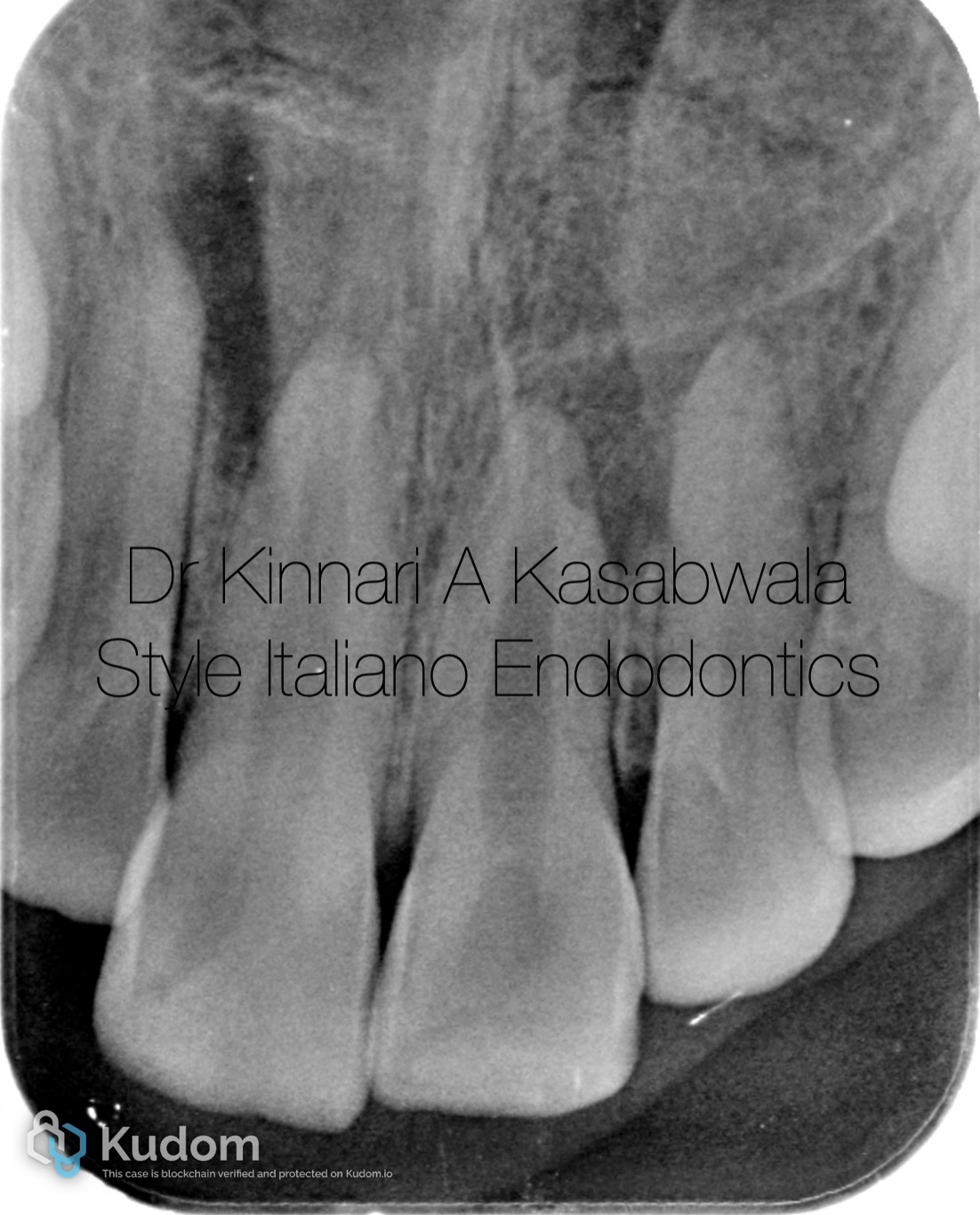
Fig. 1
32 year old female patient reported to the dental office with severe, throbbing pain in upper front teeth region. Patient did experience pain before, however suppressed it with painkillers.
Patient had a history of fall with focussed impact of trauma on her upper front teeth when she was 10 years old.
On examination:
Teeth 11,21 are tender on percussion and palpation
Teeth 12,22 slightly tender
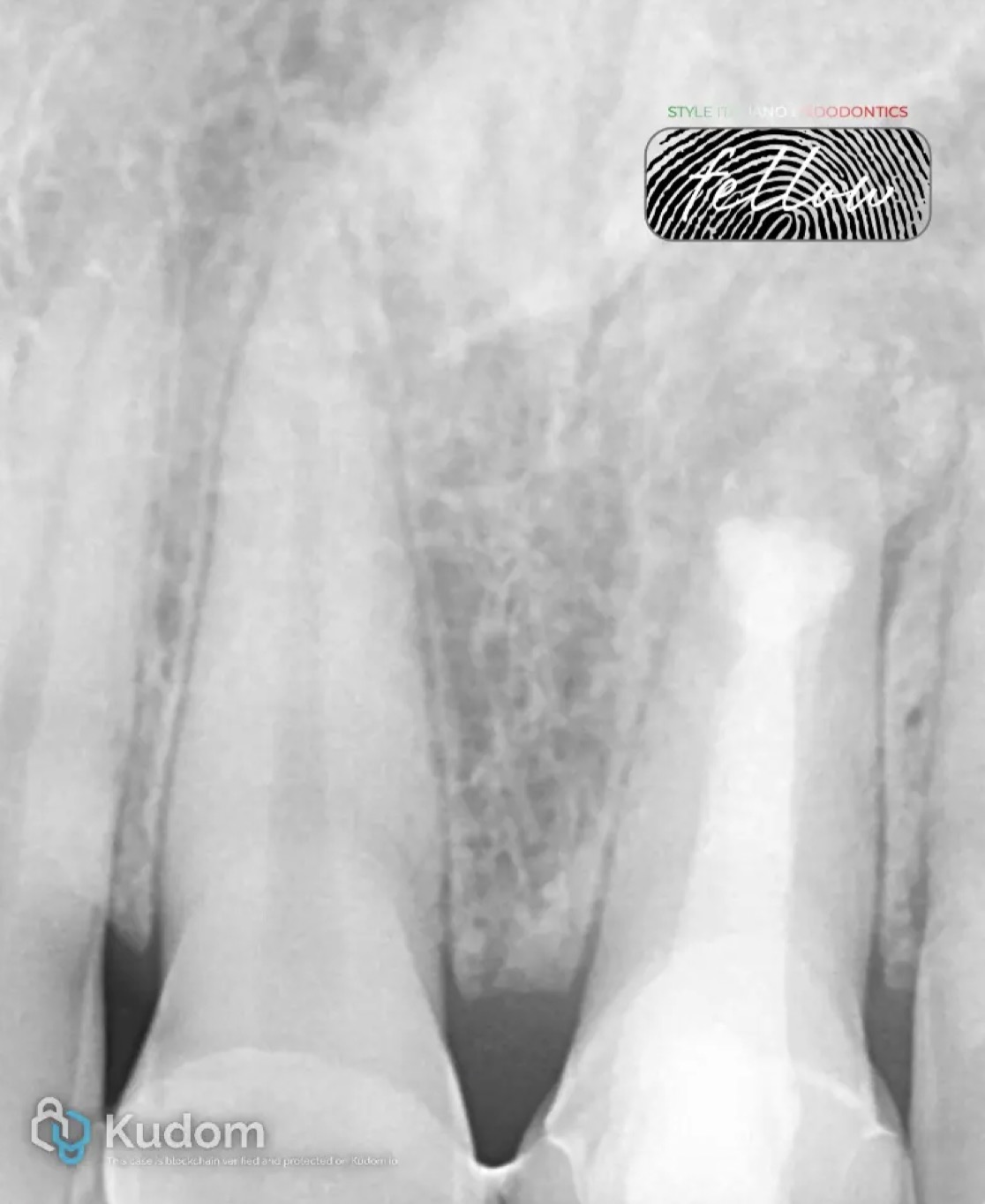
IOPA shows large periapical lesion in upper anterior region.
Patient was referred for CBCT scan of upper front region.
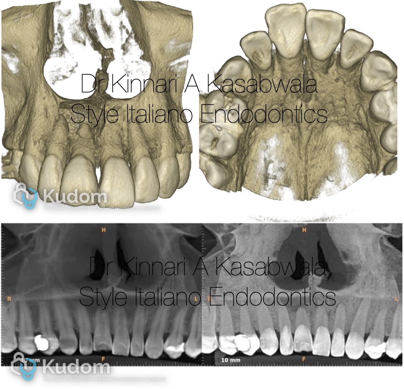
Fig. 2
Pre-op CBCT 3D reconstruction showing perforation of buccal cortical plate in 11,12 region
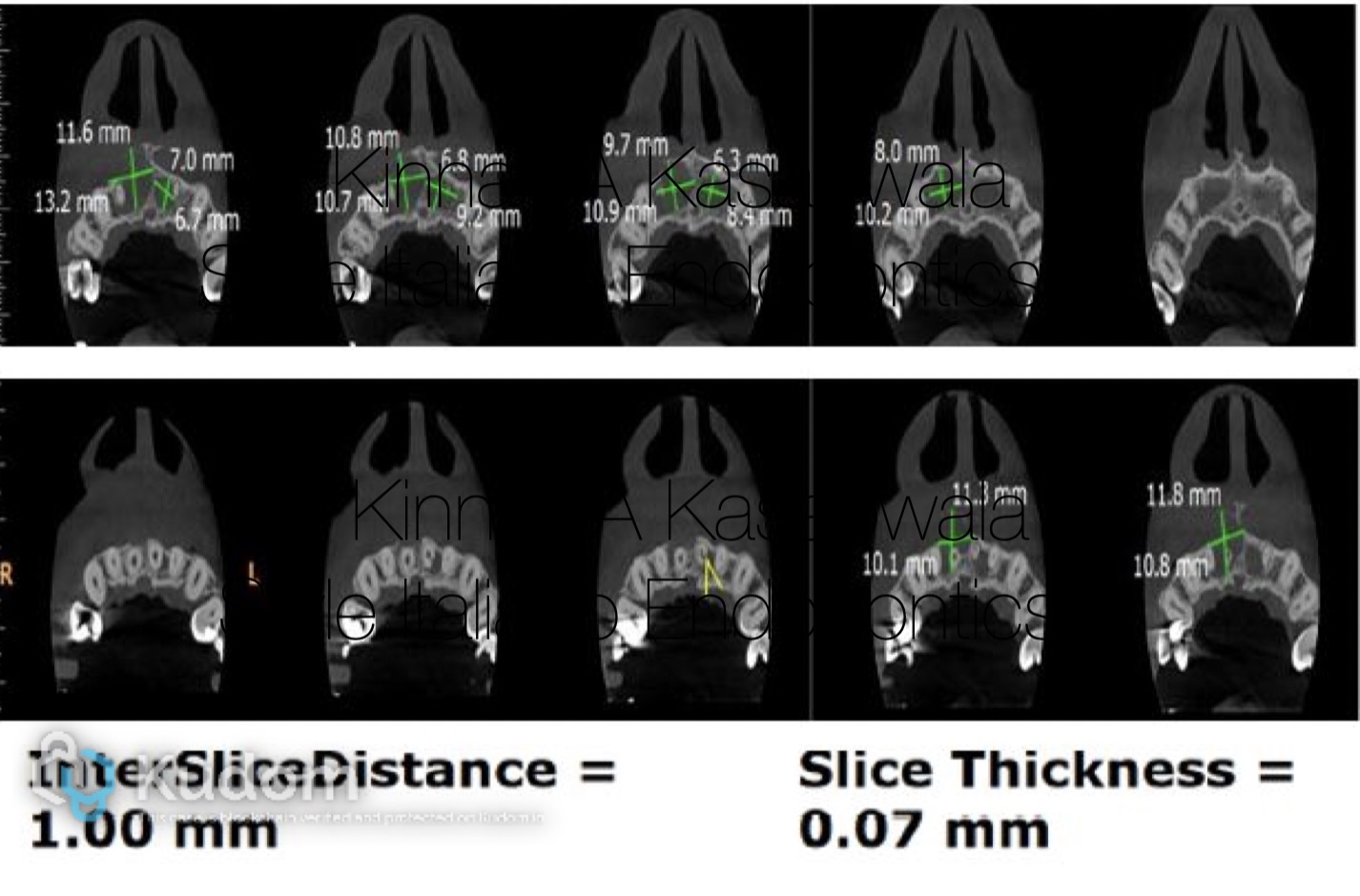
Fig. 3
AXIAL VIEW CBCT
Periapical lesion seen involving teeth 11,12,21,22 with perforation of buccal cortical plate in apical region of teeth 11,12.
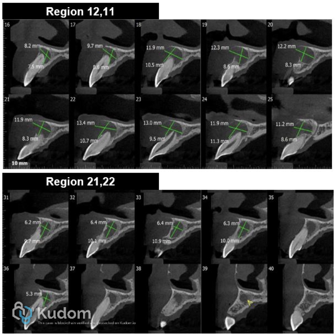
Fig. 4
SAGITTAL VIEW CBCT
Shows perforation of buccal cortical plate in 11,12 region with lesion approximating the floor of the nasal cavity in the same region.
Thinning of palatal cortical plate seen in 11 region
Tooth 11 also shows an open apex
Lesion seen extending from distal portion of tooth 12 extending till mesial portion of 11 in a continuous fashion.
Separate periapical lesion seen wrt teeth 21,22, approximating nasal floor in tooth 21 region.
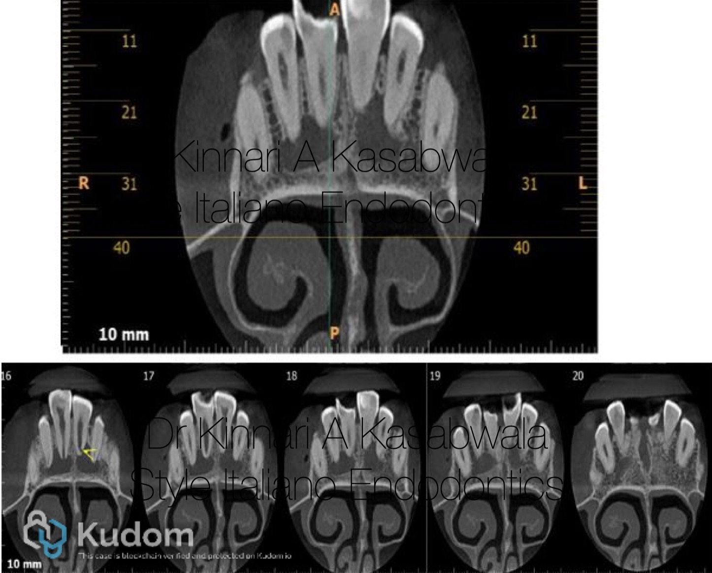
Fig. 5
CORONAL VIEW CBCT
Shows 2 large and separate periapical lesions
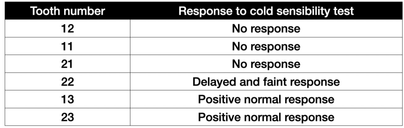
Fig. 6
Before access opening pulp sensibility test was performed using cold test using Endo frost (Roeko) to determine the course of treatment.
Following the test, it was decided that teeth 12,22,21,22 would undergo endodontic treatment.
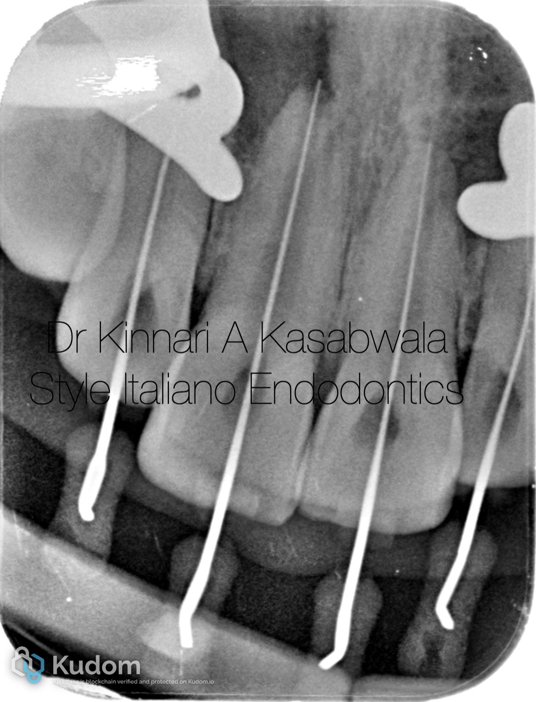
Fig. 7
Access opening was done under rubber dam isolation for all 4 teeth simultaneously.
Arbitrary working was determined using apex locator and was confirmed with IOPA.
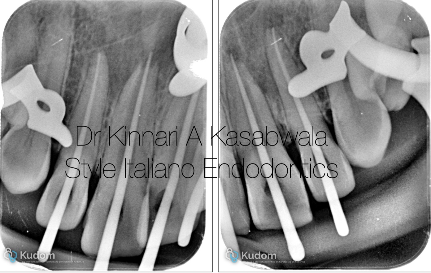
Fig. 8
After shaping and cleaning, master cone IOPA was taken.
Teeth 12,22 were shaped till 30,6% and teeth 11,21 were shaped till 40,6% as master apical file sizes.
Corresponding size GP cones were chosen.
Apexification procedure was planned in tooth 11 owing to the open apex.
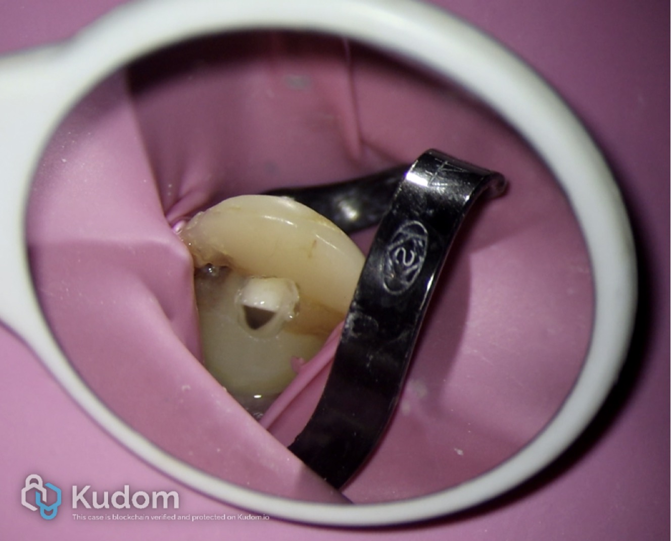
Fig. 9
Irrigation done with 5.25% sodium hypochlorite activated with Endoactivator (Dentsply)- 40 sec cycles x 4 cycles per tooth
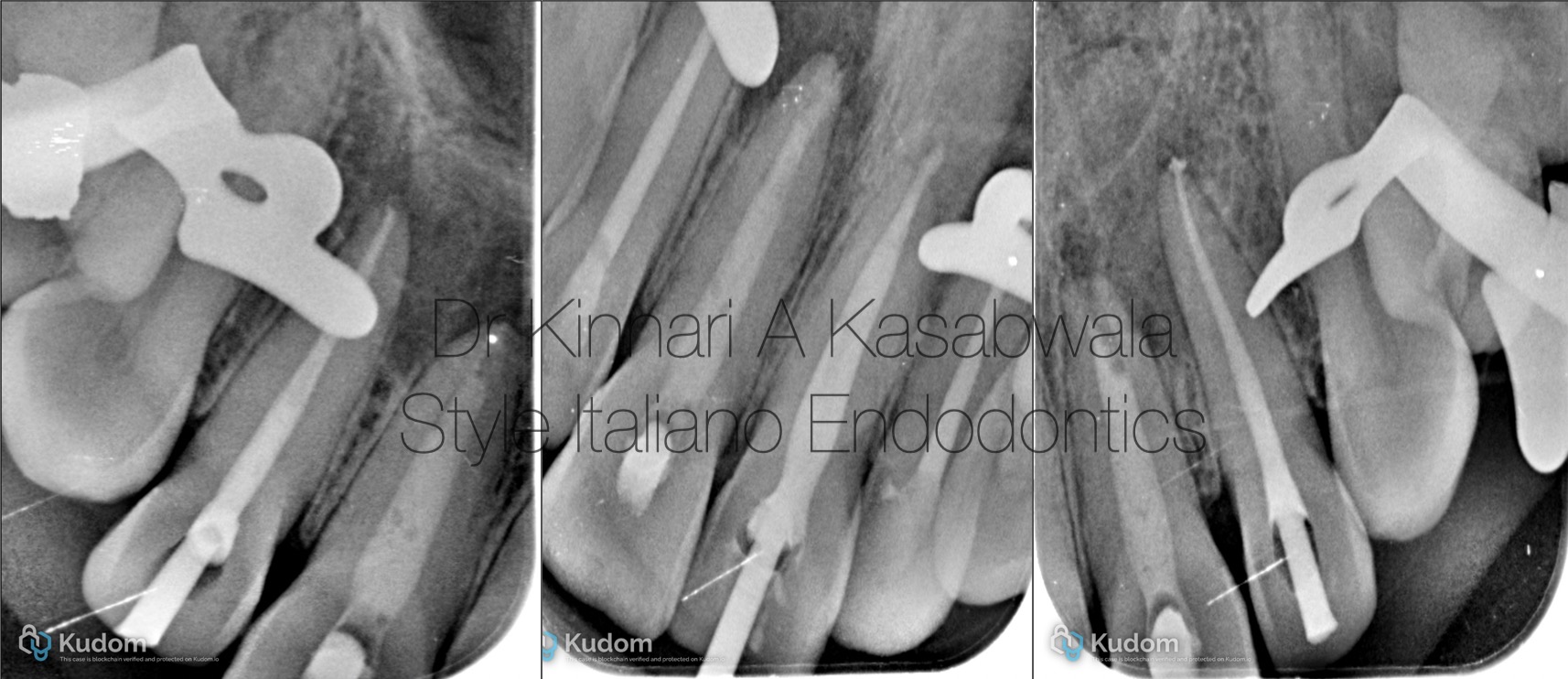
Fig. 10
Calcium hydroxide dressing was given for all 4 teeth for 2 weeks.
Dry canals were obtained in teeth 12,22 after one calcium hydroxide dressing (Cleanical- Maruchi).
Following this, obturation was performed using Ceraseal bioceramic sealer (Meta Biomed) and modified downpack technique.
Tooth 21, required 2 calcium hydroxide dressings of 2 weeks each before obturation.
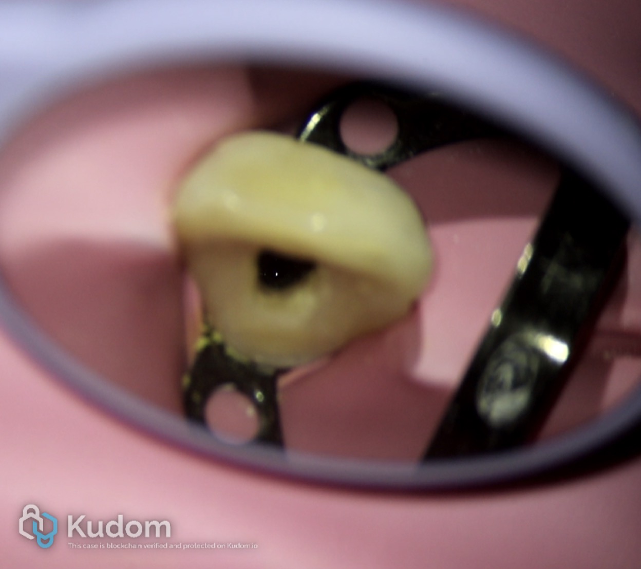
Fig. 11
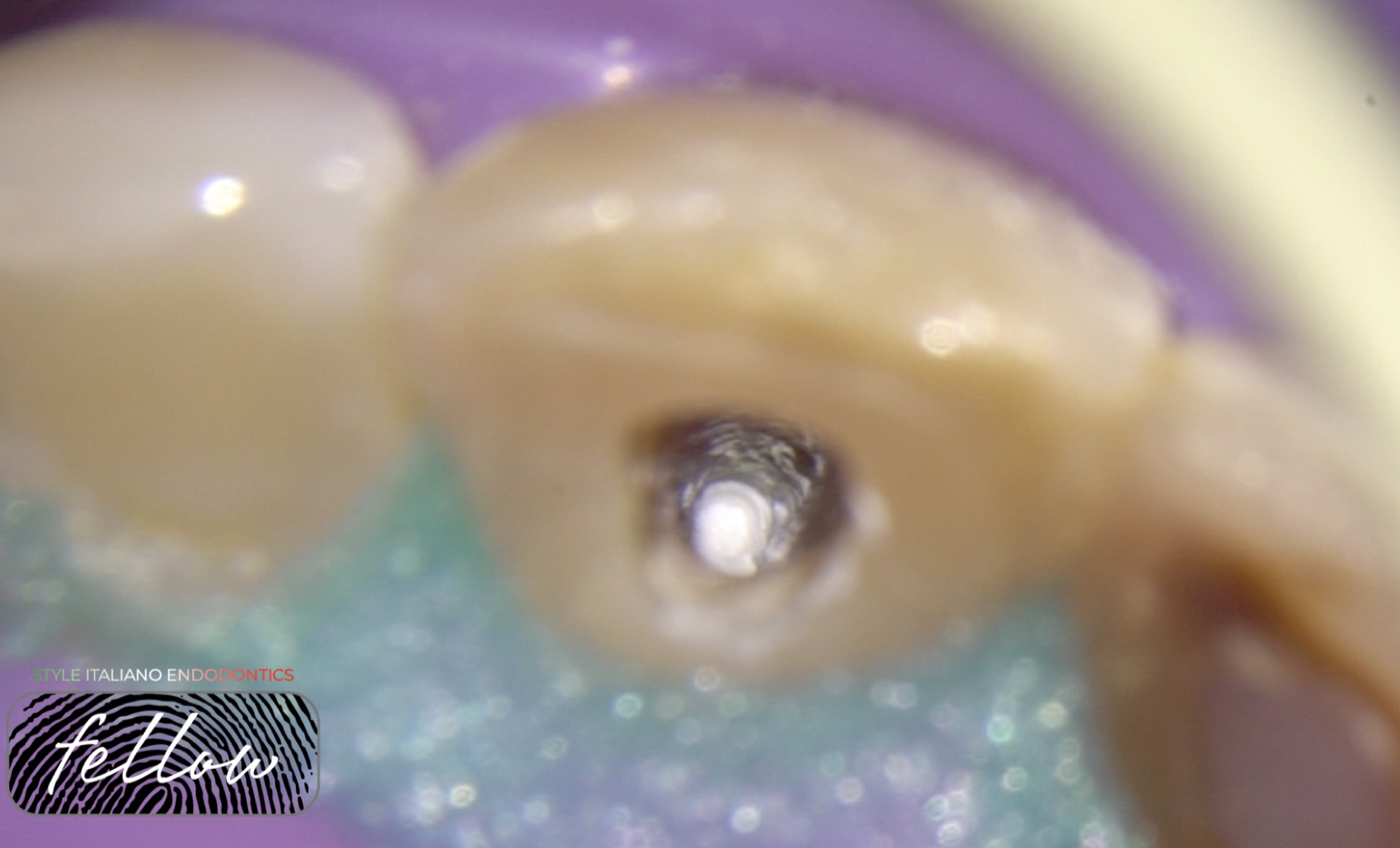
Weeping canal in tooth 11- periapical pus exudate extruding from the periapical lesion to inside the canal
Tooth 11 also had a blunderbuss canal/ open apex.
Image shows weeping canal at 25x magnification
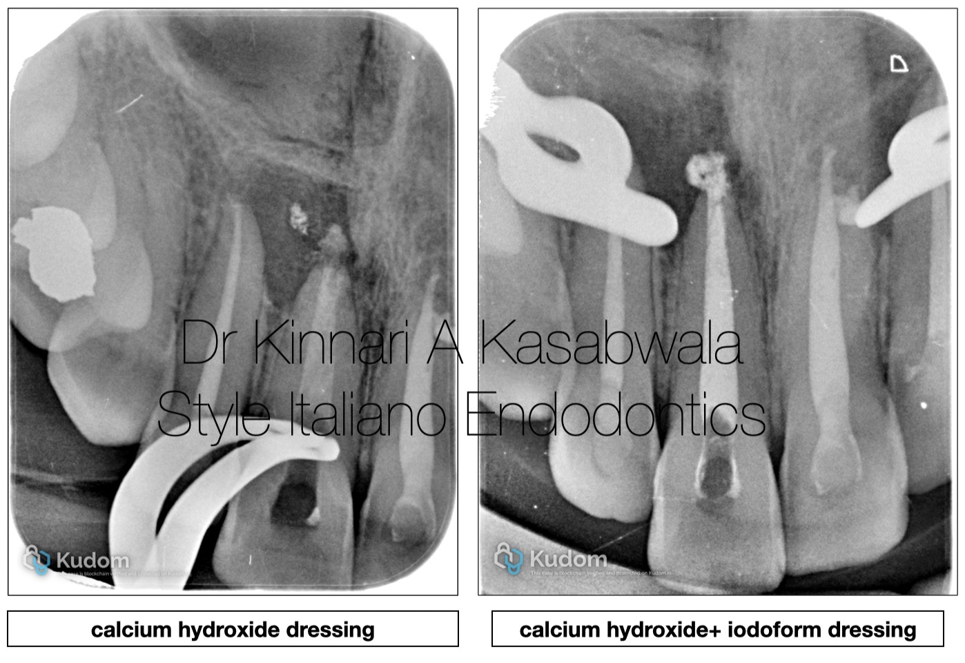
Fig. 12
In tooth 11, 3 calcium hydroxide dressings (Cleanical- Maruchi) of 2 weeks each were given followed by a long term calcium hydroxide+ iodoform dressing (Diapex plus- Diadent) for 1 month due to continuous weeping due to an appreciably large periapical lesion.
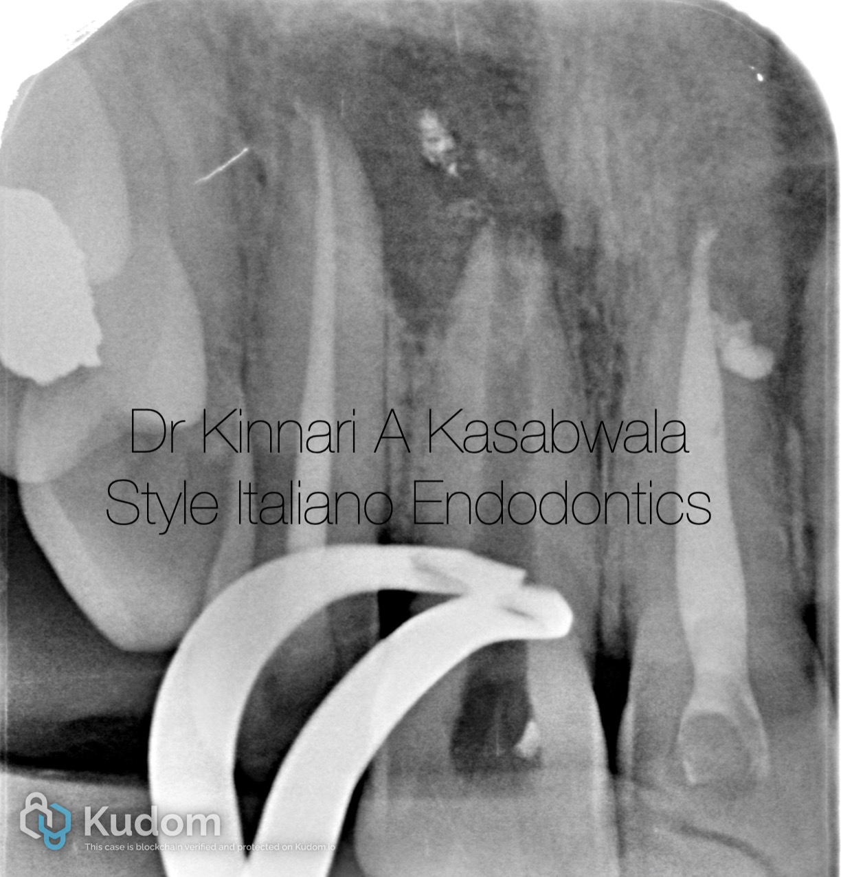
Fig. 13
Following of removal of calcium hydroxide+ iodoform dressing after 1 month, weeping had ceased. Apexification procedure was planned owing to the open apex.
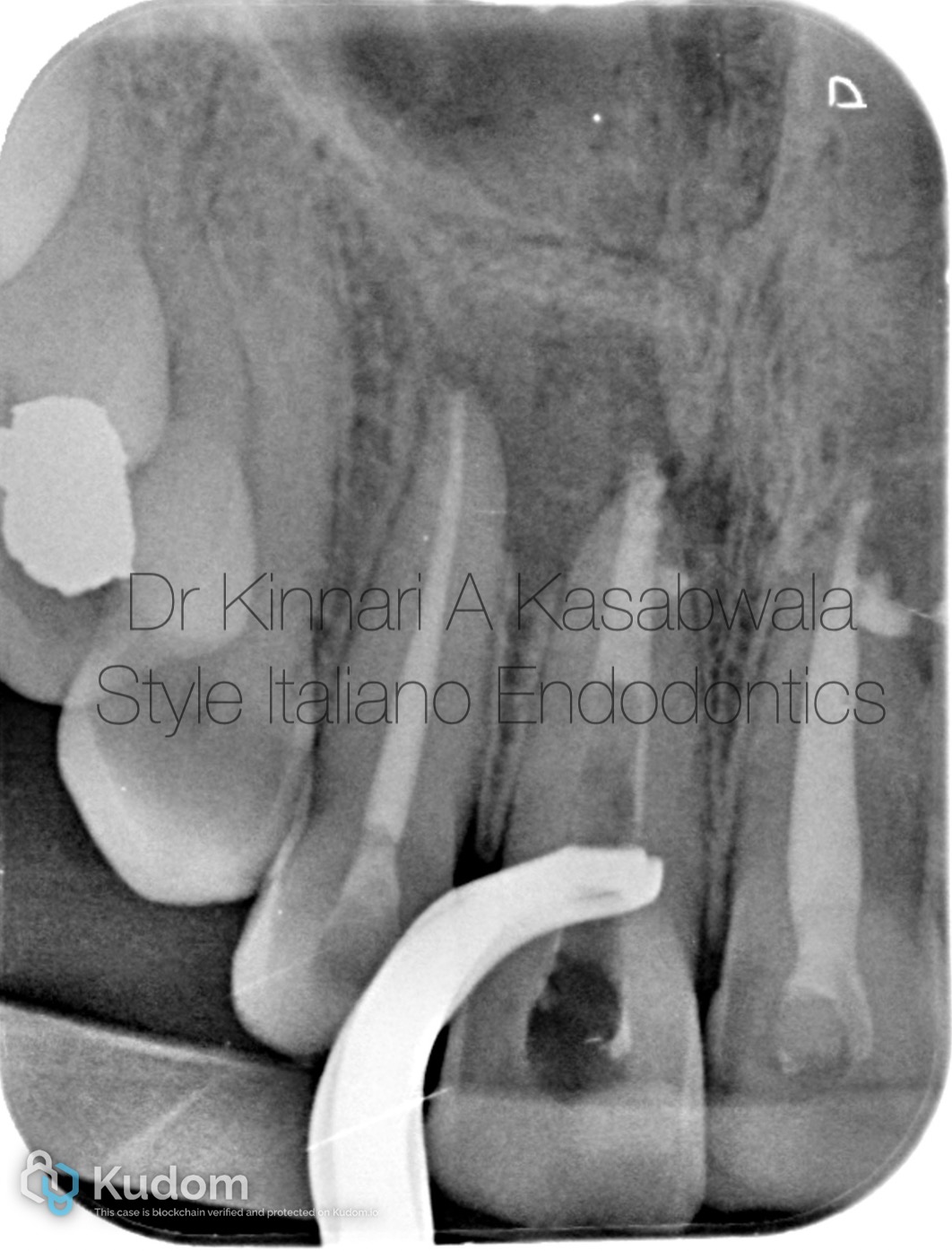
Fig. 14
5 mm apical plug was created using white MTA (Angelus) to seal the open apex.
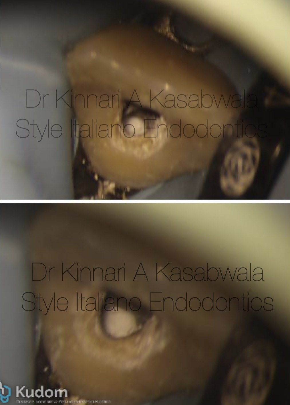
Fig. 15
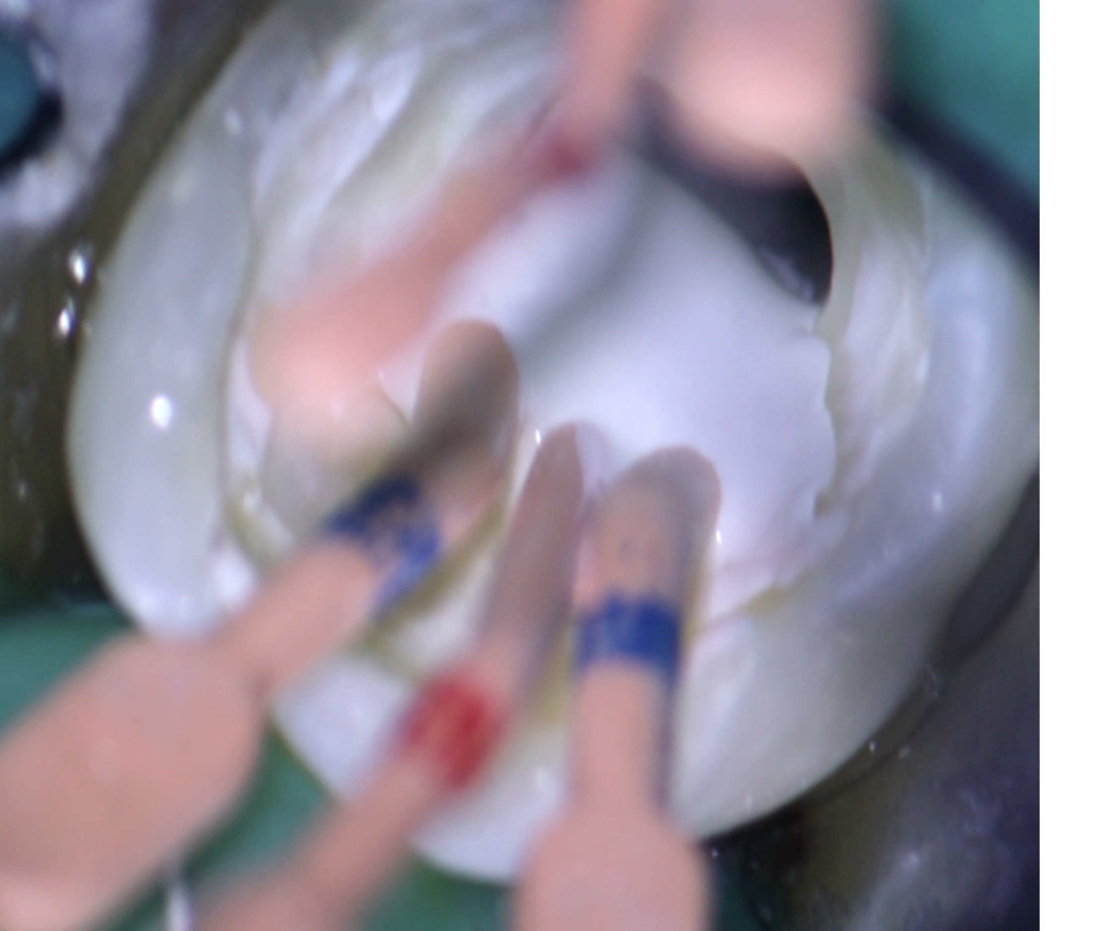
5 mm Apical MTA plug was made by incrementally placing small amounts of MTA using MTA carrier and compacting it with a predetermined size plugger with rubber stopper adjusted at the occlusal reference point.
MTA apical plug as seen at 16x and 25x magnification in image.
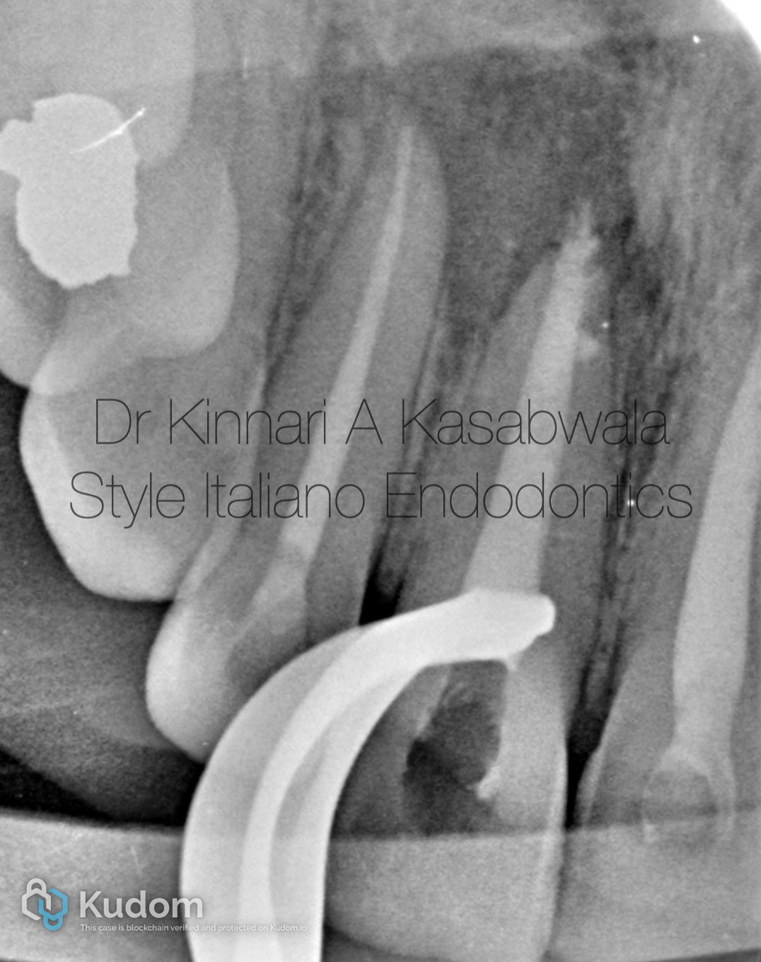
Fig. 16
Following satisfactory setting of the MTA apical plug, the canal was incrementally backfilled with thermoplastic GP (EQV- Meta Biomed) till the CEJ, while simultaneously condensing each increment with appropriate sized pluggers to avoid void formations.
IOPA for Immediate post op after back fill.
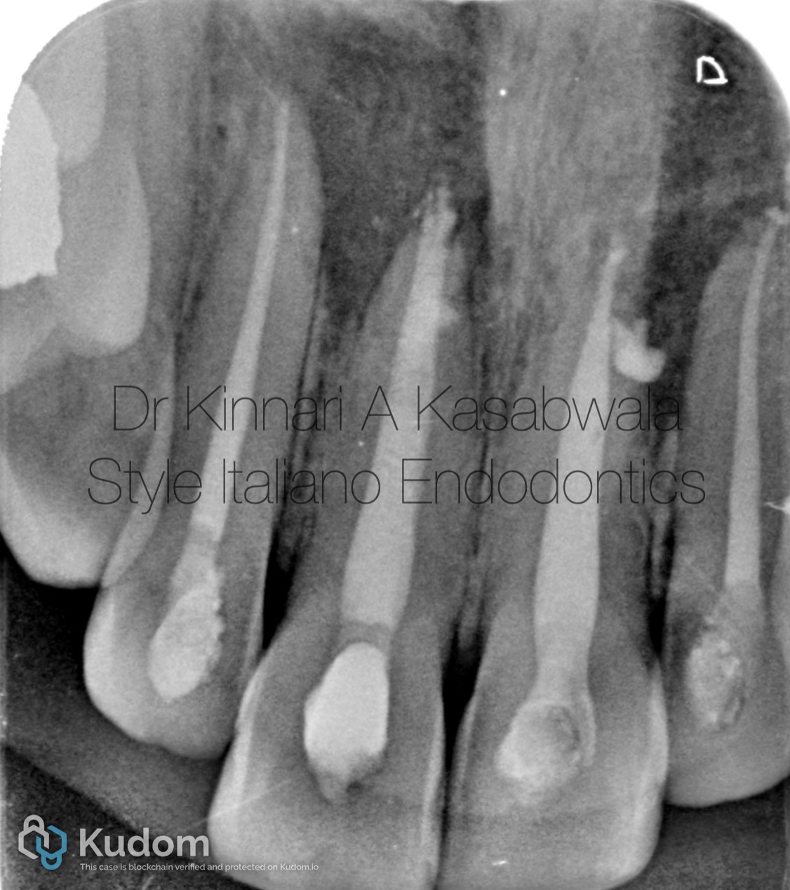
Fig. 17
Immediate post-op IOPA
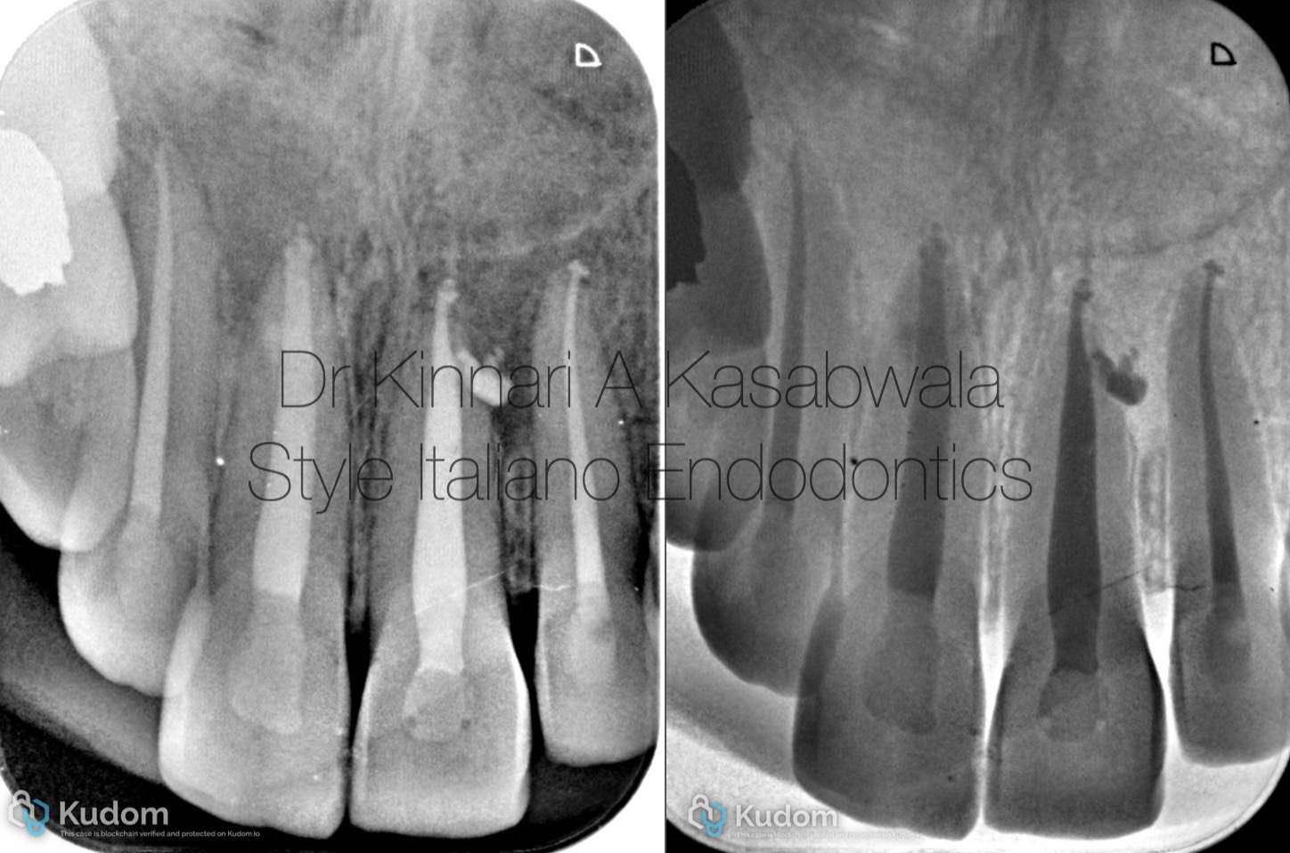
Fig. 18
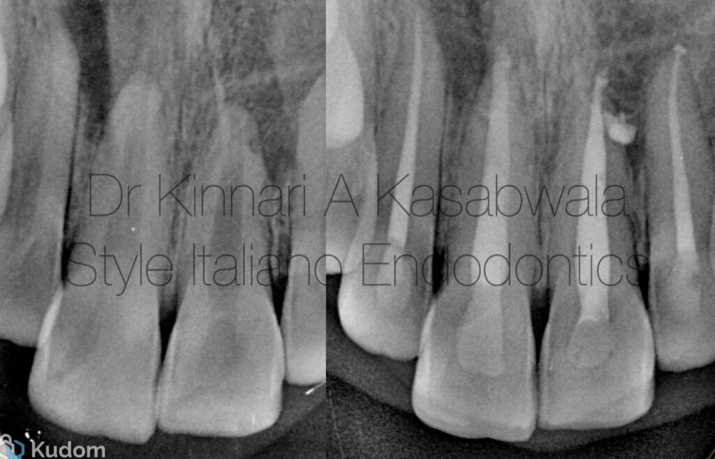
6 months recall IOPA – shows complete healing in 21,22 region and healing lesion in 11,12 region
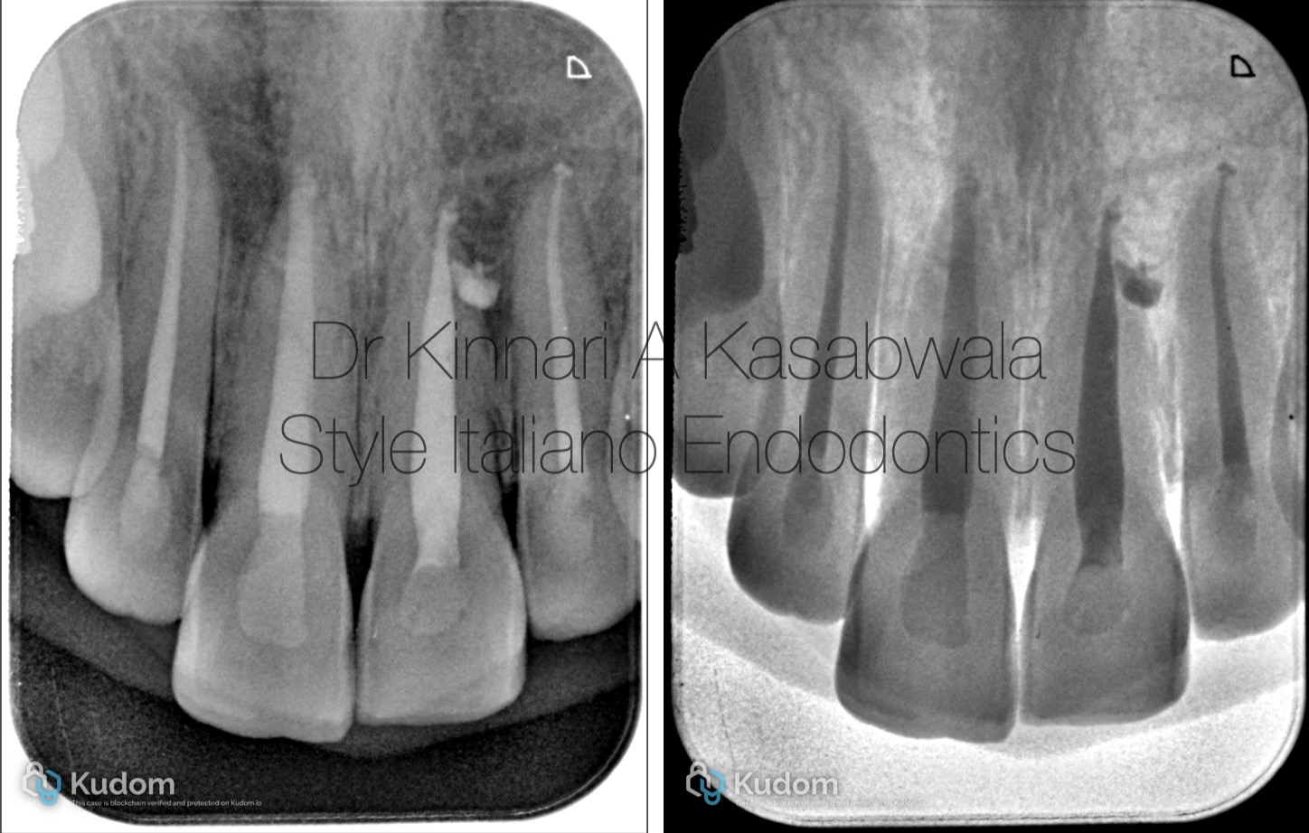
Fig. 19
1 year recall IOPA- shows improvement in the bone formation in 11,12 region with decreased radiolucency; and maintenance of healed bone status in 21,22 region.
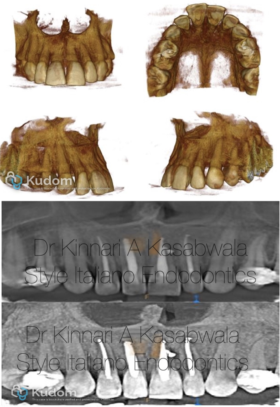
Fig. 20
Recall CBCT taken 1 year post-op
3D reconstruction shows healed perforation in buccal cortical plate in 11,12 region.
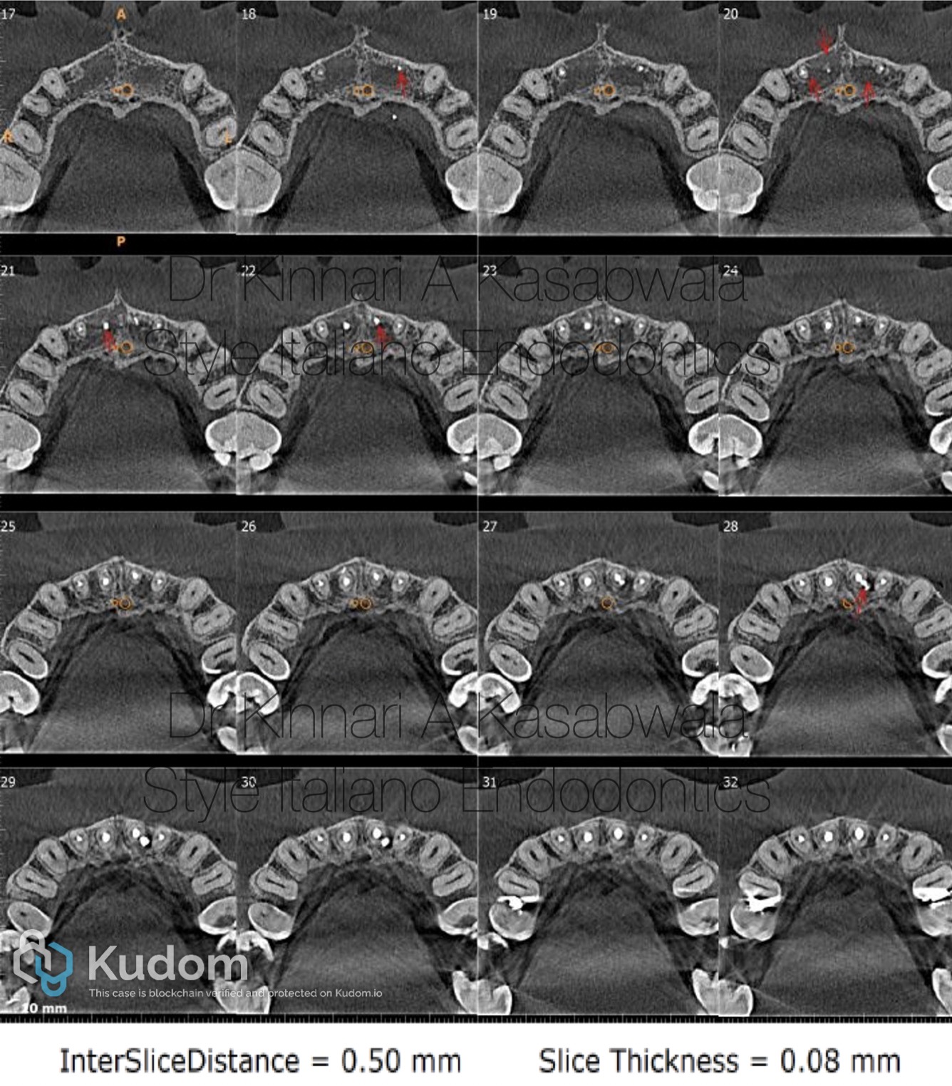
Fig. 21
AXIAL VIEW CBCT
Shows appreciable resolution of periapical lesion wrt teeth 11,12 and complete resolution wrt teeth 21,22
Complete healing of previously perforated buccal cortical plate seen
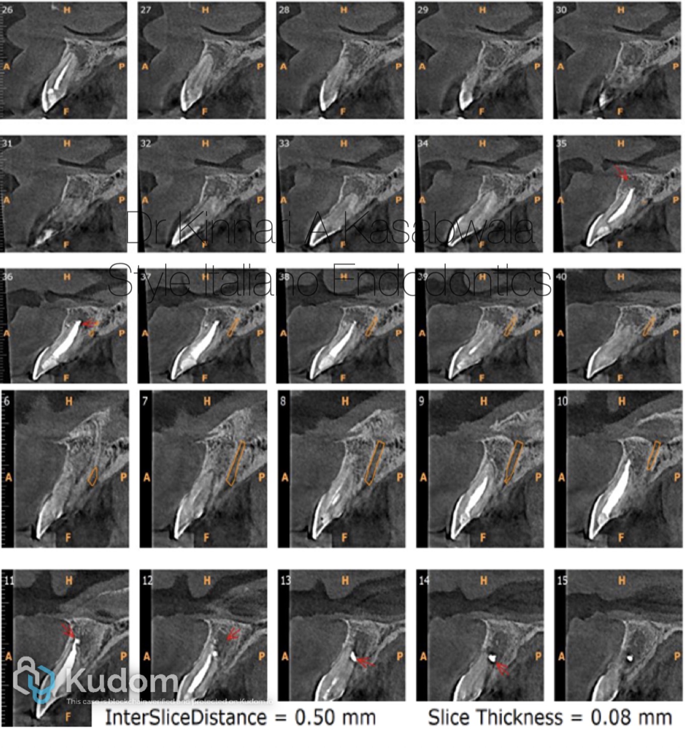
Fig. 22
SAGITTAL VIEW CBCT
Shows marked bone deposition in periapical region of 11,12 and confirmation of a fully formed healthy buccal cortical plate.

Fig. 23
About the author
- D.S. from Nair hospital dental college, Mumbai (2011-2016)
- D.S. specializing in Conservative dentistry and Endodontics from Meenakshi Ammal dental college and hospital, MAHER University, Chennai (2017-2020)
She is awarded with the following:
- "Emerging star 2019" for being the outstanding postgraduate by Conservative dentistry and endodontics association of Tamil Nadu
- Selected as top finalist at the National level in Endodontics for "Young Achiever award 2020" by Indian association of conservative dentistry and endodontics
She is a patent holder of "Indigenous cost-effective biofilm flow cell model and bubble trap" under Intellectual property India, Government of India.
She has authored and co-authored several publications in National and International journals.
She is inducted as a national key opinion leader with Bombay dental surgical.
She is inducted as a fellow with Style Italiano endodontics forum from May 2023.
She has conducted various lectures and hands-on workshops in association with team Bombay dental surgical and Style Italiano Endodontics, India.
Dr. Kinnari Kasabwala is the co-owner of Dr Kasabwala dental speciality clinic in Mumbai which has 2 branches. She is currently practising as a private practitioner, specializing in micro-endodontics. Her interests are Endodontics, dental aesthetic procedures and multi-disciplinary dentistry. She is the co-owner of Kasabwala dental academy in Andheri, Mumbai.
Conclusions
The regression of large periapical lesions without surgical intervention is possible when root canal treatment is carried out with careful adherence to endodontic principles, including aseptic management, comprehensive shaping and cleaning, effective irrigation and disinfection, and the use of intracanal medicaments such as calcium hydroxide.
Historically, numerous periapical lesions were considered cystic in origin and were therefore managed surgically. Recent insights into lesion pathogenesis, aided by three-dimensional imaging and molecular biology, have clarified their predominantly inflammatory etiology. In addition, innovations in calcium silicate-based biomaterials and regenerative endodontic procedures have strengthened the scientific rationale for non-surgical management, contributing to its growing clinical acceptance and success.
Bibliography
Ramachandran Nair PN, Pajarola G, Schroeder HE. Types and incidence of human periapical lesions obtained with extracted teeth. Oral Surg Oral Med Oral Pathol Oral Radiol Endod 1996; 81(1): 93-102.
Nair PNR. On the causes of persistent apical periodontitis: A review. Int Endod J 2006; 39(4): 249-81.
Ng YL, Mann V, Gulabivala K. A prospective study of the factors affecting outcomes of nonsurgical root canal treatment: Part 1: Periapical health. Int Endod J 2011; 44(7): 583-609.
Gulabivala K, Ng YL. Factors that affect the outcomes of root canal treatment and retreatment : A reframing of the principles. Int Endod J 2023; 56(S2)(Suppl. 2): 82-115.
Ramachandran Nair PN. Non‐microbial etiology: foreign body reaction maintaining post‐treatment apical periodontitis. Endod Topics 2003; 6(1): 114-34.
Del Fabbro M, Taschieri S, Testori T, Francetti L, Weinstein RL. Surgical versus non‐surgical endodontic re‐treatment for periradicular lesions. Cochrane Database Syst Rev 2007; 18(3): CD005511.
Bucchi C, Rosen E, Taschieri S. Non‐surgical root canal treatment and retreatment versus apical surgery in treating apical periodontitis: A systematic review. Int Endod J 2023; 56(S3)(Suppl. 3): 475-86.
Ricucci D, Pascon EA, Pitt Ford TR, Langeland K. Epithelium and bacteria in periapical lesions. Oral Surg Oral Med Oral Pathol Oral Radiol Endod 2006; 101(2): 239-49.
Ricucci D, Siqueira JF Jr. Biofilms and apical periodontitis: Study of prevalence and association with clinical and histopathologic findings. J Endod 2010; 36(8): 1277-88.
Siqueira JF Jr, Rôças IN, Ricucci D, Hülsmann M. Causes and management of post-treatment apical periodontitis. Br Dent J 2014; 216(6): 305-12.