Beyond Canal Number: Anatomical and Clinical Challenges in Maxillary Premolars – A Case Series
24/08/2026
Warning: Undefined variable $post in /home/styleendo/htdocs/styleitaliano-endodontics.org/wp-content/plugins/oxygen/component-framework/components/classes/code-block.class.php(133) : eval()'d code on line 2
Warning: Attempt to read property "ID" on null in /home/styleendo/htdocs/styleitaliano-endodontics.org/wp-content/plugins/oxygen/component-framework/components/classes/code-block.class.php(133) : eval()'d code on line 2
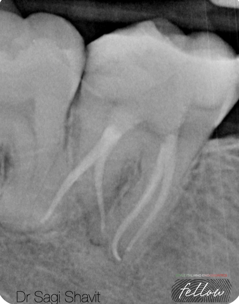
Maxillary premolar cases highlight how anatomy and technical factors can turn expected root canal treatment into a clinical challenge.
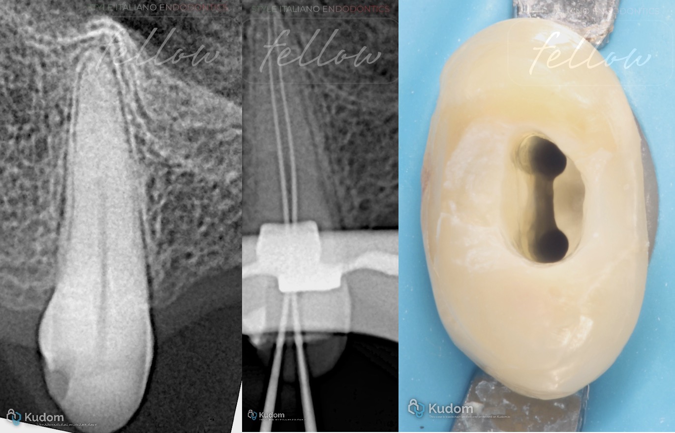
Fig. 1
Expected Anatomy with Narrow Canals
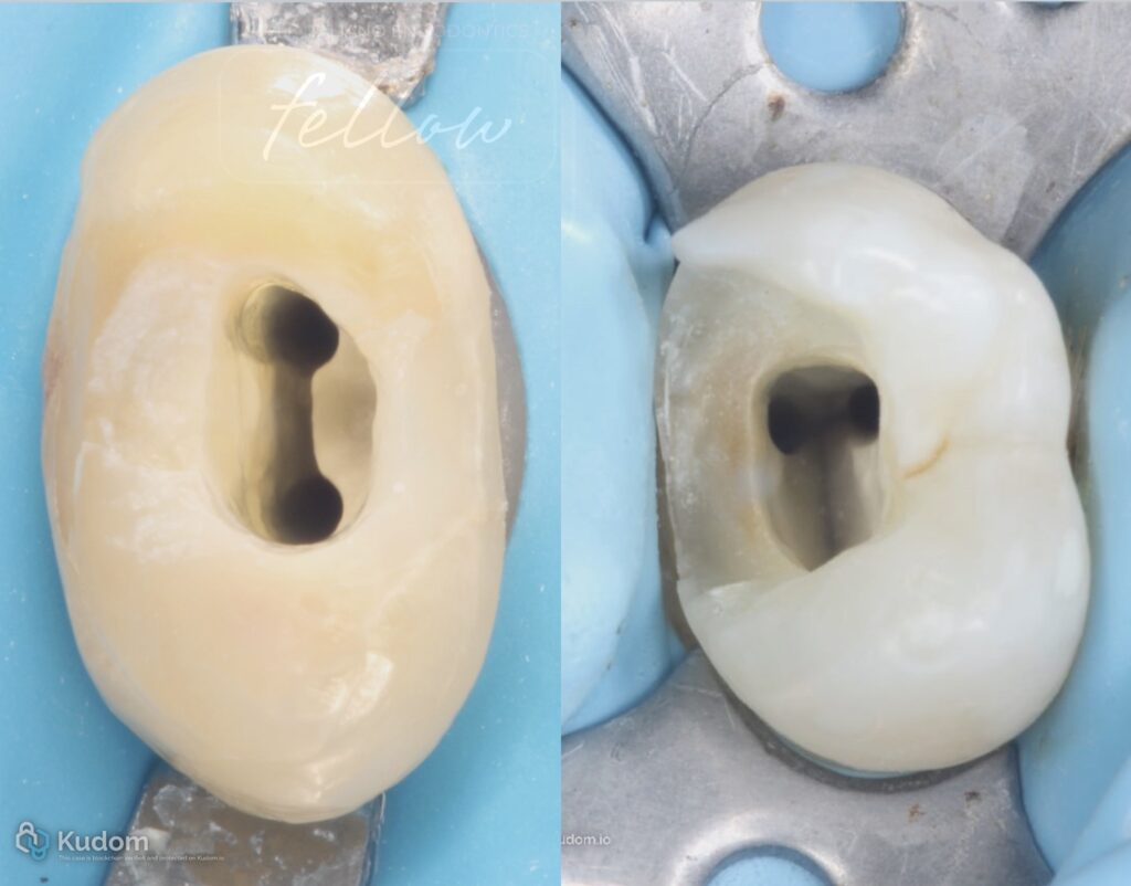
A maxillary premolar presented with the expected two-canal configuration. Despite the conventional anatomy, both canals were markedly narrow, making negotiation and instrumentation more challenging.
Careful exploration using small hand files under magnification allowed progressive negotiation while maintaining a conservative approach.
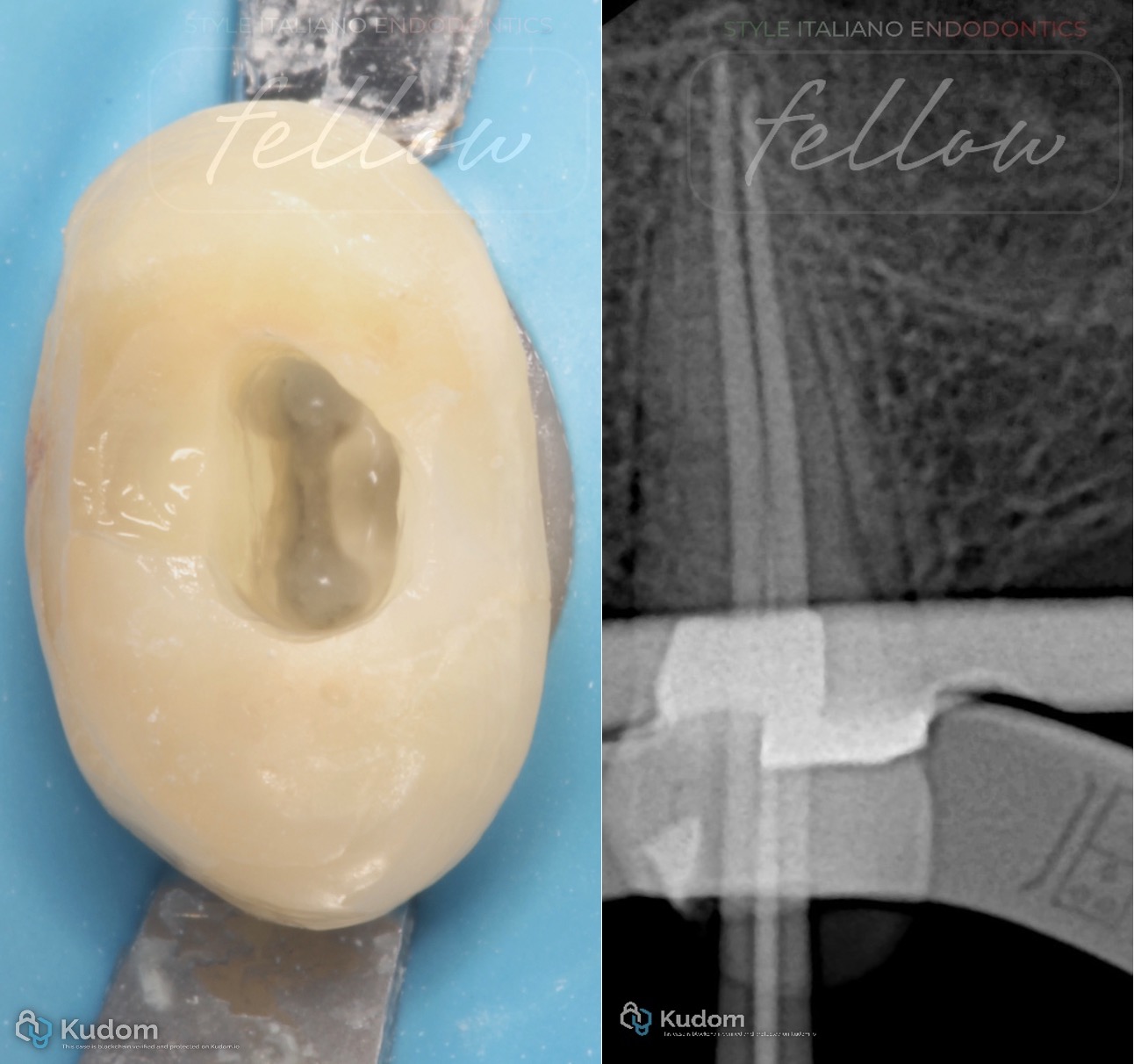
Fig. 2
(A) Clinical view during irrigant activation. (B) Master cone radiograph confirming the fit and position of both gutta-percha cones.
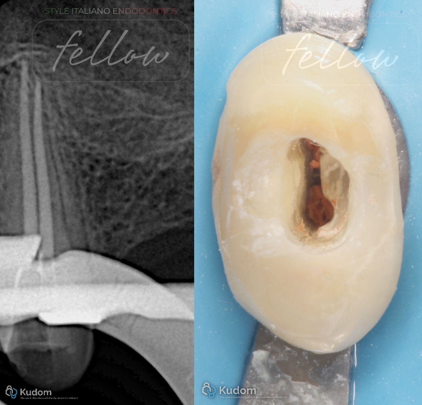
Fig. 3
A) Postoperative periapical radiograph demonstrating the final obturation of both canals. (B) Clinical view of the pulp chamber following obturation.
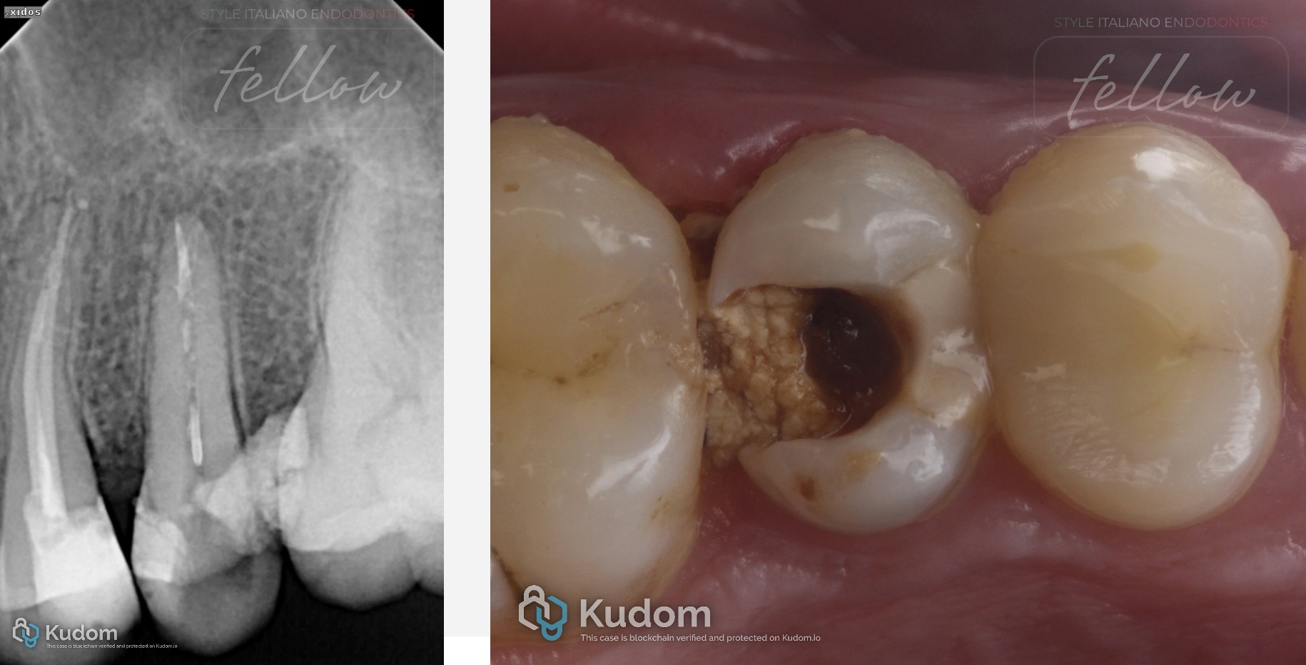
Fig. 4
Single Canal with a Deep Split
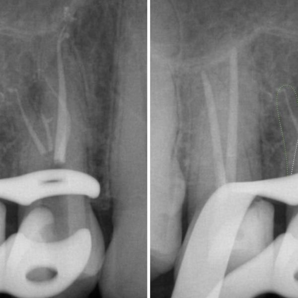
The premolar initially presented as a single canal extending through a considerable portion of the root. Deeper exploration revealed bifurcation into two separate canal pathways.
Deep canal divisions may be particularly difficult to detect because the coronal anatomy initially suggests a simple single-canal configuration [2,5]. (A) Preoperative periapical radiograph showing an apparently single canal in the coronal portion of the root. (B) Initial clinical view of the access cavity showing a single canal orifice.
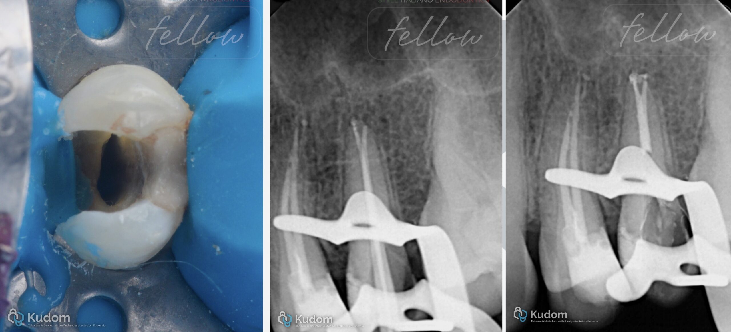
Fig. 5
Figure 5. (A) Clinical view following access refinement. (B, C) Master cone radiographs demonstrating the deep bifurcation of the initially single canal into two separate canal pathways.
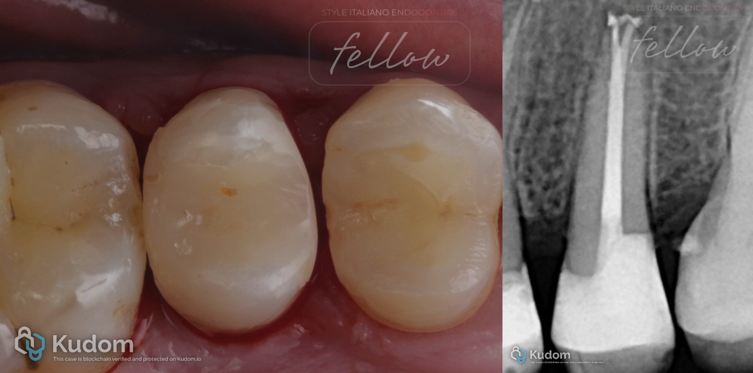
Fig. 6
Figure 6. (A) Postoperative clinical view. (B) Final periapical radiograph demonstrating the initially single canal dividing deeply into two separate, obturated canal pathways.
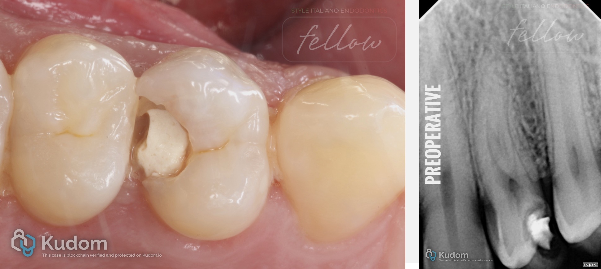
Fig. 7
Molarized Premolar
Clinical and radiographic examination revealed an unusual maxillary premolar with three separate roots and three canals arranged in a molar-like configuration. Three-rooted maxillary premolars are uncommon [1,2]. When arranged as two buccal roots and one palatal root, the morphology resembles that of a miniature maxillary molar and has been described as a “small molar” or “radiculous molar” [4,6]. Identification of all three canals required careful assessment of the preoperative anatomy and exploration of the pulp chamber floor.
(A) Preoperative clinical view of the maxillary premolar. (B) Preoperative periapical radiograph revealing an unusual root outline suggestive of complex root anatomy.
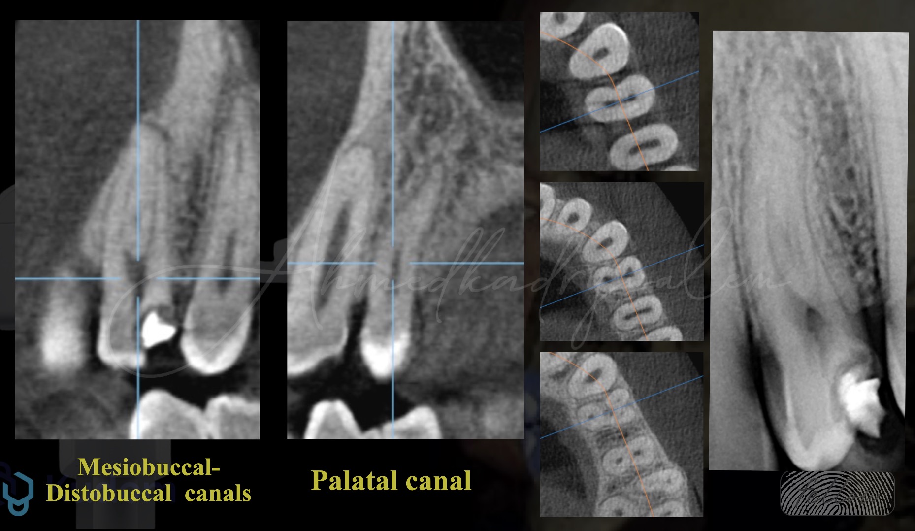
Fig. 8
CBCT evaluation confirming three separate roots and three canals: mesiobuccal, distobuccal, and palatal.
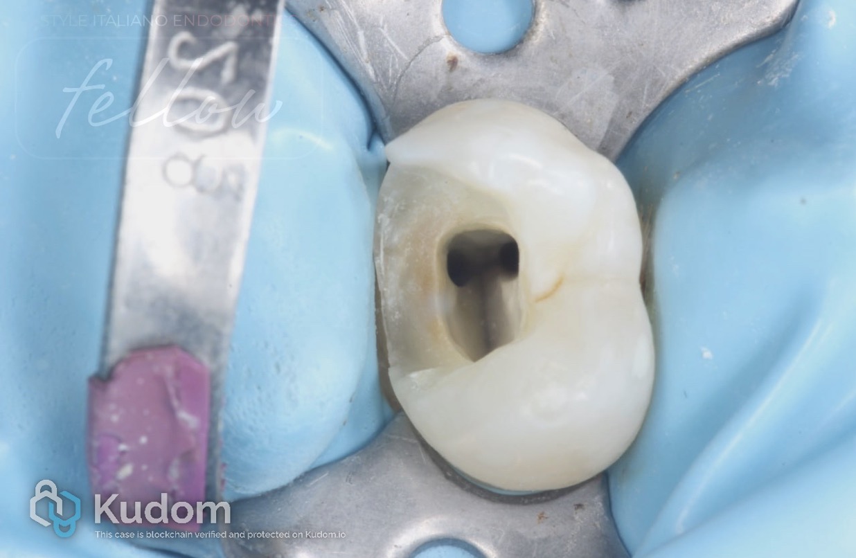
Fig. 9
Clinical view of the access cavity demonstrating the two buccal canal orifices: mesiobuccal and distobuccal.
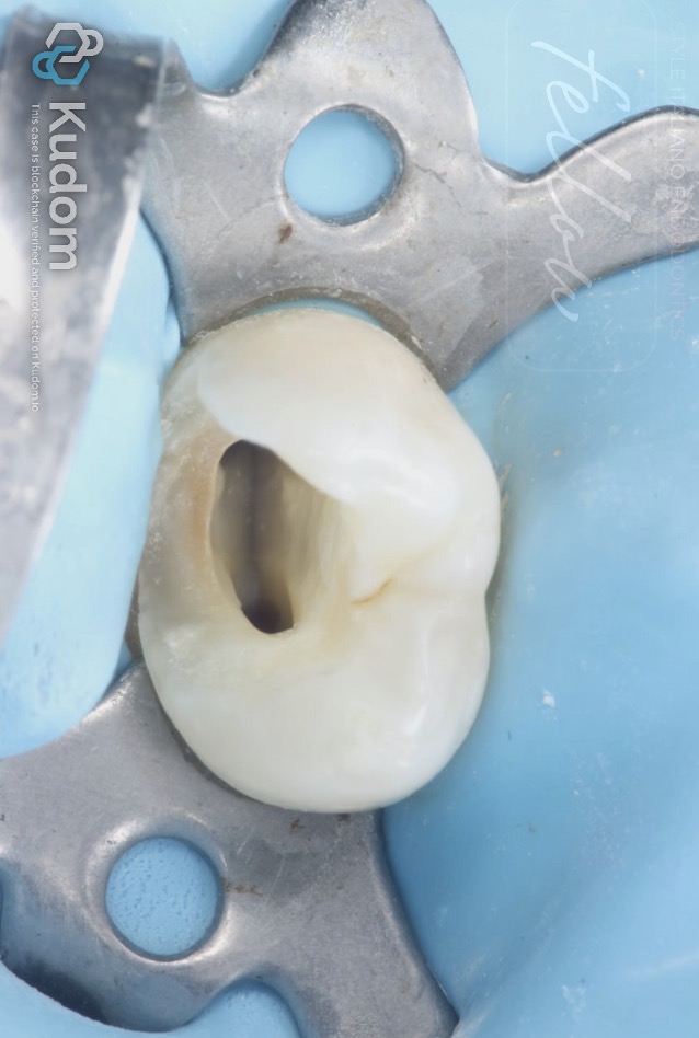
Fig. 10
Clinical view demonstrating the palatal canal orifice.
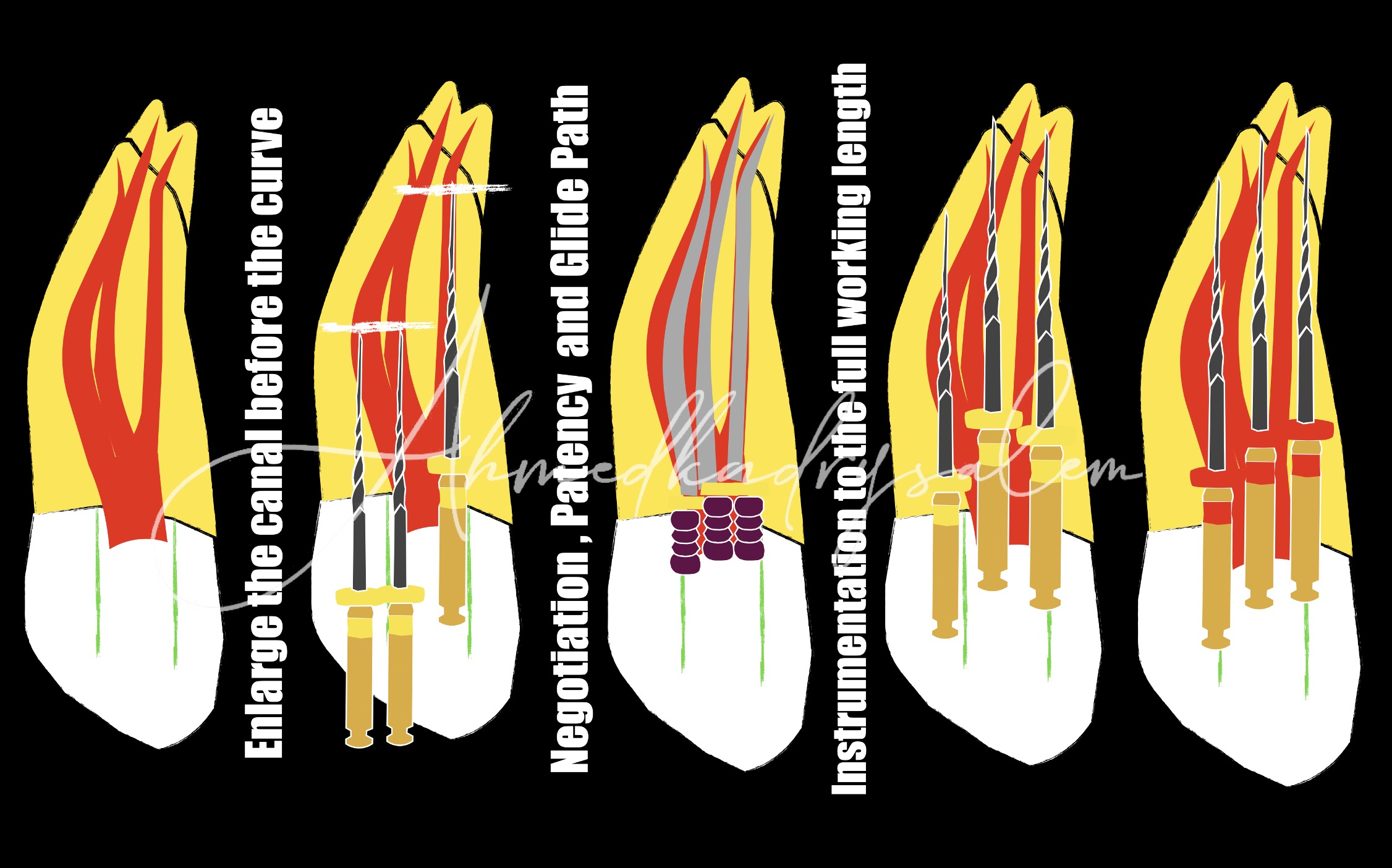
Fig. 11
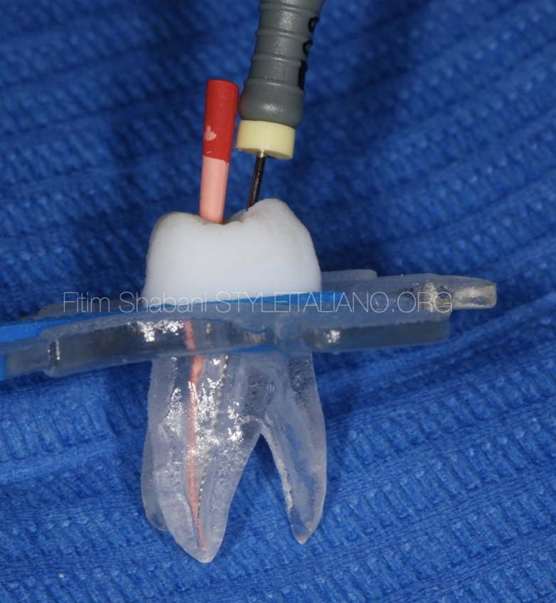
Schematic illustration of the negotiation, glide-path establishment, and instrumentation of the three-canal system in the molarized maxillary premolar
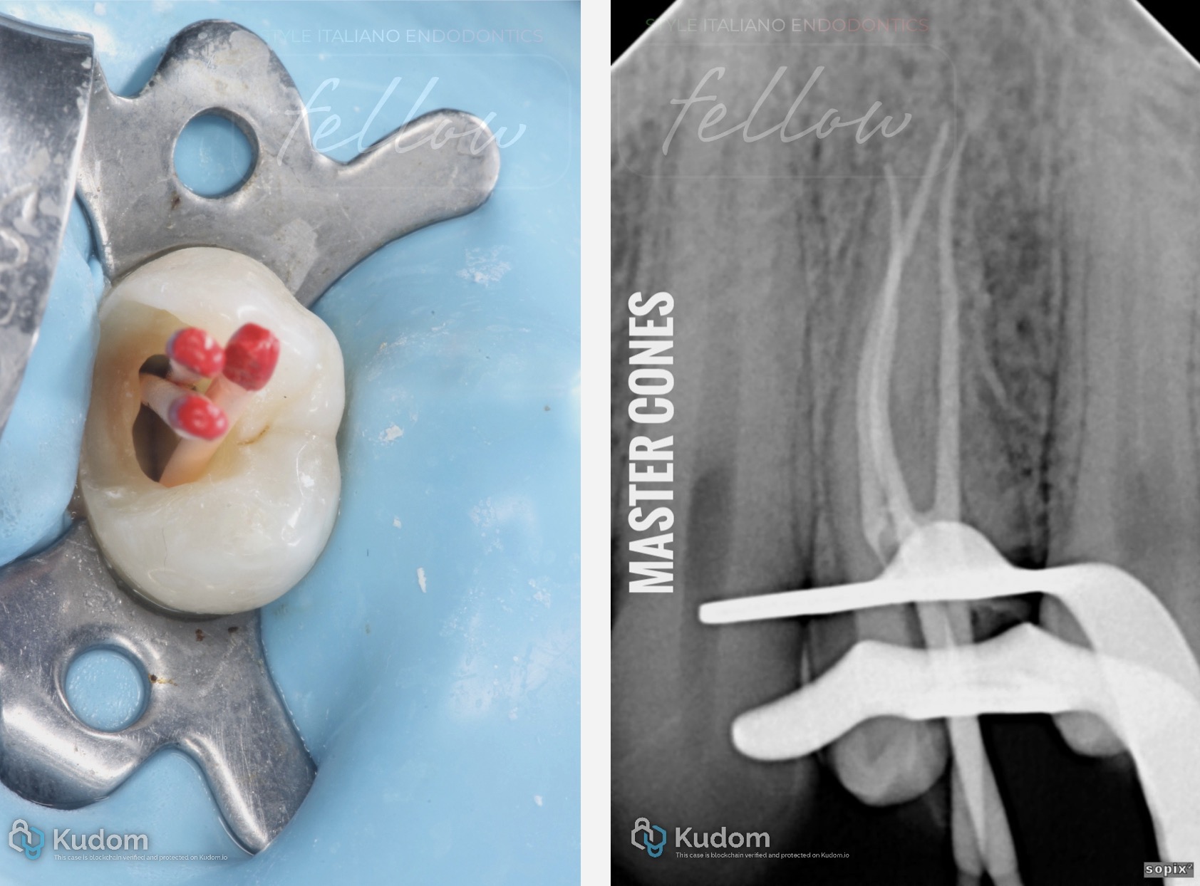
Fig. 12
. (A) Clinical view of the three master gutta-percha cones positioned in the mesiobuccal, distobuccal, and palatal canals. (B) Master cone radiograph confirming their position within the three separate canal pathways.
Video of the procedure.
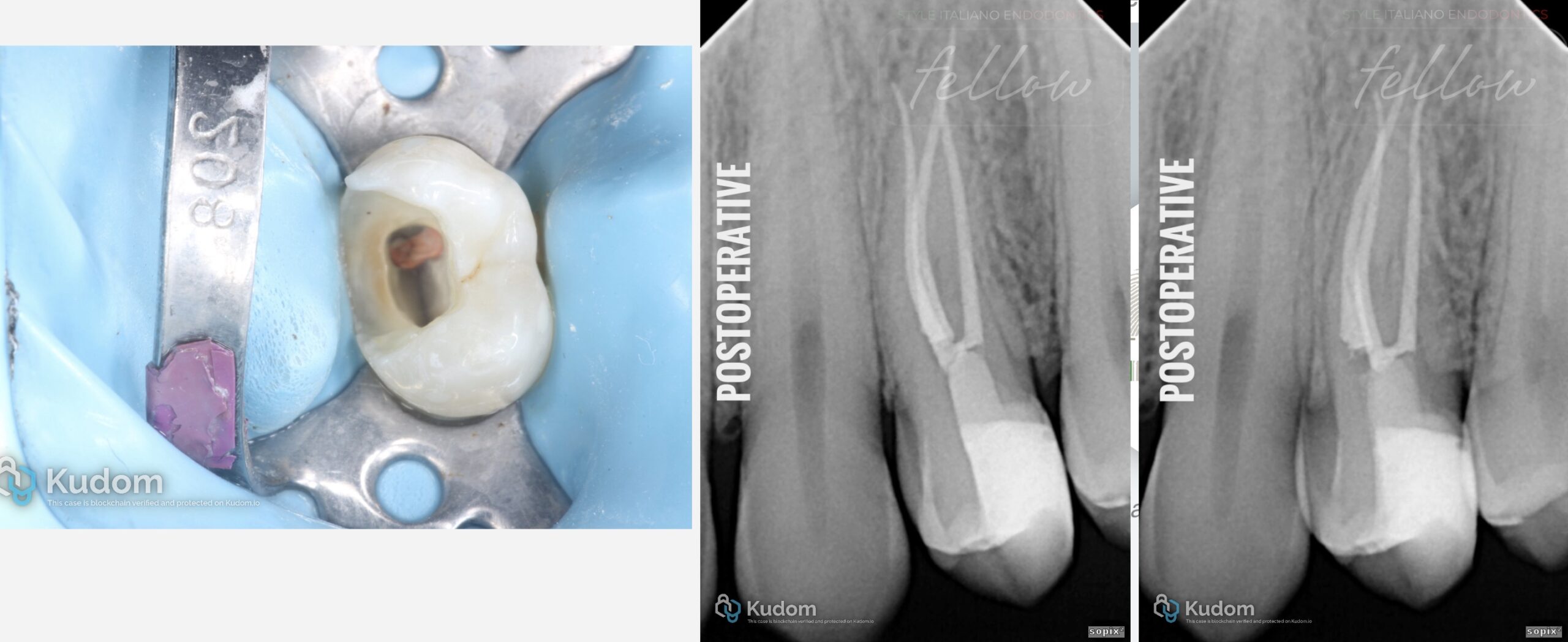
Fig. 13
(A) Clinical view following obturation. (B, C) Postoperative periapical radiographs at different horizontal angulations demonstrating final obturation of the three separate canals.

Fig. 14
About Dr. Ahmed Kadry Salem
- BDS - Pharos University
- Msc in Endodontic - Alexandria University
- T.A - AAST Alamein Branch
- T.A - Alexandria National University
- Fellow of Style Italiano Endodontics
- Member of Royal College of Edinburgh
- Founder of Kadry Dental Care
- Founder of Dental Products Review Group
Conclusions
The presented cases demonstrate that the difficulty of maxillary premolar endodontics is multifactorial and extends beyond the number of roots and canals. Even expected anatomy may present challenges due to narrow canals, while apparently simple canal systems may exhibit deep bifurcations. At the other end of the spectrum, rare three-rooted, three-canal configurations may closely resemble the anatomy of a maxillary molar [1–6]. Importantly, treatment difficulty may also arise from technical rather than anatomical factors. A straight and wide canal may become challenging when the orifice is difficult to locate or following previous iatrogenic intervention. In such situations, adequate magnification and illumination, together with controlled ultrasonic exploration, can facilitate conservative canal localization and management [7,8]. Collectively, these cases emphasize that expected anatomy should serve as a guide rather than an assumption. Careful radiographic assessment, magnification, conservative access, appropriate instrumentation, and continuous reassessment of the canal anatomy are essential for predictable endodontic treatment of maxillary premolars.
Bibliography
[1] Martins JNR, Worldwide Anatomy Research Group, Versiani MA, et al. Worldwide assessment of the root and root canal characteristics of maxillary premolars: a multi-center cone-beam computed tomography cross-sectional study with meta-analysis. Journal of Endodontics. 2024;50(1):31–54. doi:10.1016/j.joen.2023.10.009.
[2] Xu M, Ren H, Liu C, Zhao X, Li X. Systematic review and meta-analysis of root morphology and canal configuration of permanent premolars using cone-beam computed tomography. BMC Oral Health. 2024;24:656. doi:10.1186/s12903-024-04419-y.
[3] Wolf TG, Kozaczek C, Campus G, Paqué F, Wierichs RJ. Root canal morphology of 116 maxillary second premolars by micro-computed tomography in a mixed Swiss-German population with systematic review. Journal of Endodontics. 2020;46(11):1639–1647. doi:10.1016/j.joen.2020.08.012.
[4] Saber SEDM, Ahmed MHM, Obeid M, Ahmed HMA. Root and canal morphology of maxillary premolar teeth in an Egyptian subpopulation using two classification systems: a cone beam computed tomography study. International Endodontic Journal. 2019;52(3):267–278. doi:10.1111/iej.13016..
[5] Jiannan C, Yangpeng Z, Huanhuan S, Qiang Z. Endodontic treatment of maxillary premolar with three roots: a case report. Case Reports in Dentistry. 2024;2024:5525349. doi:10.1155/2024/5525349.
[6] Al Shaikhly B, Harrel SK, Umorin M, Augsburger RA, Jalali P. Comparison of a dental operating microscope and high-resolution videoscope for endodontic procedures. Journal of Endodontics. 2020;46(5):688–693. doi:10.1016/j.joen.2020.01.013