Periapical osteoperiostitis: CBCT diagnosis and healing following endodontic retreatment
27/08/2026
Warning: Undefined variable $post in /home/styleendo/htdocs/styleitaliano-endodontics.org/wp-content/plugins/oxygen/component-framework/components/classes/code-block.class.php(133) : eval()'d code on line 2
Warning: Attempt to read property "ID" on null in /home/styleendo/htdocs/styleitaliano-endodontics.org/wp-content/plugins/oxygen/component-framework/components/classes/code-block.class.php(133) : eval()'d code on line 2
The close anatomical relationship between the roots of maxillary posterior teeth and the floor of the maxillary sinus creates a potential pathway for the extension of endodontic infection beyond the periapical tissues. Infection originating within the root canal system may therefore induce inflammatory changes not only in the apical periodontium but also in the cortical floor, periosteum, and mucosa of the maxillary sinus. The American Association of Endodontists (AAE) defines maxillary sinusitis of endodontic origin (MSEO) as sinusitis secondary to periradicular disease of endodontic origin and emphasizes that recognition and elimination of the endodontic source are fundamental to successful management.
The clinical presentation of MSEO can be diagnostically challenging. Patients may present predominantly with sinonasal complaints, including unilateral nasal congestion, rhinorrhea, postnasal drainage, facial pressure or pain, and unpleasant odor, while characteristic dental symptoms may be mild or entirely absent. In teeth with pulpal necrosis or previously unsuccessful endodontic treatment, thermal symptoms are frequently absent, and periapical tenderness or intraoral swelling may also be minimal. Consequently, the absence of toothache does not exclude an endodontic source of maxillary sinus disease.
An important manifestation of this endodontic–sinus relationship is periapical osteoperiostitis (PAO). According to the AAE Position Statement on MSEO, PAO develops when apical periodontitis situated adjacent to the cortical floor of the maxillary sinus induces an inflammatory reaction in the sinus periosteum. Expansion and elevation of the periosteum into the sinus are followed by reactive periosteal osteogenesis, with deposition of new bone along the internal surface of the elevated periosteum. Radiographically, this process produces a characteristic thin, dome-shaped hard-tissue elevation of the sinus floor, frequently described as a radiopaque “halo” lesion adjacent to the involved root apex. With persistent inflammation, the reactive bone may become progressively thicker and extend farther into the sinus. PAO may occur with or without clinical symptoms and may coexist with varying degrees of sinus mucosal edema or fluid accumulation.
PAO should be distinguished from periapical mucositis (PAM), another characteristic manifestation of endodontic inflammation involving the maxillary sinus. Whereas PAO represents a predominantly periosteal and osseous reactive response, PAM represents inflammatory edema and thickening of the sinus mucosa and is typically visualized on CBCT as localized or dome-shaped soft-tissue thickening directly adjacent to the affected root apex. Both conditions may represent different expressions of the same endodontic inflammatory process and, if the source persists, may progress toward partial or complete sinus involvement.
Cone-beam computed tomography (CBCT) is particularly important in this diagnostic process. Conventional two-dimensional radiography may fail to demonstrate periapical lesions in the posterior maxilla because of superimposition of the maxillary sinus and zygomatic structures and may inadequately depict the relationship between the root apices and the sinus floor. CBCT provides three-dimensional visualization of the tooth–bone–sinus complex and can reveal periapical bone loss, cortical involvement, PAO, mucosal changes, untreated canals, and the direct anatomical relationship between the endodontic lesion and the maxillary sinus.
The present case demonstrates periapical osteoperiostitis associated with endodontic infection of a maxillary posterior tooth and its subsequent resolution following endodontic treatment. The case highlights the importance of recognizing PAO on CBCT as a potential marker of endodontic involvement of the maxillary sinus and llustrates how elimination of the intracanal infectious source can result in healing not only of the periapical tissues but also of the associated sinus-floor inflammatory changes.
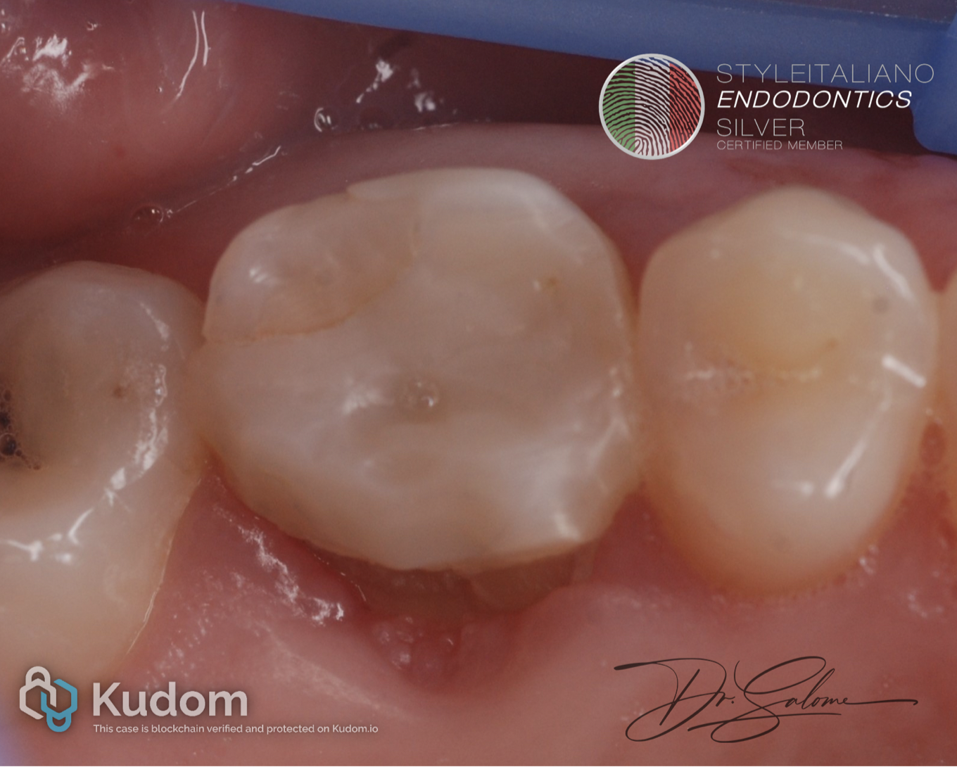
Fig. 1
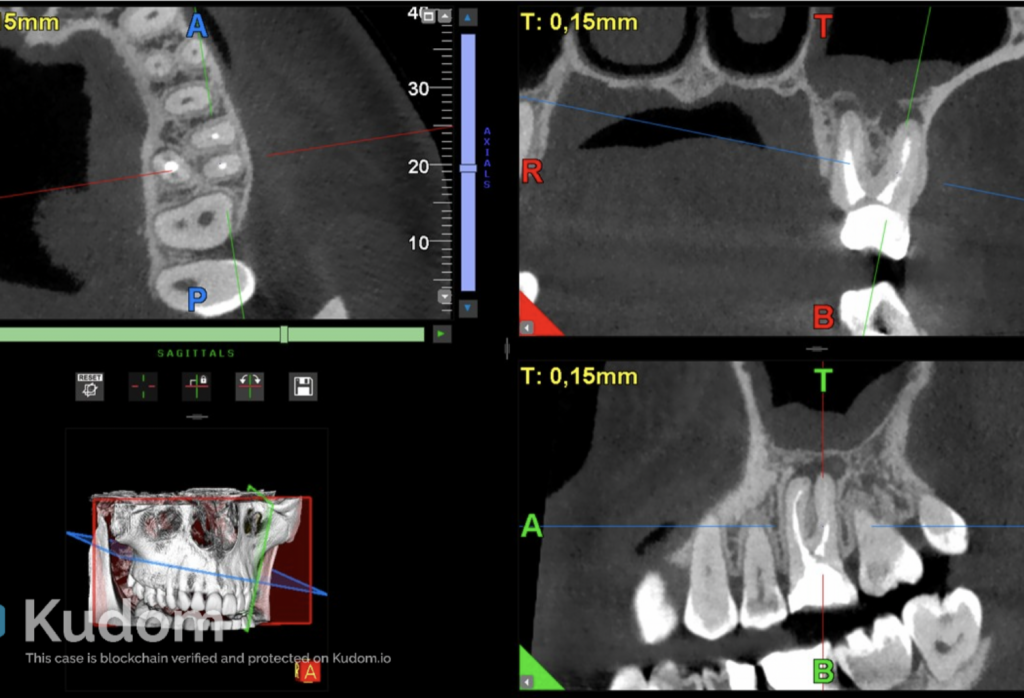
A young female patient presented with a fractured coronal restoration of a previously endodontically treated maxillary posterior tooth. The tooth was completely asymptomatic. however, medical history revealed several episodes of recurrent unilateral sinusitis during the preceding two years, without associated dental pain, which was treated by ENT with antibiotics.
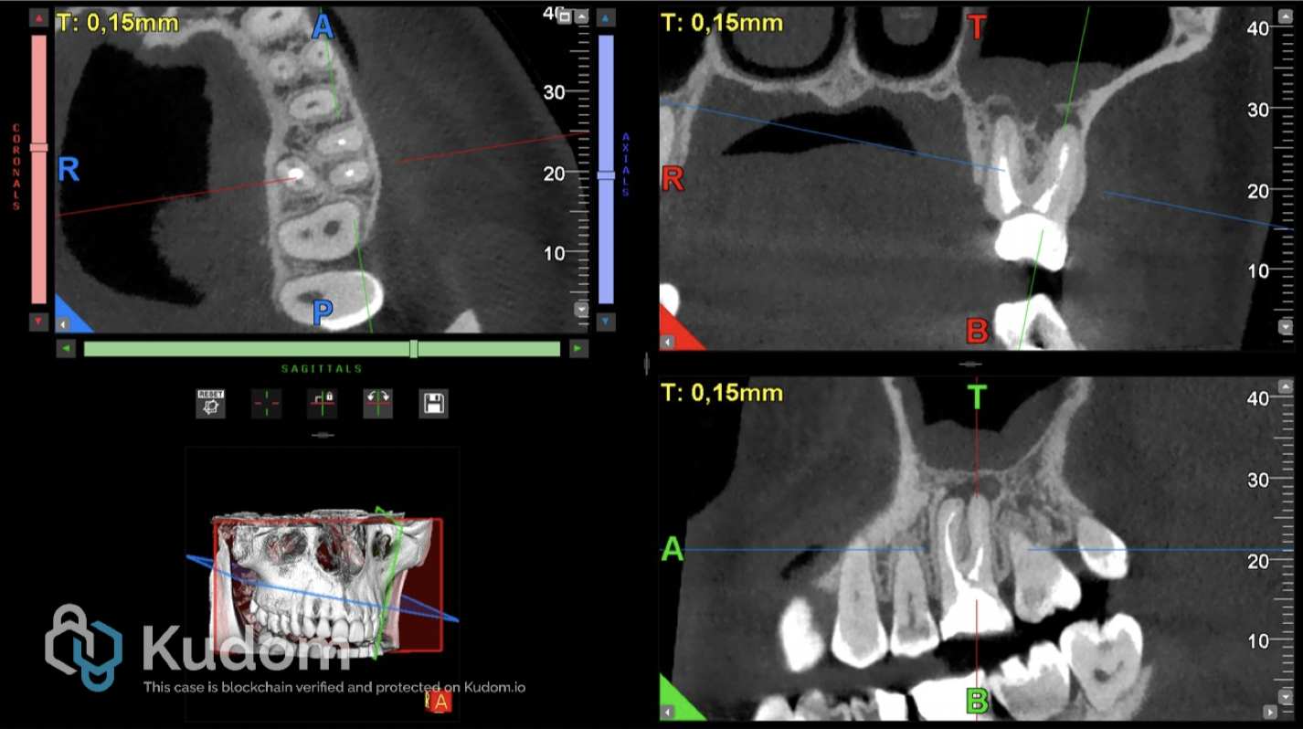
Fig. 2
CBCT revealed persistent apical periodontitis extending to the sinus floor, with reactive periosteal and adjacent mucosal thickening, consistent with periapical osteoperiostitis.
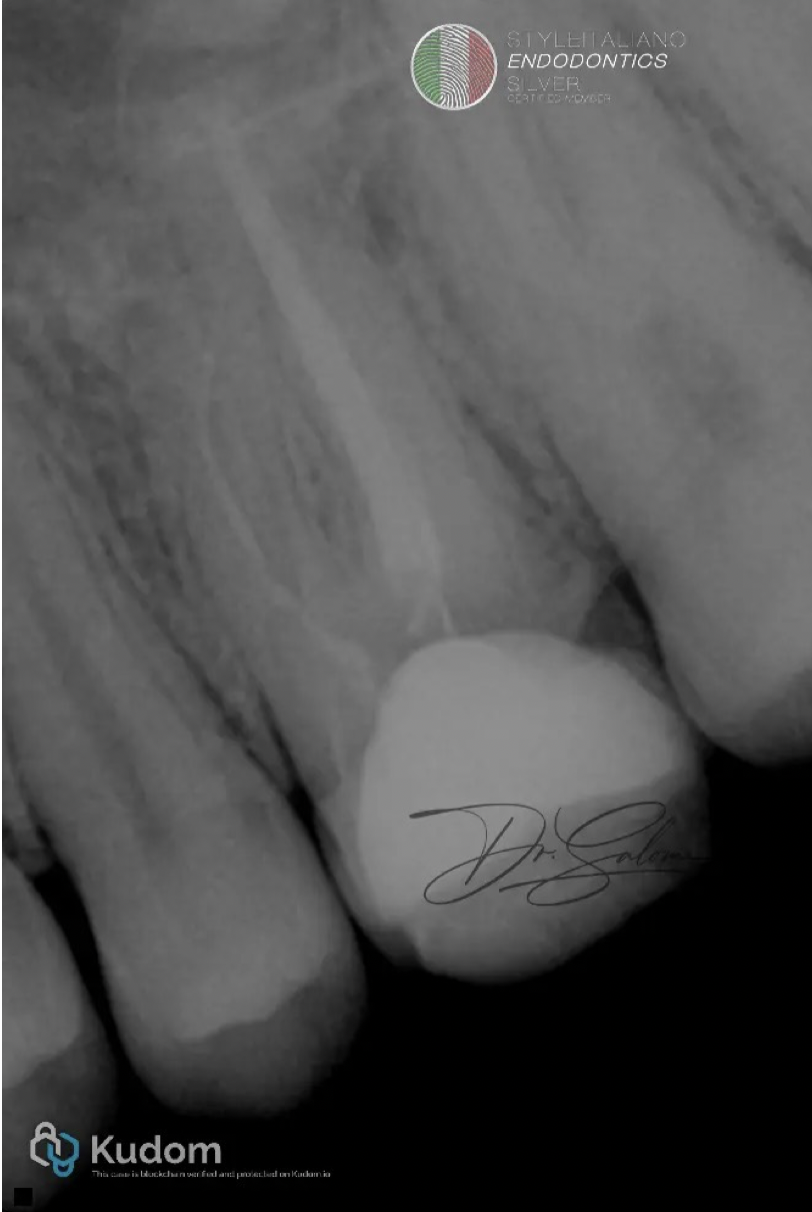
Fig. 3
Primary X-ray of affected tooth.
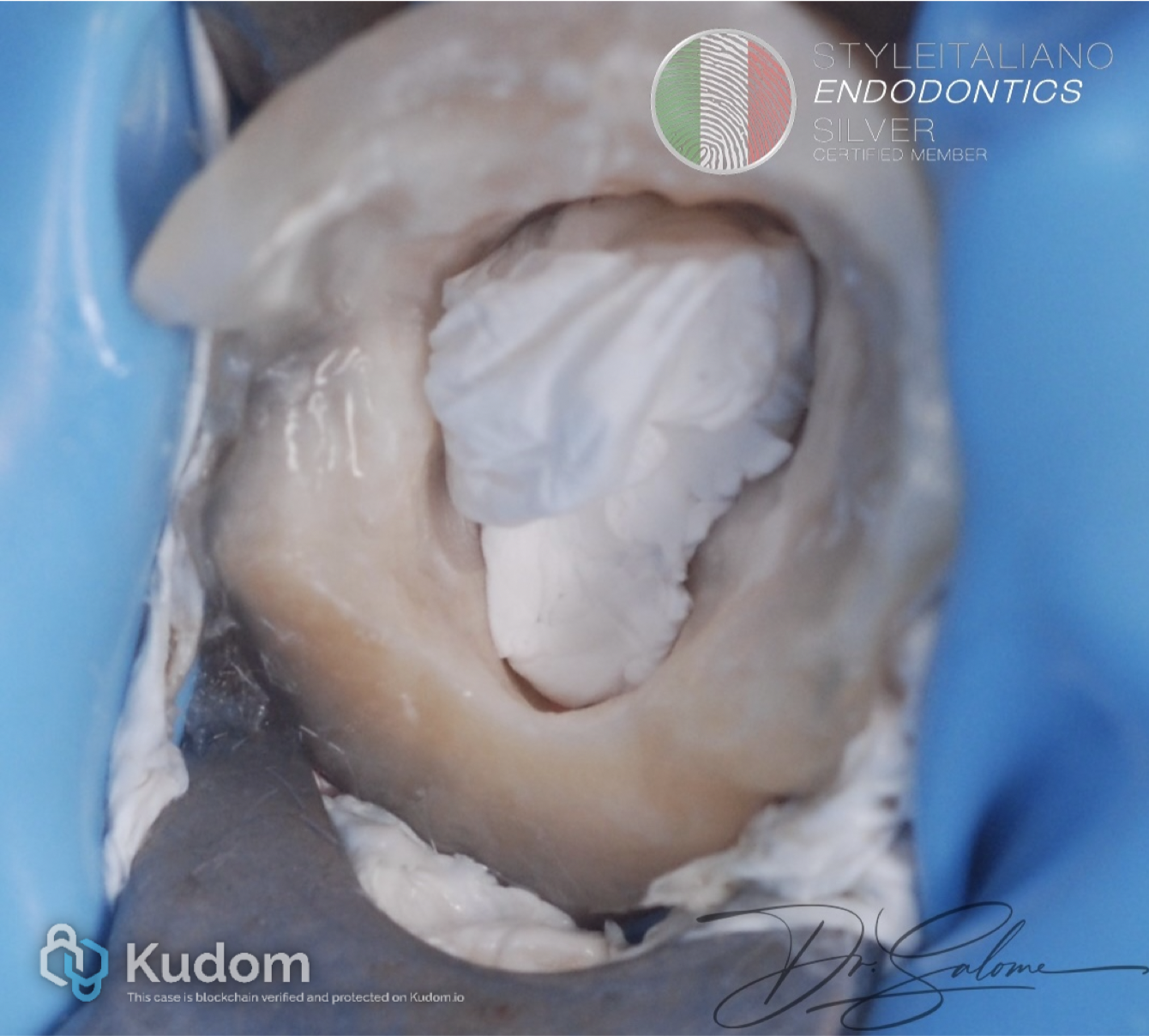
Fig. 4
The tooth was isolated under rubber dam, and the existing restoration and all carious tissue were completely removed .
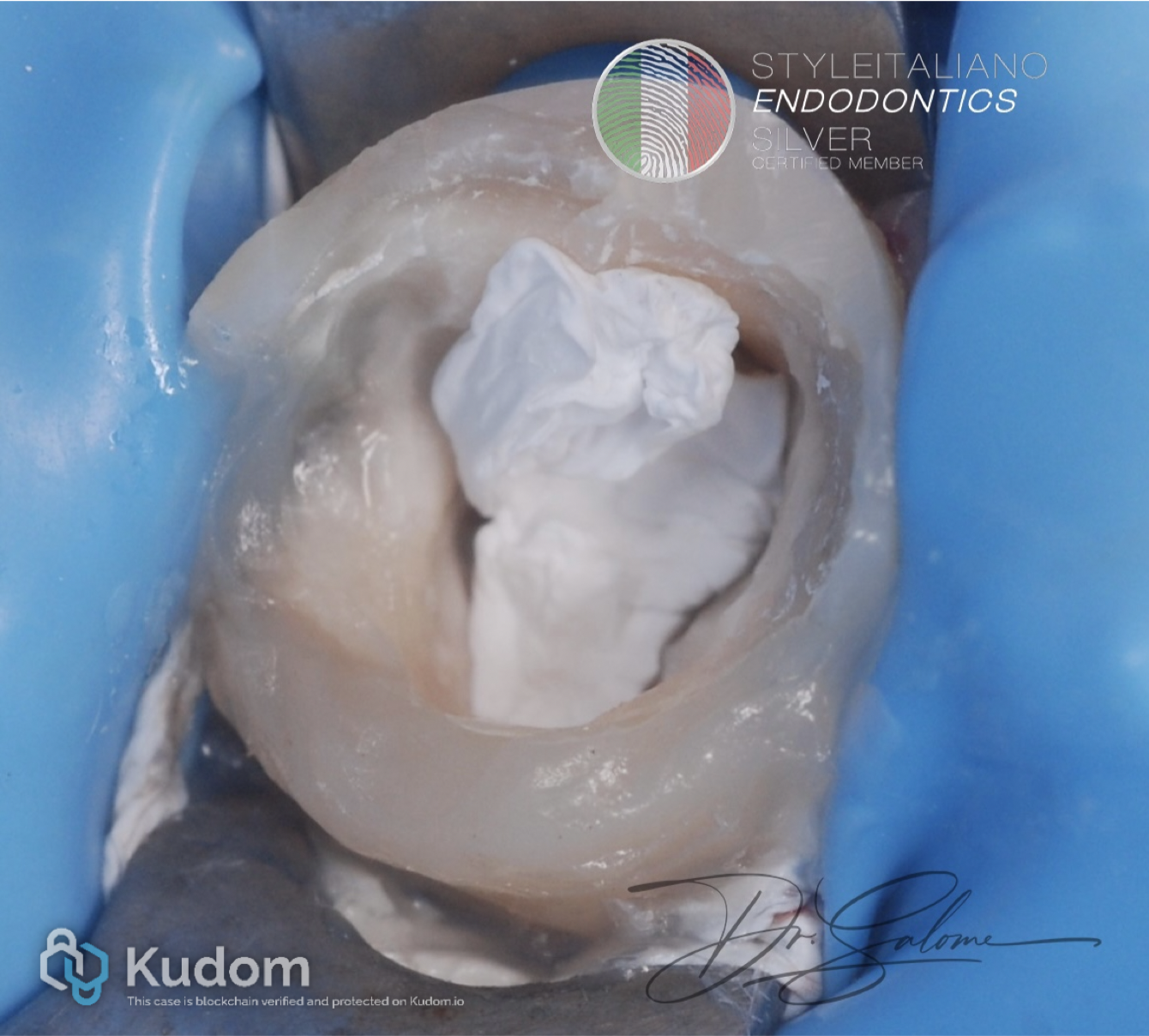
Fig. 5
Pre endodontic build-up was done
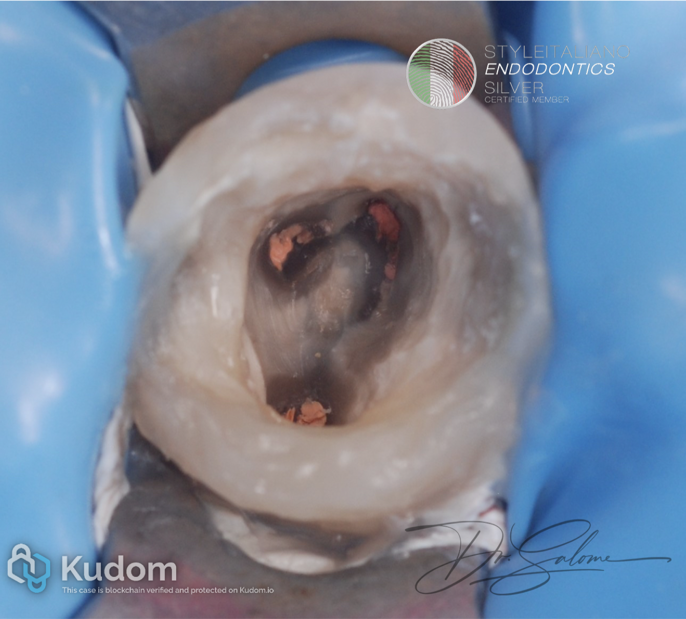
Fig. 6
The next step involved thorough cleaning of the pulp chamber and removal of the existing root canal obturation material to allow complete reassessment and disinfection of the root canal system.
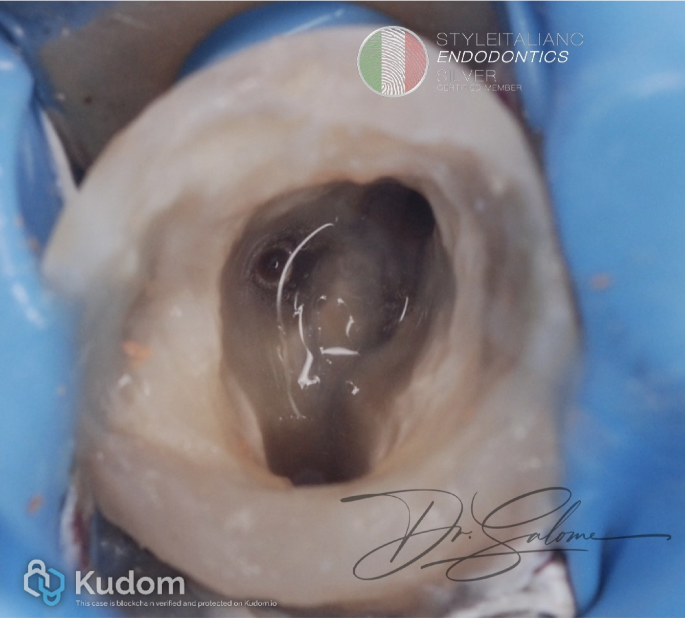
Fig. 7
Retreatment was performed using the ReTreaty system, with copious sodium hypochlorite (NaOCl) irrigation.
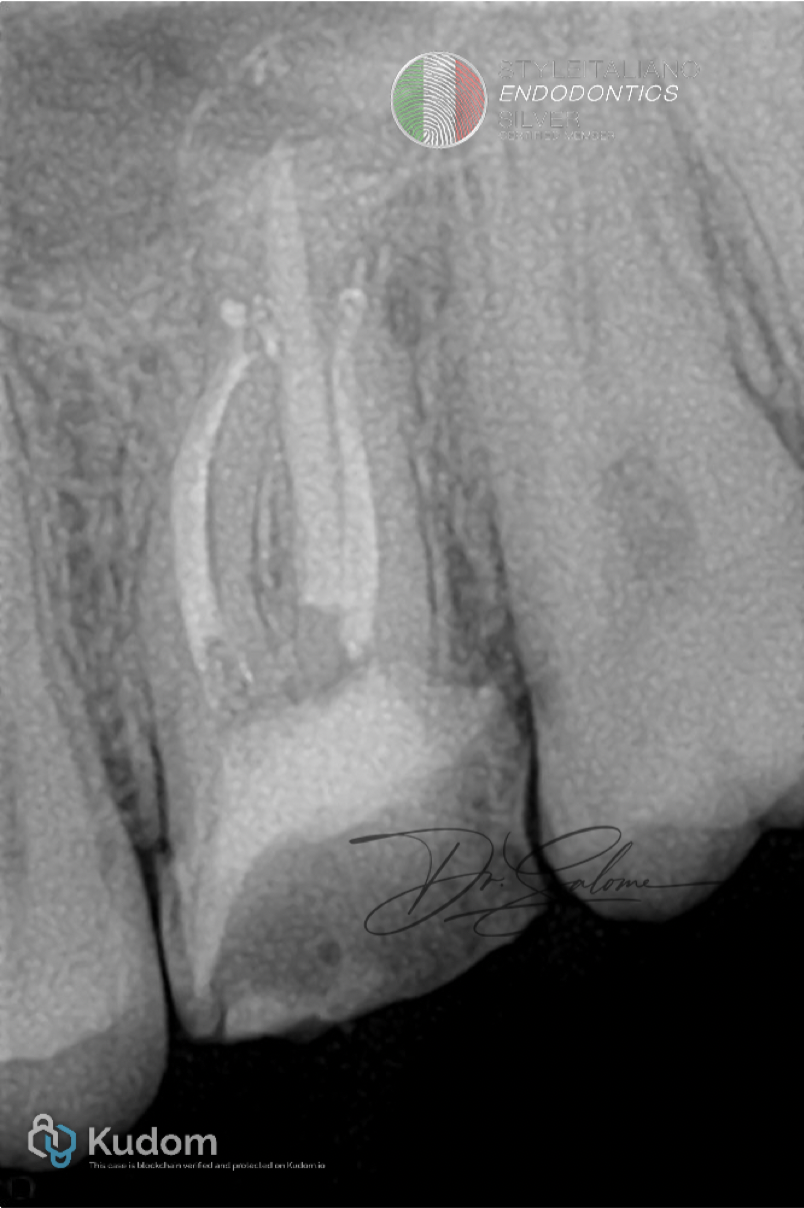
Fig. 8
Obturation revealed two apical POE on mesial root. Obturation was done with Bioceramic sealer and warm gutta-percha.
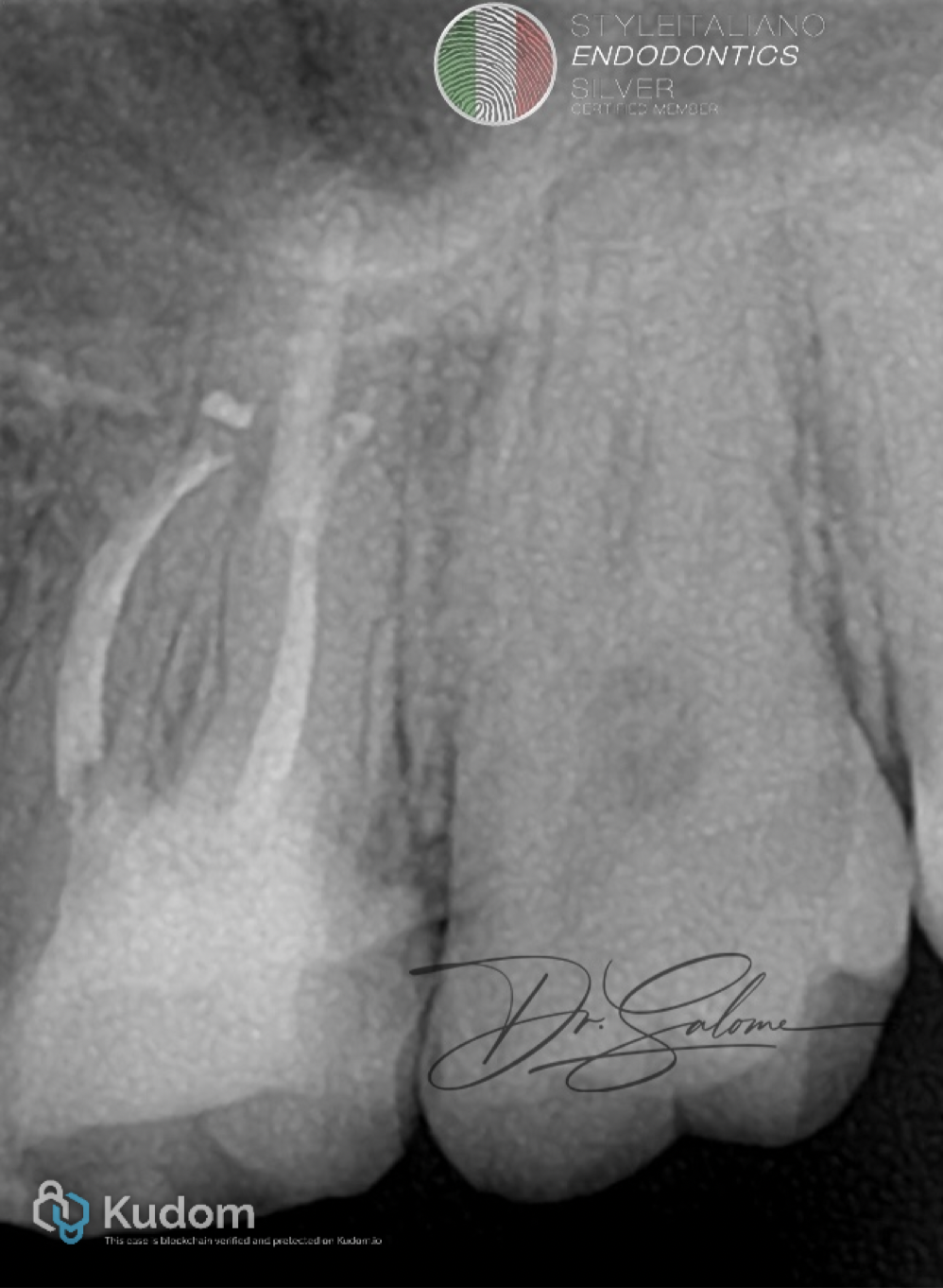
Fig. 9
Follow-up X-ray.
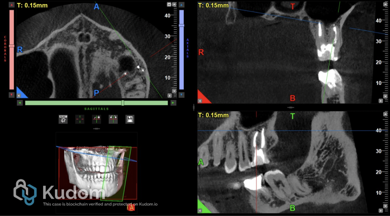
Fig. 10
Follow-up CBCT after 10 months demonstrated complete resolution of the apical pathosis, with healing of the periapical tissues and regression of the associated osteoperiosteal and sinus-floor changes. No symptoms or signs of Sinusitis was observed in this follow up period.
Conclusions
This case highlights the importance of accurate diagnosis in identifying the true source of pathology and demonstrates the effectiveness of appropriate endodontic treatment in achieving complete healing.
Bibliography
- American Association of Endodontists. AAE Position Statement: Maxillary Sinusitis of Endodontic Origin. 2018.
- American Association of Endodontists and European Society of Endodontology. Periapical Diagnoses. Updated diagnostic terminology, 2025
- Siqueira JF Jr, Lenzi R, Hernández S, et al. Effects of endodontic infections on the maxillary sinus: a case series of treatment outcome. J Endod. 2021;47(7):1166–1176. doi:10.1016/j.joen.2021.04.002.