Three-Year Clinical Follow-Up of Quadrant Restoration After Removal of a Fractured Root Canal Instrument: A Case Report
31/07/2026
Fellow
Warning: Undefined variable $post in /home/styleendo/htdocs/styleitaliano-endodontics.org/wp-content/plugins/oxygen/component-framework/components/classes/code-block.class.php(133) : eval()'d code on line 2
Warning: Attempt to read property "ID" on null in /home/styleendo/htdocs/styleitaliano-endodontics.org/wp-content/plugins/oxygen/component-framework/components/classes/code-block.class.php(133) : eval()'d code on line 2
A substantial portion of clinical dental practice comprises endodontic and restorative procedures. Among these, quadrant rehabilitation frequently presents as a common and complex clinical challenge. During quadrant rehabilitation, the replacement of extensive existing restorations often uncovers structural complications such as cracks and significant loss of tooth structure. In such cases, contemporary indirect restorative techniques offer effective solutions. Advances in digital dentistry and chairside systems enable the completion of these restorations in a single appointment.
The long-term success of endodontically treated teeth is predominantly dependent on the quality and integrity of the coronal restoration. In teeth with previous root canal treatment, retreatment may be necessary prior to replacement of the restoration if periapical pathology or coronal leakage is detected. Sometimes, root canal therapy appears successful for many years but fails due to coronal microleakage, necessitating retreatment. Additional challenges, such as fractured instruments within the canal, often complicate management. However, with the advancements in magnification tools, technological innovations, and clinical expertise, these challenges can now be effectively addressed.
This case report presents the retreatment of a root canal therapy that remained asymptomatic for approximately 12 years but contained a fractured endodontic file. It will also describe the retrieval of the broken file and the full rehabilitation of the quadrant using a single-visit indirect restoration approach.
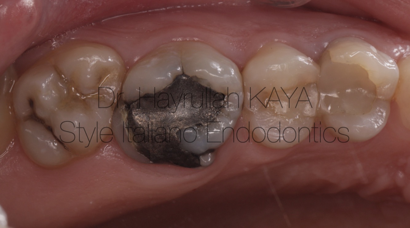
Fig. 1
Preoperative view of the teeth to be treated in the second quadrant
Clinical view following the removal of the previous amalgam restoration
Clinical view after removal of previous restorative materials and caries removal
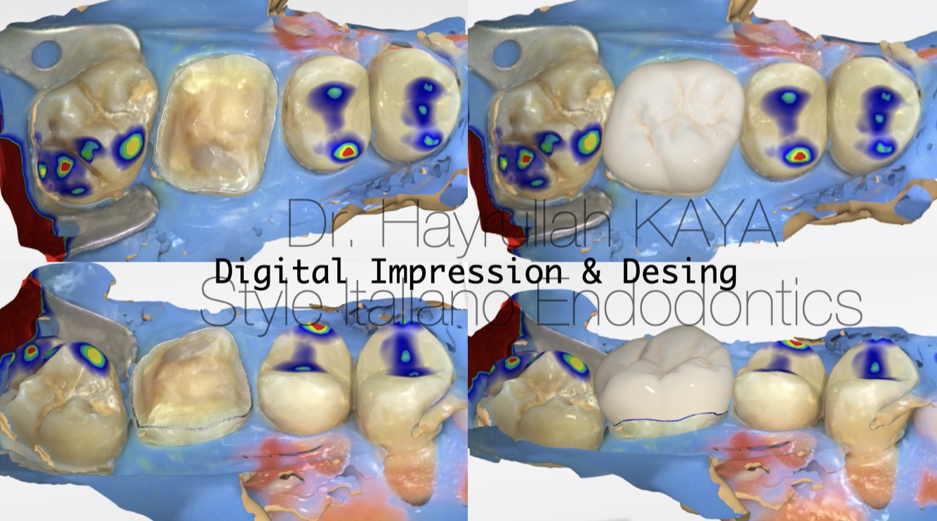
Fig. 2
Digital scanning with an intraoral scanner under rubber dam isolation, margin definition, restoration design, and design evaluation
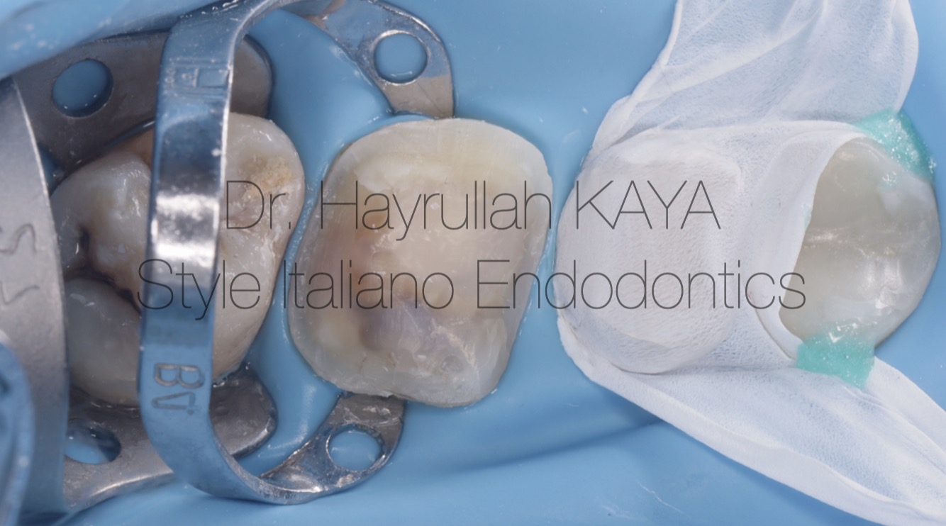
Fig. 3
During pre-cementation preparation, teflon tape was applied to prevent resin cement from bonding to adjacent teeth.
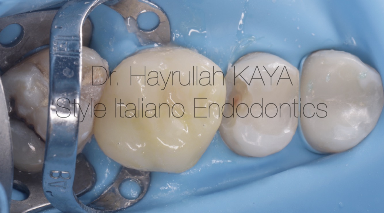
Fig. 4
Post-cementation view. Performing restorations on adjacent teeth after cementation allows for improved proximal contacts
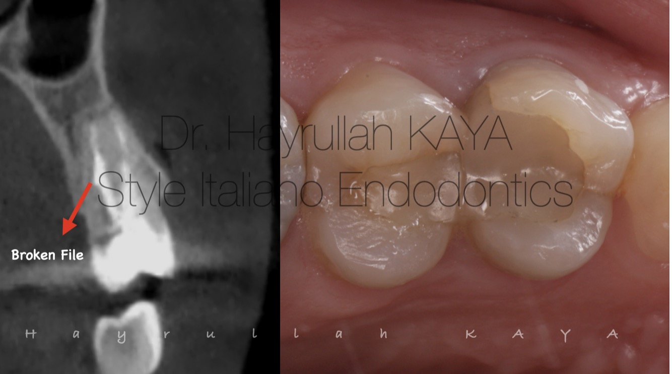
Fig. 5
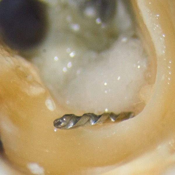
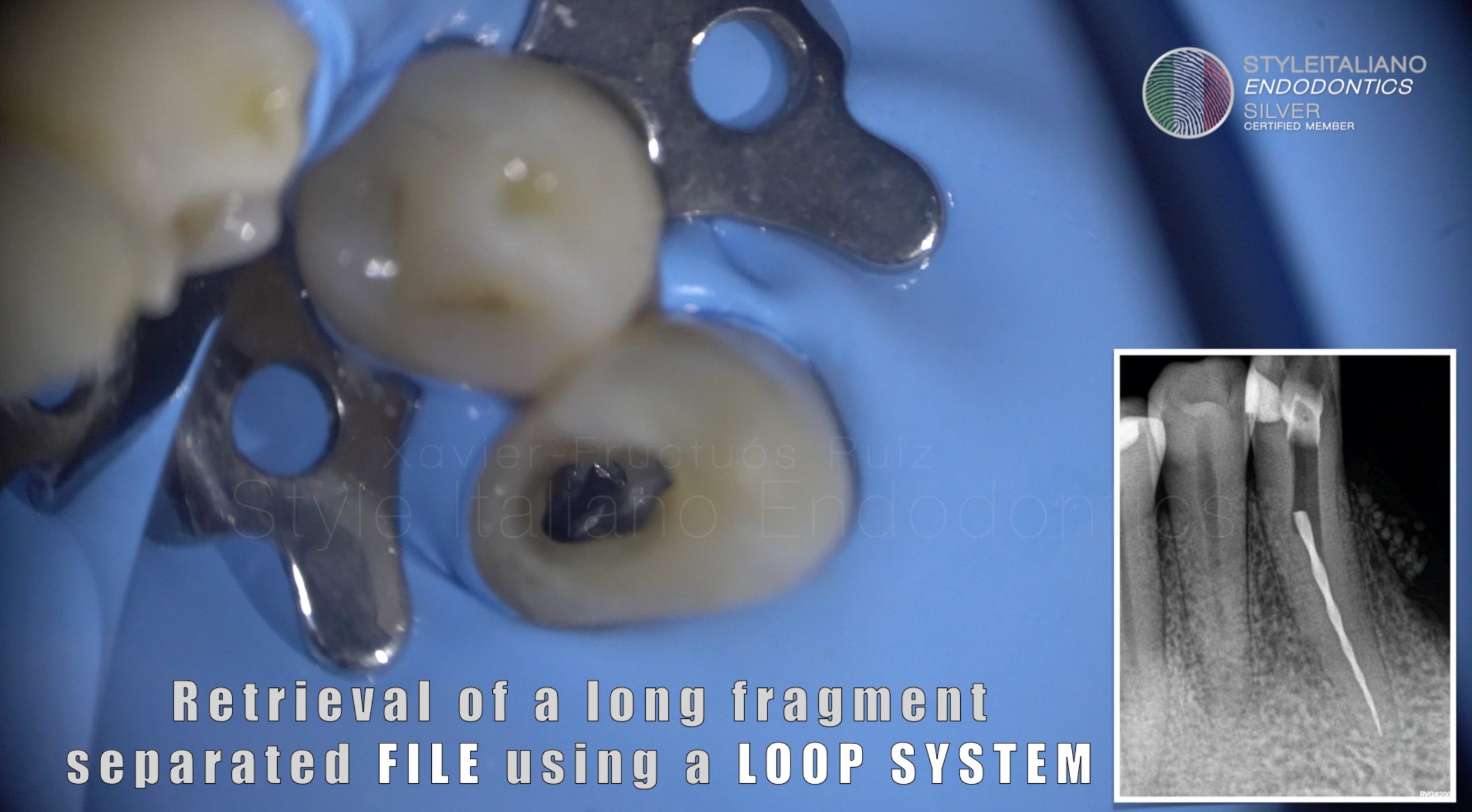
After completion of the treatment of the molar tooth, it was decided to initiate the treatment of the premolar teeth. The examined premolar tooth had undergone root canal treatment and coronal restoration 12 years prior. Despite the presence of a fractured instrument within the root canal, the patient had remained asymptomatic for 12 years. However, deciding to retreat the root canal should not be based solely on radiographic evaluation. After complete removal of the previous restoration, careful inspection for any leakage around the gutta-percha is essential.
The appearance of the gutta-percha shown in this video indicated leakage after complete removal of all coronal restorations, leading to the decision to perform retreatment.
Removal of a fractured root canal instrument using a custom-made loupe and ultrasonic assistance
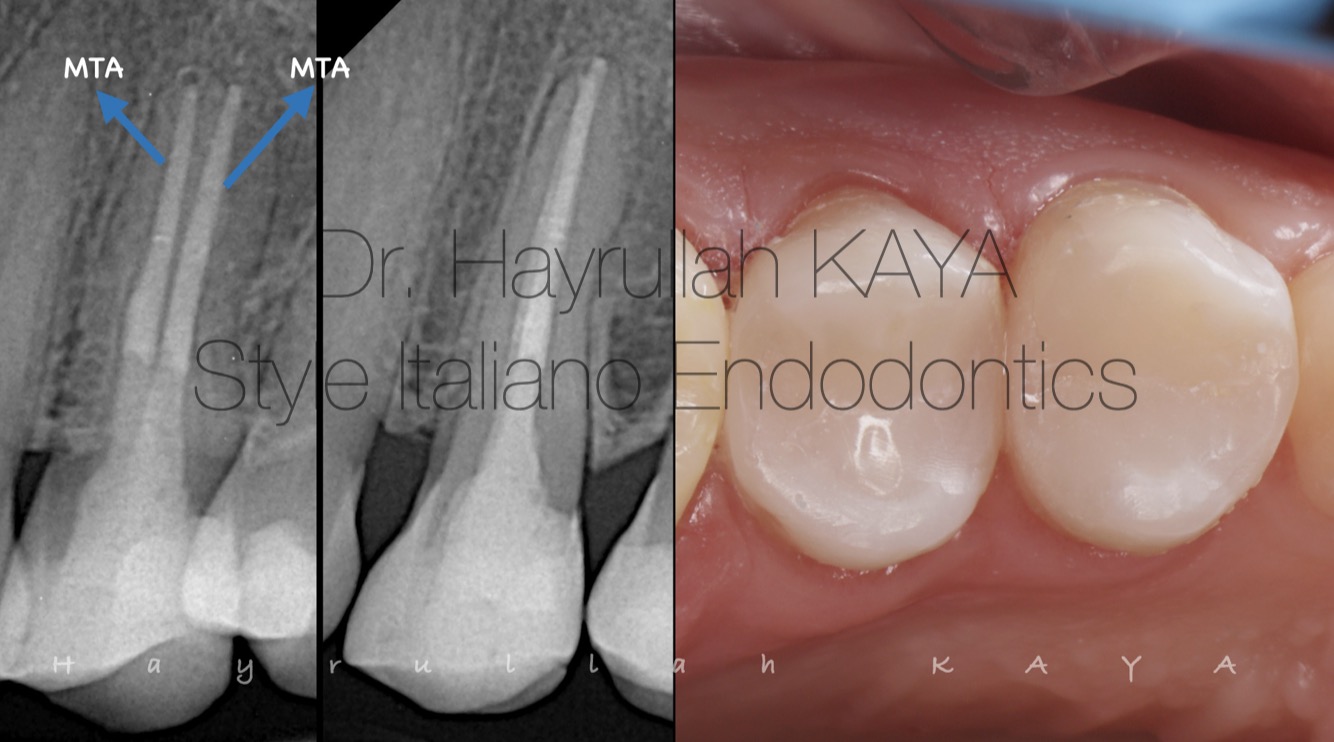
Fig. 6
Following the removal of the fractured endodontic instrument, the root canals were obturated with Mineral Trioxide Aggregate (MTA), and the coronal restoration was subsequently completed using direct composite resin.
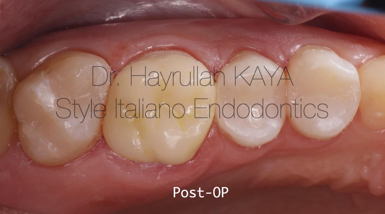
Fig. 7
Postoperative quadrant intraoral image following the completion of all restorations. The restorations include:
Tooth 27 with a direct restoration,
Tooth 26 with an indirect restoration,
Tooth 25 with an indirect restoration and
Tooth 24 with a direct restoration.
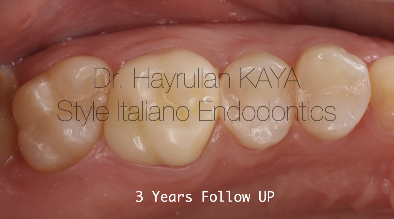
Fig. 8
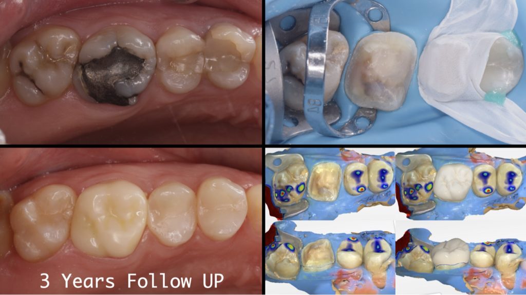
Three-years follow-up
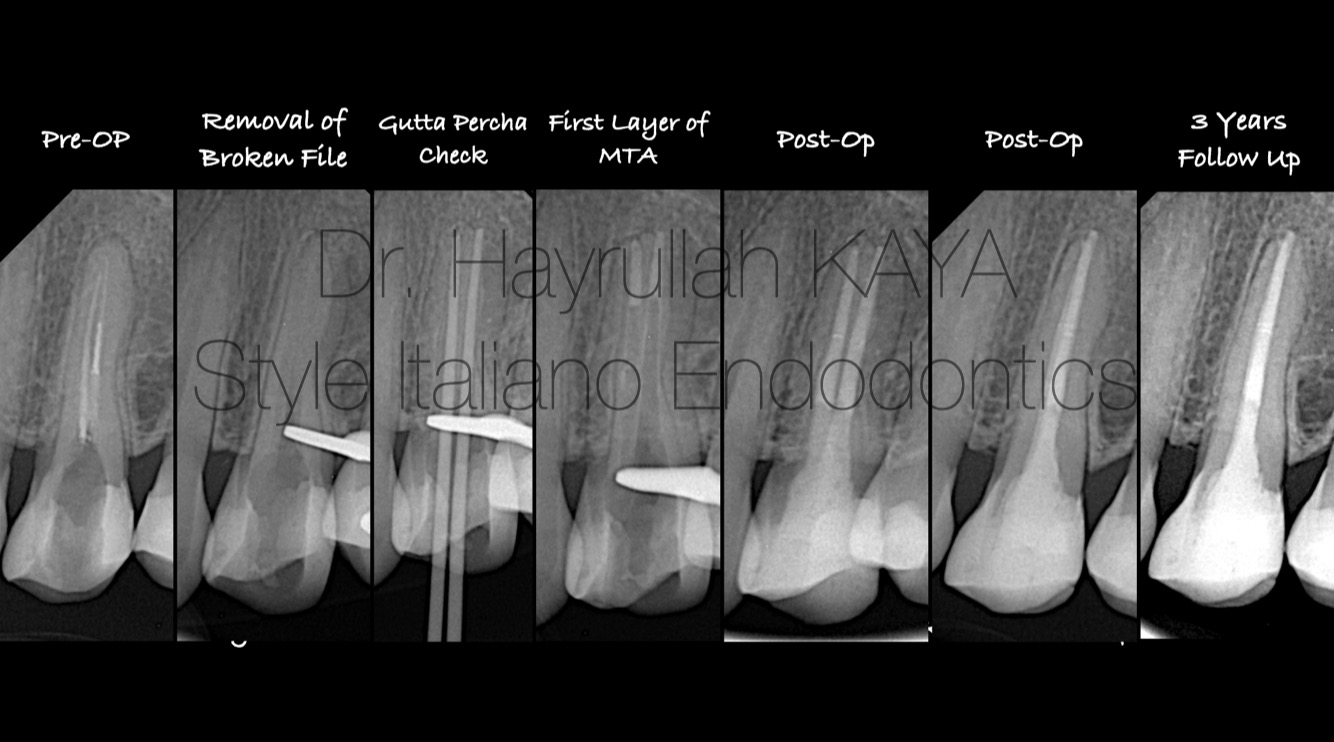
Fig. 9
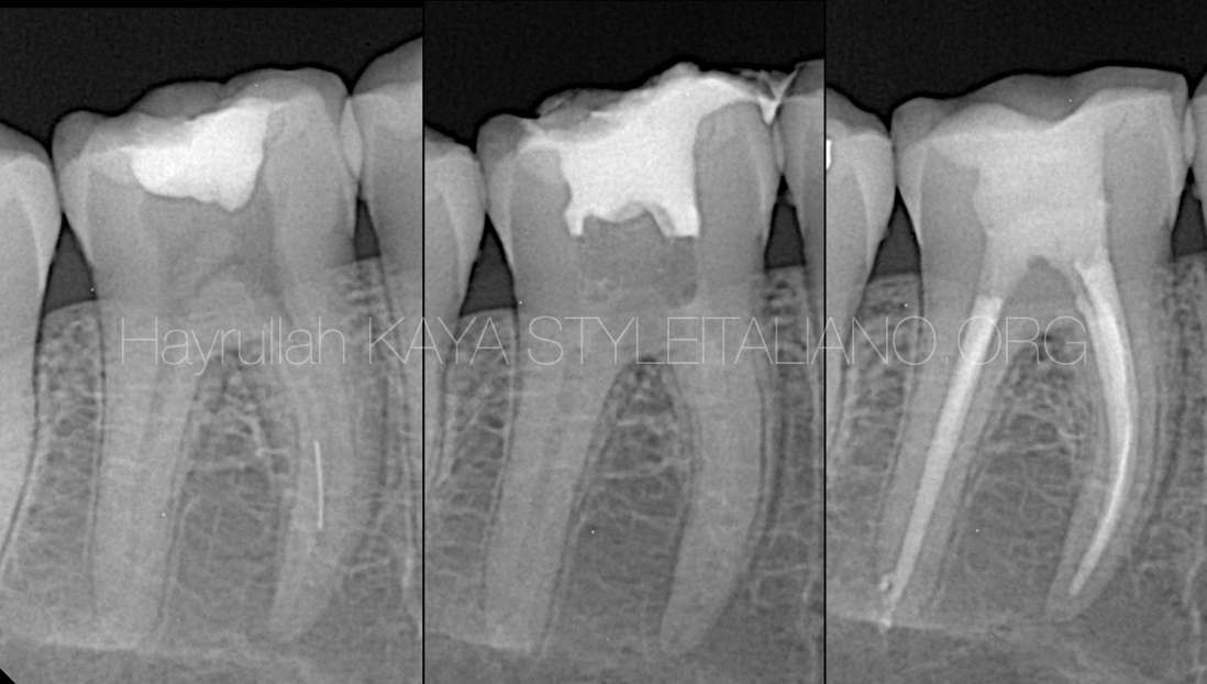
Radiographic image of the three-year follow-up : Retreatment case involving the removal of a fractured root canal instrument.

Fig. 10
Hayrullah Kaya graduated from Atatürk University Faculty of Dentistry . He has been
continuing his professional life in private clinics since his graduation. His areas of
interest include MTA applications, endodontic treatments under microscope, dental
isolation systems, digital and adhesive dentistry.
Hayrullah teaches at courses about Endodontics in Dentsply Sirona as Key Opinion
Leader (KOL).
He is also involved in teaching courses on Rubber Dam and MTA applications and
offering personalized one-on-one training sessions at clinics.
He still continues his professional career in his own clinic in Istanbul.
Conclusions
The most important thing the dentist must take into account when removing any broken instrument inside the root is to know how to deal with the root and to reduce the amount of dentin removal to maintain a longer life of the tooth, in addition to knowing when to stop and do not forget that the availability of the required equipment and experience is a factor very important.
Bibliography
- Periapical status of endodontically treated teeth in relation to the technical quality of the root filling and the coronal restoration, H A Ray 1 , M Trope
- Fracture strength of bovine incisors after intra-radicular treatment with MTA in an experimental immature tooth model, E A Bortoluzzi 1 , E M Souza, J M S N Reis, R M Esberard, M Tanomaru-Filho
- Madarati AA, Hunter MJ, Dummer PM. Management of intracanal separated instruments. J Endod 2013;39:569–81.
- Cuj e J, Bargholz C, Hulsmann M. The outcome of retained instrument removal in a specialist practice. Int Endod J 2010;43:545–54.
- Ward JR, Parashos P, Messer HH. Evaluation of an ultrasonic technique to remove fractured rotary nickel-titanium endodontic instruments from root canals: clinical cases. J Endod. 2003b;29(11):764–7
- Endodontic retreatment: a rational approach to non-surgical root canal the- raphy of immature teeth, Lieberman N, Trombridge C, Klein A, Levy S.