Management of Two Broken Intruments in an Upper Right Canine – A Clinical Approach
16/08/2026
Warning: Undefined variable $post in /home/styleendo/htdocs/styleitaliano-endodontics.org/wp-content/plugins/oxygen/component-framework/components/classes/code-block.class.php(133) : eval()'d code on line 2
Warning: Attempt to read property "ID" on null in /home/styleendo/htdocs/styleitaliano-endodontics.org/wp-content/plugins/oxygen/component-framework/components/classes/code-block.class.php(133) : eval()'d code on line 2
Instrument separation is one of the most common complications encountered during root canal treatment. Although fracture of the instrument itself may not directly cause treatment failure, retained fragments within the root canal system can hinder adequate cleaning and shaping of the canal.
With the advent of the dental operating microscope and the introduction of specialized ultrasonic tips for the management of separated instruments, the success rate of treating these cases has increased significantly, while the risk of procedural complications has been considerably reduced.
Currently, two principal approaches are used for the management of fractured instruments: bypassing and retrieval. The decision to bypass or remove a separated instrument depends on several factors, including the location, length, type, and material of the fragment, as well as the tooth involved and canal anatomy.
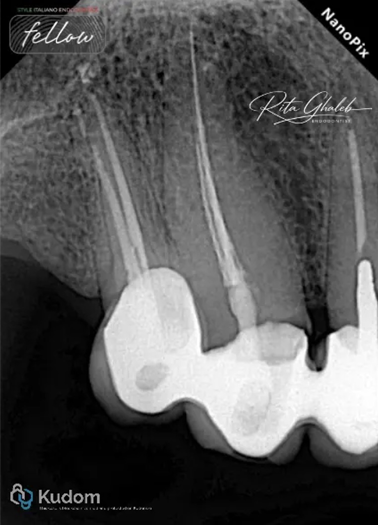
Fig. 1
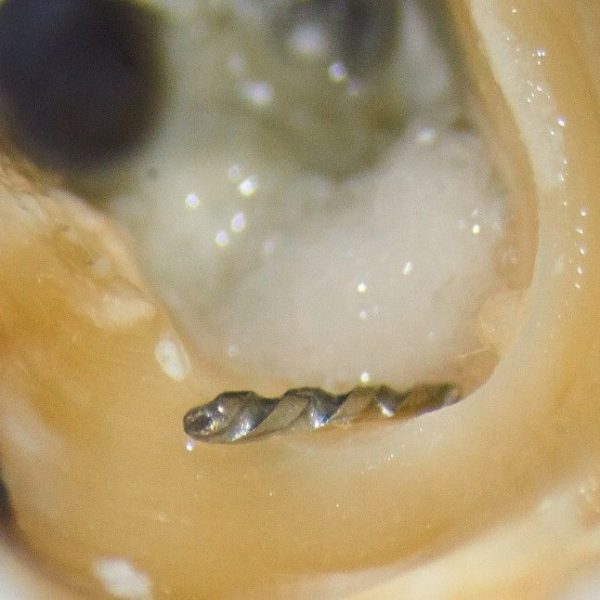
A 64-year-old patient presented to our clinic complaining of pain following root canal treatment of the maxillary right first premolar and canine performed by a general practitioner. Clinical and radiographic examinations revealed a short obturation in the palatal canal of the premolar and the presence of two separated instruments in the canine. One long, large fragment lodged apically and a second thinner, shorter fragment, both embedded within a mass of gutta-percha.
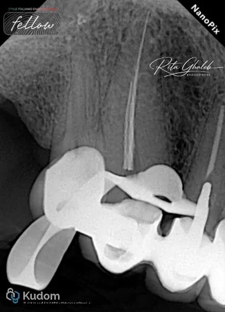
Fig. 2
As part of the endodontic treatment plan, a non-surgical approach under a dental operating microscope was selected for both teeth through the existing crowns, as the patient refused prosthetic crown removal because of an upcoming trip and limited time for restorative procedures.
The patient was informed about the benefits, limitations, and possible complications of the procedure, and informed consent was obtained.
For the premolar, gutta-percha was removed using a 25/.06 shaping file system, and canal patency was successfully regained
However, the main challenge in this case was establishing an appropriate strategy for managing the two separated instruments. The difficulty lay in determining which fragment should be approached first and how both fragments could be disengaged from the surrounding gutta-percha.
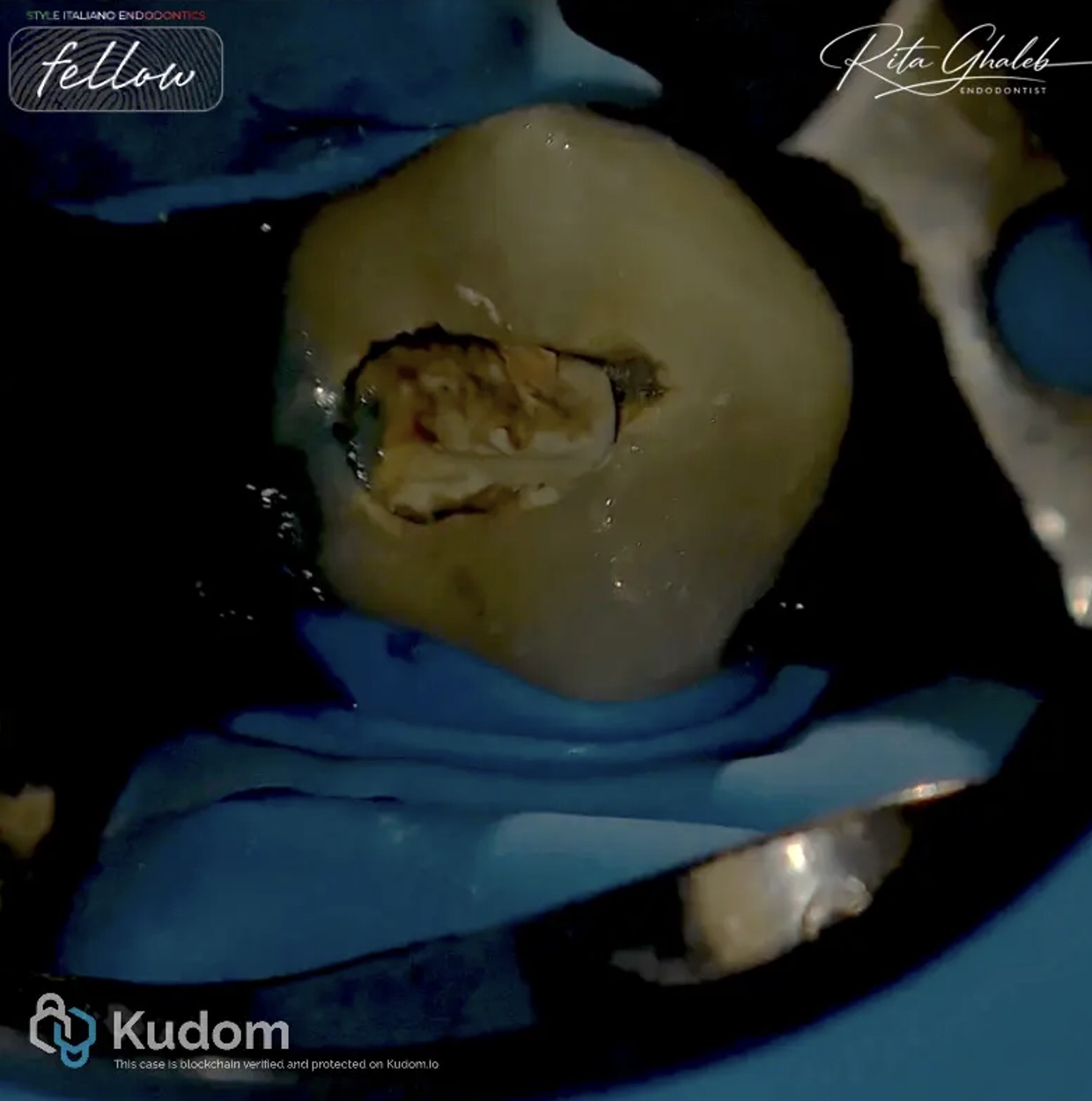
Fig. 3
Initially, the access cavity was refined. The strategy then involved removing as much of the gutta-percha mass as possible from the access cavity and canal walls using ultrasonics.
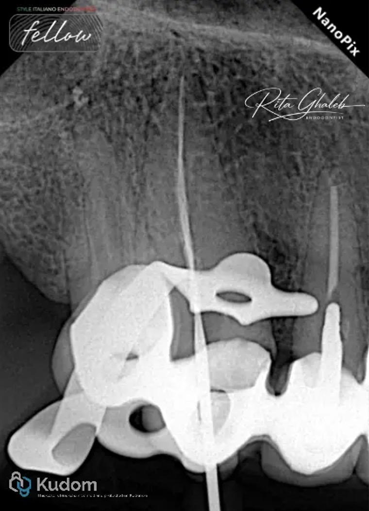
Fig. 4
This was followed by bypassing the broken instruments mesially, distally, buccally, and palatally, thereby separating the fragments from the gutta-percha adhered to the canal walls. As a result, both fragments became loosened and demonstrated increased mobility.
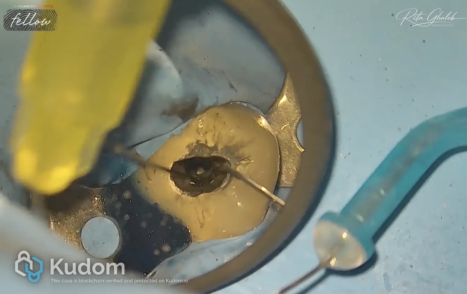
Fig. 5
Although the larger and longer instrument was more visible and initially appeared easier to retrieve, the smaller broken instrument was removed first in order to facilitate access to and liberation of the larger fragment.
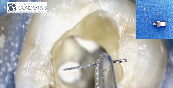
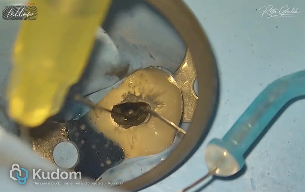
The tip of an irrigation needle was inserted between the smaller fragment and the canal wall in order to straighten the broken instrument and create sufficient space for loop placement.
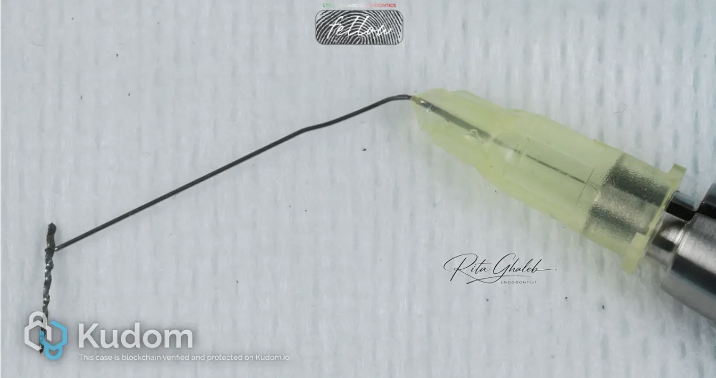
Fig. 6
The smaller fragment was then successfully retrieved using the loop technique with the BTR Pen.
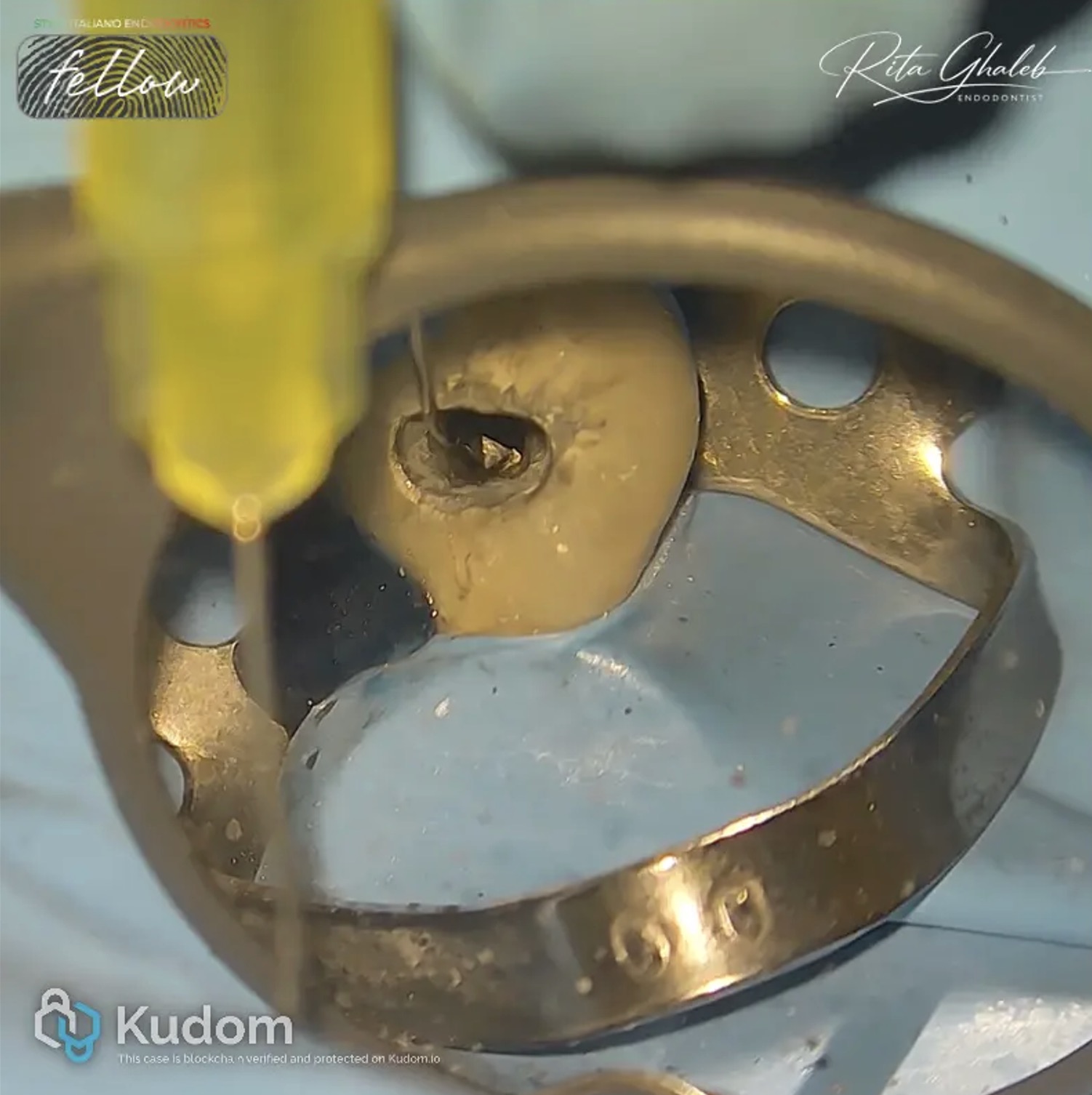
Fig. 7
Following removal of the smaller fragment, the second instrument was also successfully retrieved using the same loop technique
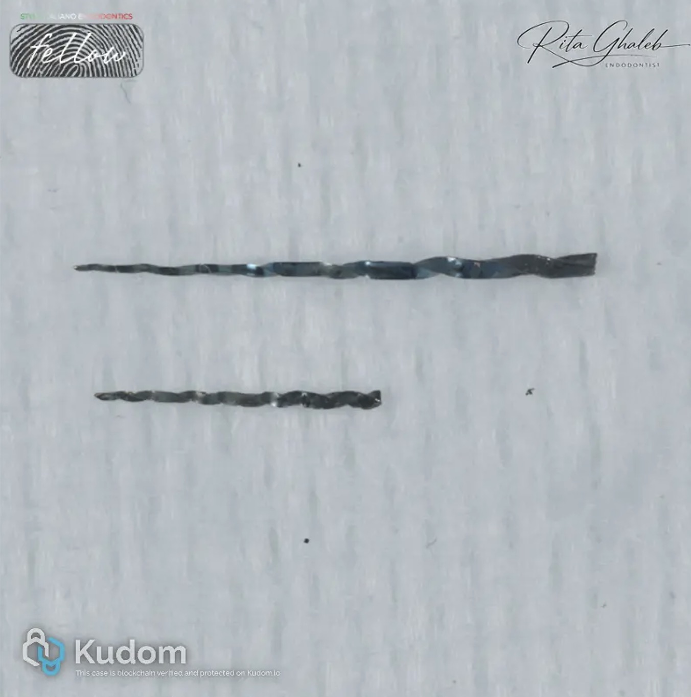
Fig. 8
removal of the two broken instruments.
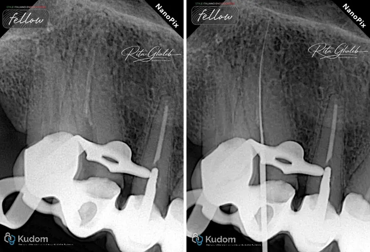
Fig. 9
Once the canal was free of broken instruments, working length determination was performed, followed by canal shaping with 25/.04 and then 25/.06 instruments
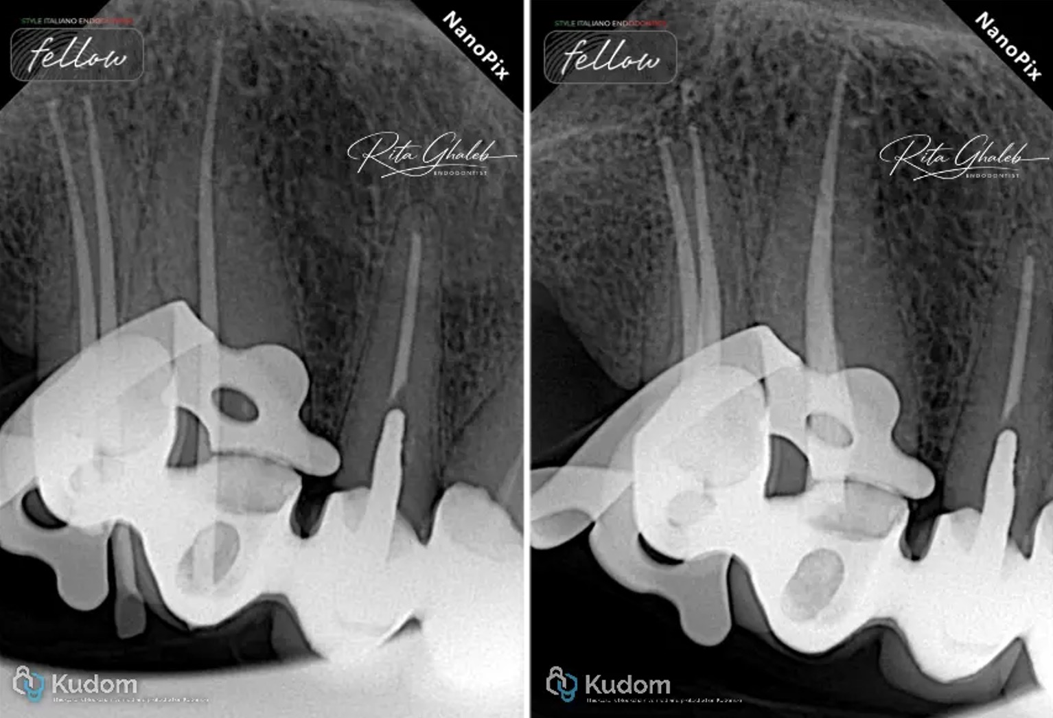
Fig. 10
Copious irrigation with sodium hypochlorite (NaOCl) was carried out throughout the procedure, followed by cone-fit verification and obturation using the warm vertical compaction technique for both teeth.
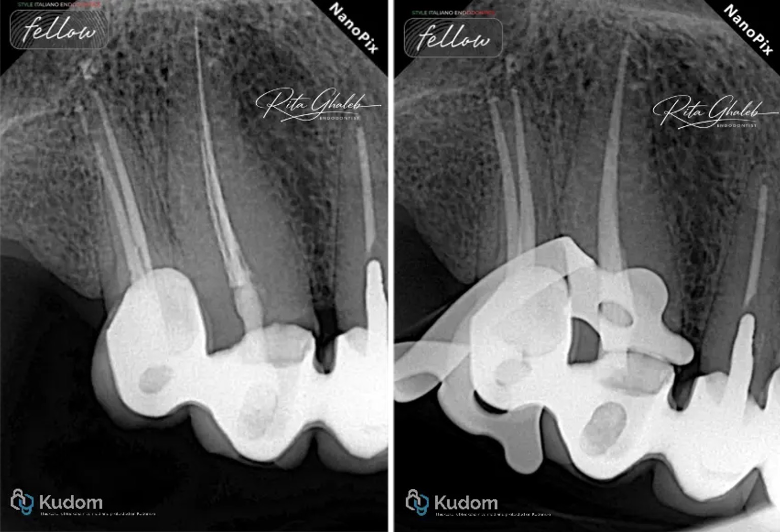
Fig. 11
Final xrays
Conclusions
Successful management of separated instruments requires careful case evaluation and strategic clinical decision-making. Although larger fragments may appear more visible under magnification, this does not necessarily make their retrieval easier. In many cases, larger fragments are more difficult to retrieve because of their increased engagement within the canal walls. Each case presents unique anatomical and mechanical challenges that require an individualized approach and sound clinical judgment.
Bibliography
1- Ruddle CJ: Ch. 25, Nonsurgical endodontic retreatment. In Pathways of the Pulp, 8th ed., Cohen S, Burns RC, eds., St. Louis: Mosby, pp. 875-929, 2002
2- Madarati AA, Hunter MJ, Dummer PM. Management of intracanal separated instruments. J Endod. 2013 May;39(5):569-81. doi: 10.1016/j.joen.2012.12.033. Epub 2013 Mar 15. PMID: 23611371.
3- Terauchi Y, Ali WT, Abielhassan MM. Present status and future directions: Removal of fractured instruments. International Endodontic Journal, 55(Suppl. 3), 685–709.
4- McGuigan M. B., Louca C., Duncan H. F. Clinical decision-making after endodontic instrument fracture. British Dental Journal. 2013;214(8):395–400. doi: 10.1038/sj.bdj.2013.379. [DOI] [PubMed] [Google Scholar]