Management of broken file in atresic canals
01/06/2023
José Conde Pais
Warning: Undefined variable $post in /home/styleendo/htdocs/styleitaliano-endodontics.org/wp-content/plugins/oxygen/component-framework/components/classes/code-block.class.php(133) : eval()'d code on line 2
Warning: Attempt to read property "ID" on null in /home/styleendo/htdocs/styleitaliano-endodontics.org/wp-content/plugins/oxygen/component-framework/components/classes/code-block.class.php(133) : eval()'d code on line 2
Fracture or separation of endodontic instruments is one of the main complications in our clinical practice in root canal treatment. This eventuality can negatively affect the outcome of treatment, as the blockage caused by the instrument fragments will prevent correct debridement and removal of pulp tissue, bacteria or smear layer. The prognosis in these cases will depend directly on the preoperative situation, being better in vital cases or in cases where prior disinfection has been performed.
When nickel-titanium rotary instruments (NITI) are used, the incidence of fracture ranges from 0.4% to 5%.
The incidence of removal of these instruments varies widely, from 48% to 95% and is directly related to the techniques employed and the condition of the instrument.
The use of ultrasonic tips and loop techniques under the surgical microscope is considered the optimal strategy for successful removal of fractured instruments.
The use of the surgical microscope provides adequate visualisation and accessibility to the fractured instrument and is a key role in instrument removal.
This article describes the treatment of a 4.6 tooth with a fractured instrument and calcified metamorphosis.
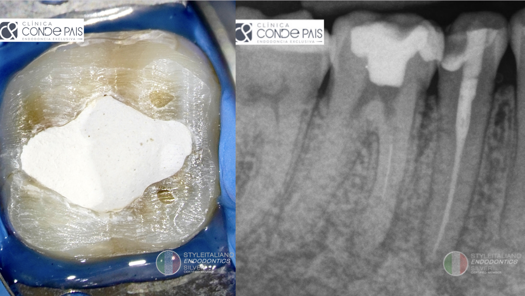
Fig. 1
A patient came to our endodontic clinic for evaluation and treatment of tooth 4.6, on which endodontic treatment had been attempted months earlier. Symptoms on examination were: positive percussion, negative palpation, negative vitality and physiological probing. Radiographic examination revealed a previously initiated root canal treatment, a fractured instrument in the mesiovestibular canal and calcific metamorphosis of the canal system. The diagnosis was symptomatic apical periodontitis, after discussing treatment options, we decided to perform orthograde treatment. The patient was informed of the pros and cons of the treatment and signed the informed consent.
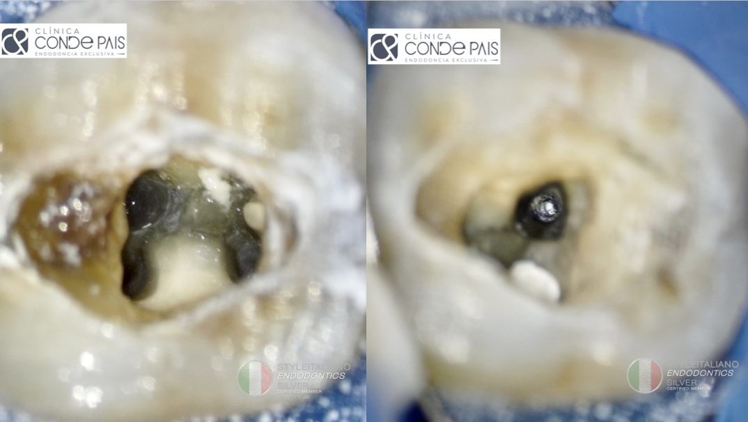
Fig. 2
After anaesthesia and isolation of the operative field, cavity access was performed. Four canals were located, using a DG16 endodontic explorer, two in the mesial root and two in the distal root. In the mesiovestibular canal we located the fractured instrument. In this phase we used an operating microscope (CJ OPTIK, Ablar, Germany) and redefined the access cavity with ultrasonic tips.
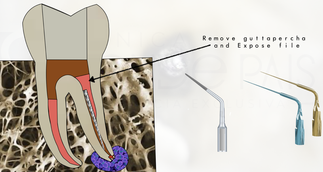
Fig. 3
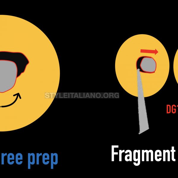
We proceed to the removal of the broken file using the loop technique following this process:
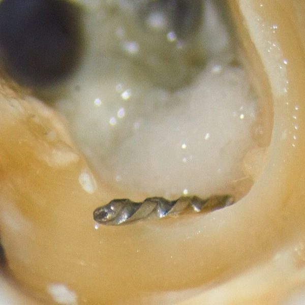
We expose the instrument with ultrasonic tips.
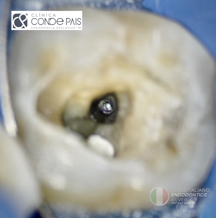
Fig. 4
File eposed
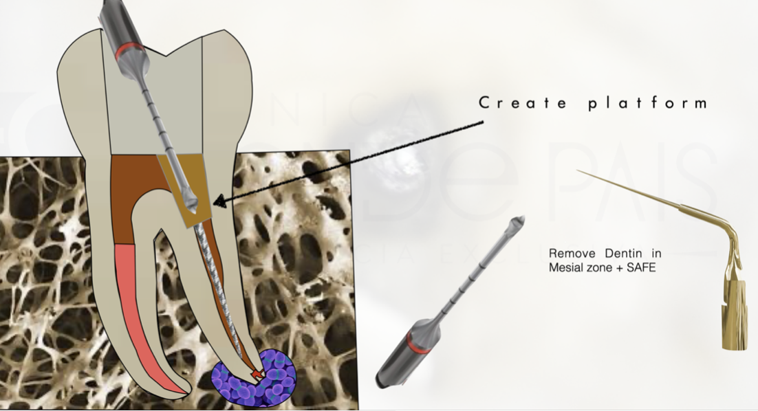
Fig. 5
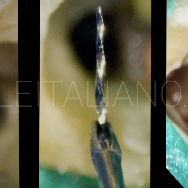
We create a working platform over the fractured instrument with small calibre ultrasonic tips and wet canal with EDTA 17% liquid.
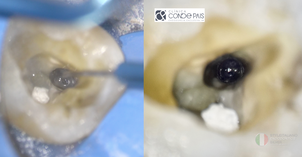
Fig. 6
Working Platform
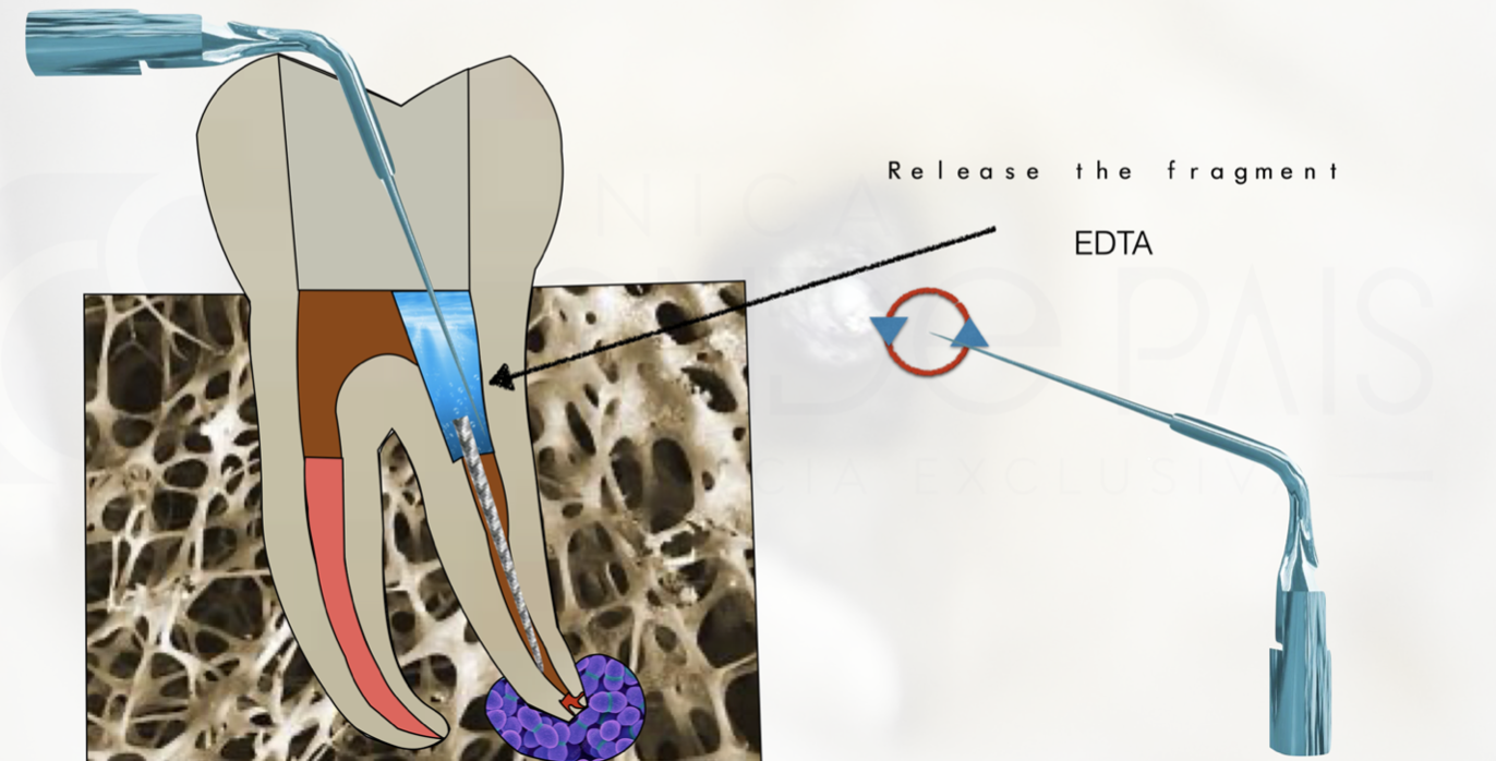
Fig. 7
We release the fractured instrument and created a space of at least 1 mm around the instrument.
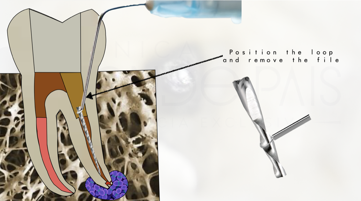
Fig. 8
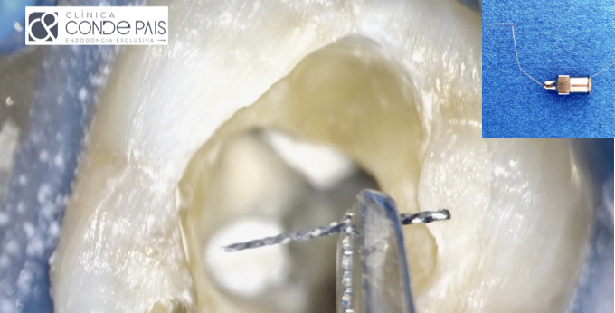
We position the loop around the instrument.
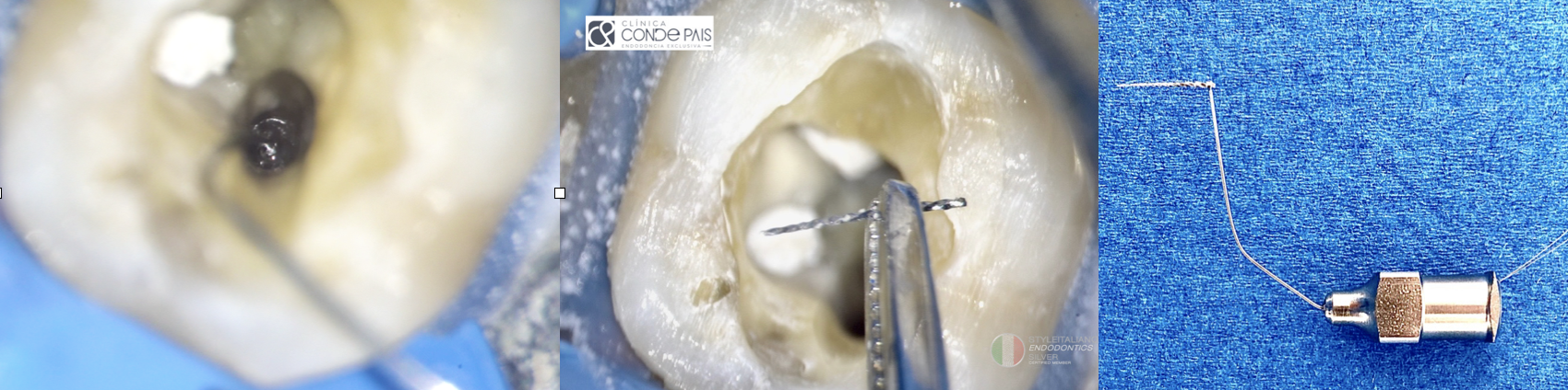
Fig. 9
Close the loop and make traction movements until the broken file is removed.
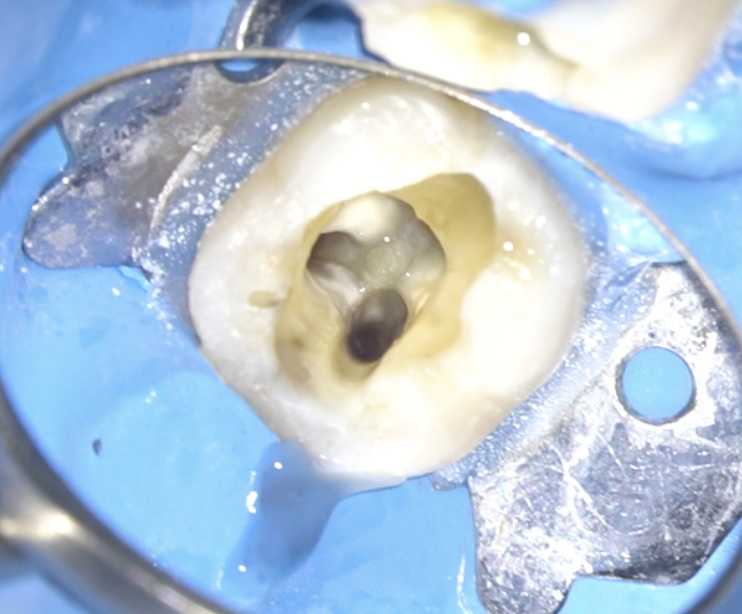
Fig. 10
Once the instrument fragment has been removed, we proceed with the endodontic treatment.
We performed an exploration of the root canals with C+ ISO 10 files and measured the working length using an electronic apex locator and checked it against parallel periapical radiographs.
Once a correct glide path was created, we proceeded to instrument the canals to (25-06). . Between file and file we used 5.25% sodium hypochlorite as irrigant, using the IrriFlex tip for its delivery.
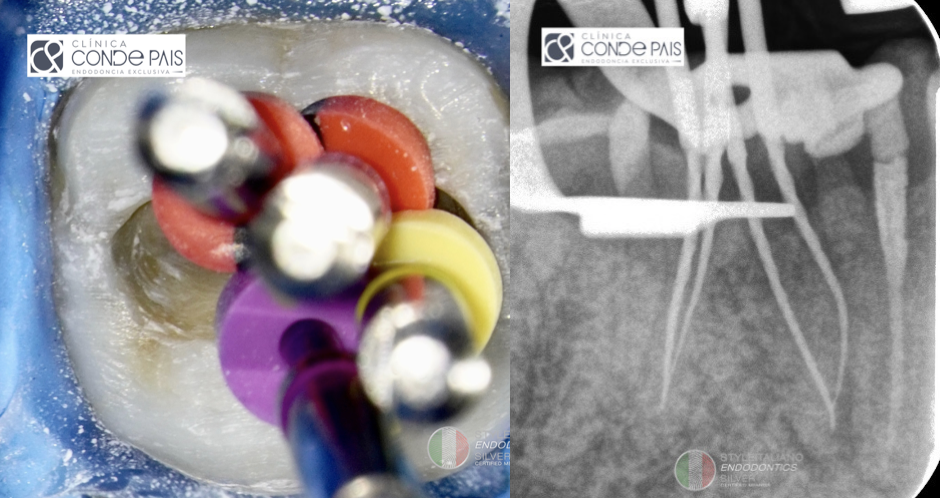
Fig. 11
We check the working length before sealing the canals.
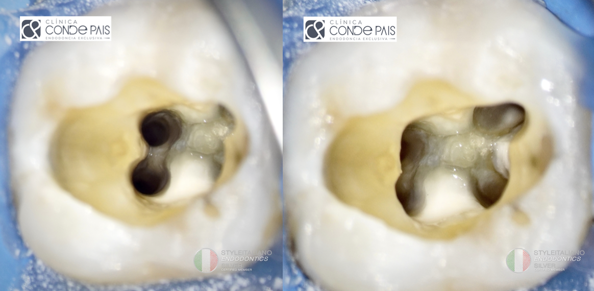
Fig. 12
Once all the canals have been instrumented, apical permeability and working length have been checked, we proceed to the final irrigation of the canal system. For this step we use 5.25% sodium hypochlorite, physiological saline solution and EDTA 17% solution.
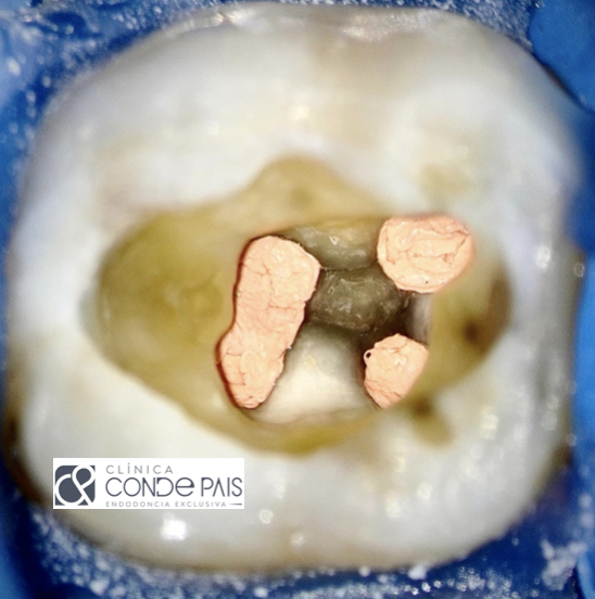
Fig. 13
After shaping and disinfection of canal system, we proceed to dry and fill using continuous wave technique, ensuring a correct 3D seal.
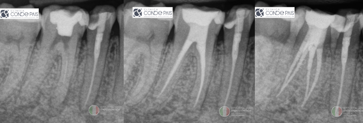
Fig. 14
Once the obturation has been made and checked to be correct, we continue with the immediate sealing with adhesive technique and Bulk Fill composite, after light curing we place provisional cement and we return to the referrer for definitive restoration.
Conclusions
These types of complications are common in our profession with the right training and equipment we can solve them. The incorporation of magnification and ultrasonic devices in our daily practice will be determining variables in this type of situation.
Bibliography
Tzanetakis GN, Kontakiotis EG, Maurikou DV, Marzelou MP. Prevalence and management of instrument fracture in the postgraduate endodontic program at the Dental School of Athens: a five-year retrospective clinical study. J Endod 2008;34:675–87.
- Cuj e J, Bargholz C, Hulsmann M. The outcome of retained instrument removal in a specialist practice. Int Endod J 2010;43:545–54.
- Fu M, Zhang Z, Hou B. Removal of broken files from root canals by using ultrasonic techniques combined with dental microscope: a retrospective analysis of treatment outcome. J Endod 2011;37:619–22.
- Gencoglu N, Helvacioglu D. Comparison of the different techniques to remove fractured
endodontic instruments from root canal systems. Eur J Dent 2009;3:90–5.
- Suter B, Lussi A, Sequeira P. Probability of removing fractured instruments from root canals. Int Endod J 2005;38:112–23.
- Madarati AA, Hunter MJ, Dummer PM. Management of intracanal separated instruments.
J Endod 2013;39:569–81.
- Terauchi Y, Sexton C, Bakland LK, Bogen G. Factors Affecting the Removal Time of Separated Instruments. J Endod. 2021;47(8):1245-1252.