This is the story of retreatment and management of tooth number 21 where a screw post was placed in the canal without performing root canal treatment. Over a decade, the […]
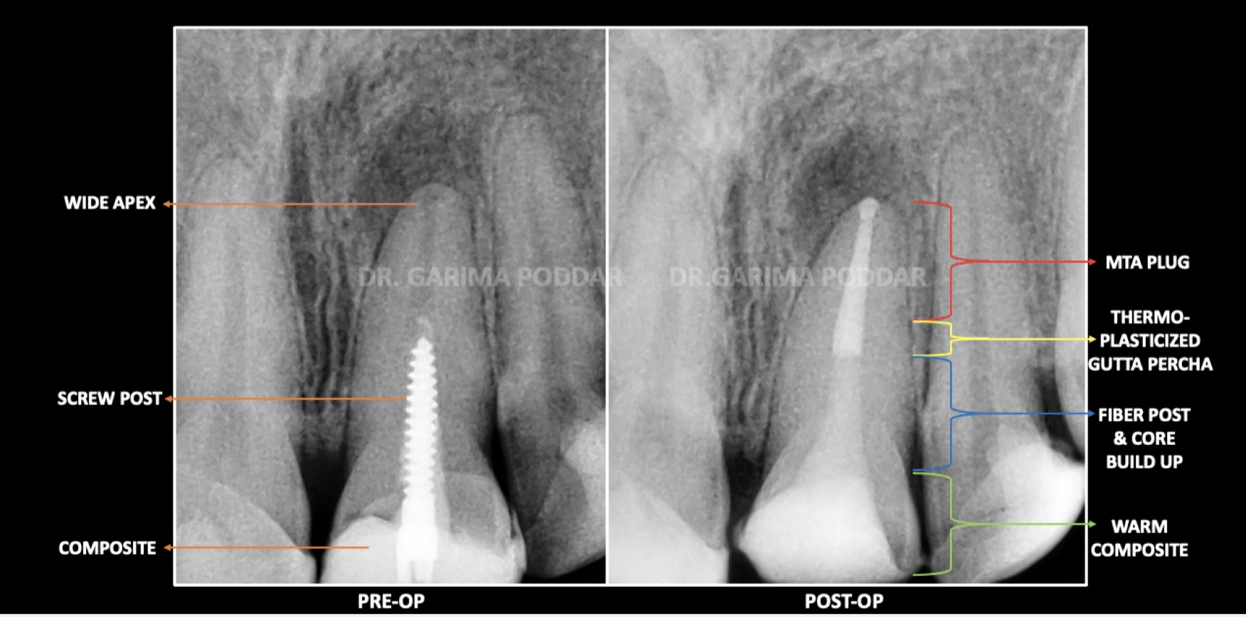 A Screw Post & Widened Apex Story
This is the story of retreatment and management of tooth number 21 where a screw post was placed in the canal without performing root canal treatment. Over a decade, the […]
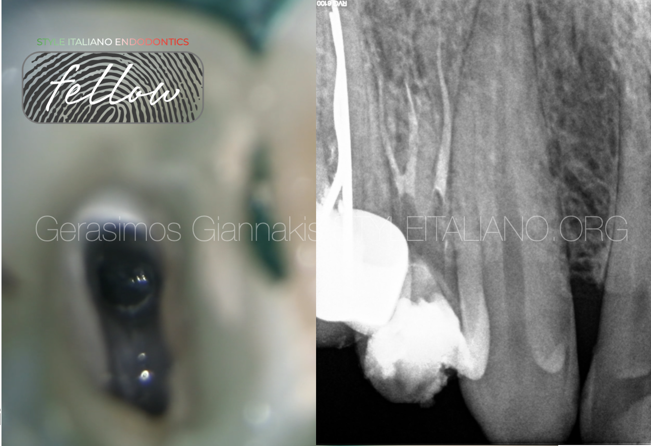 Three roots in a first maxillary premolar: A case report
How often is this anatomical variation of maxillary first premolars? How can we detect it? How to treat such anatomy? In this article I will try to address these considerations.
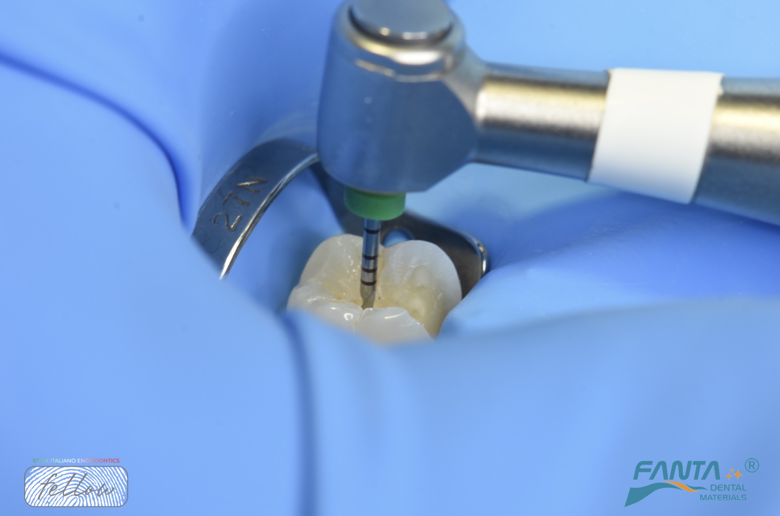 Lower molar with narrow mesial canals
The root canal treatment of a lower molar with narrow canals is a complex and delicate dental procedure aimed at removing infected or damaged pulp tissue while preserving the tooth's […]
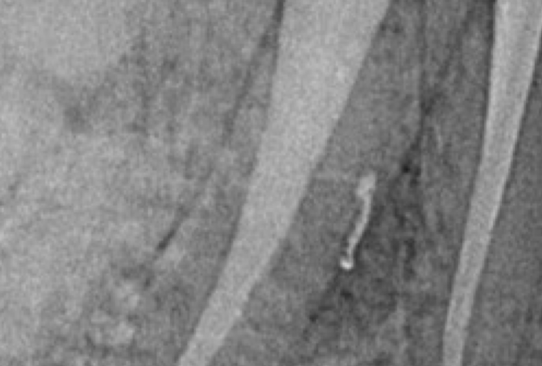 Do lateral canals matter?
The importance of treating lateral canals has been a long lasting debate between endodontists. From Schilder and Wein debates in the 80s to nowadays debates between Castellucci and Ricucci, the […]
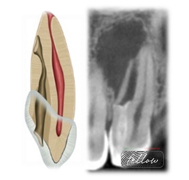 Case report Management of Dens Invaginatus type IIIA
This article showing the Management of Dens Invagenatus of upper lateral incisor with large periapical lesion.
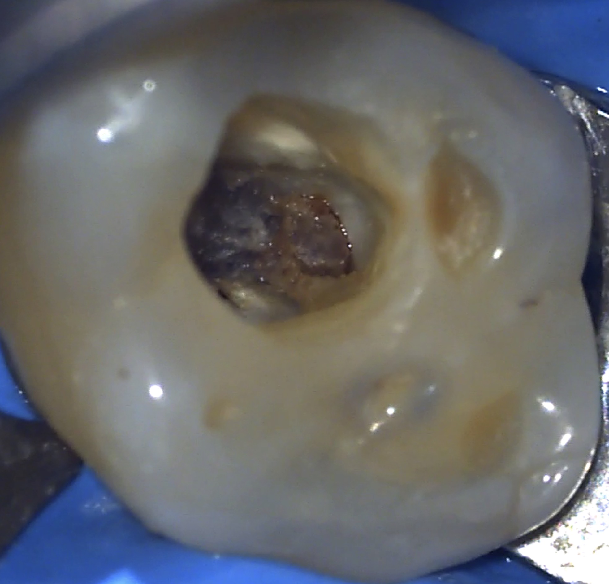 Pulp calcifications, a nightmare for the endodontist
Pulp calcifications are one of the main challenges faced by endodontic professionals. Pulp stones are defined as calcified foci that are observed in the coronal or, less frequently, radicular pulp […]
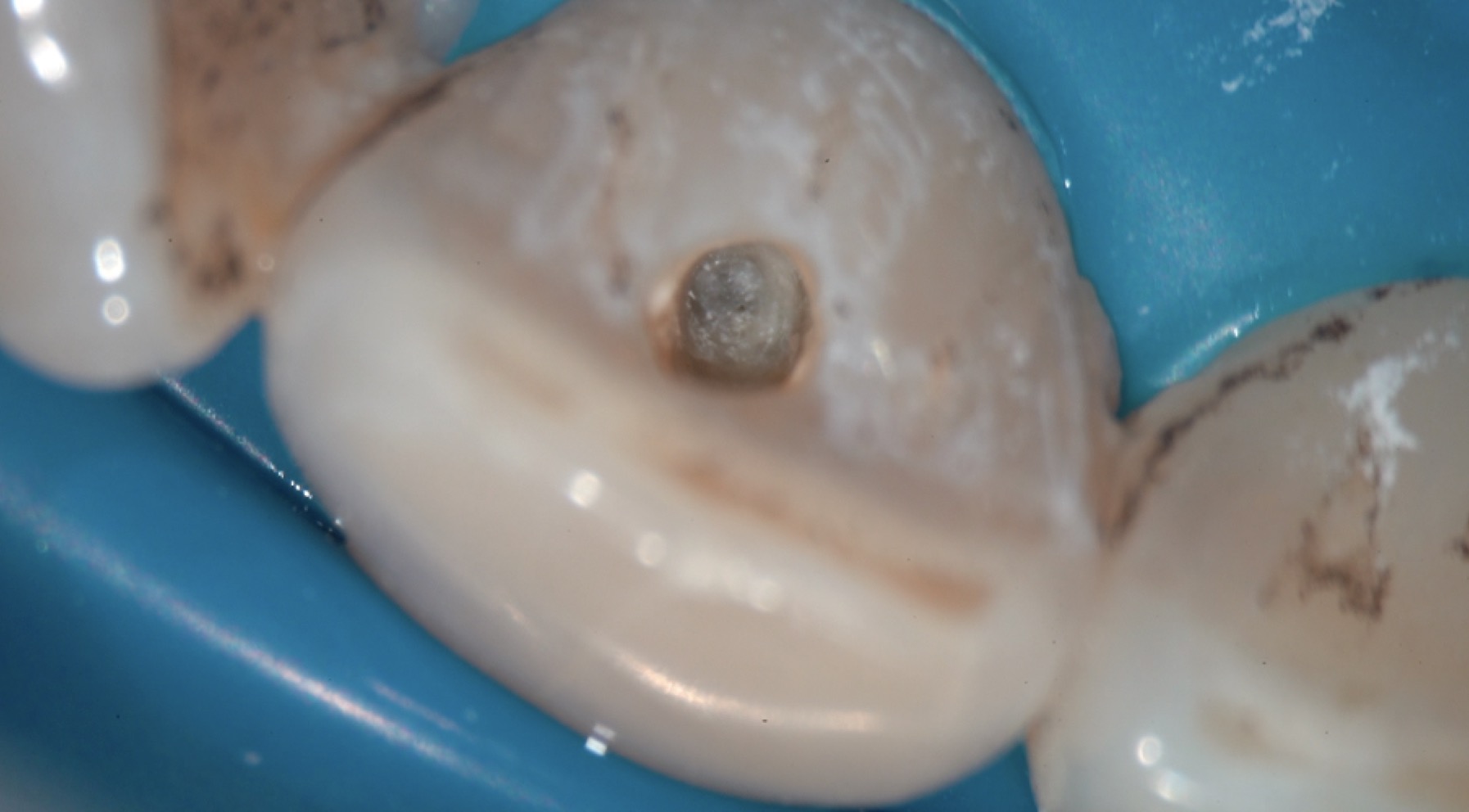 Treat the invisible canal: a clinical challenge
During primary treatment, even in simple cases, many root canal anatomy alterations and errors can occur such as access cavity over enlargement, perforations, blocage or file breakage. Sometimes, many iatrogenic […]
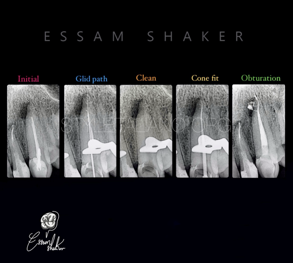 Complexity of the root canal system anatomy
The main goal of root canal treatment is to eliminate the infection in the complex root canal system for the long-term preservation of a functional tooth. Proper debridement of the […]
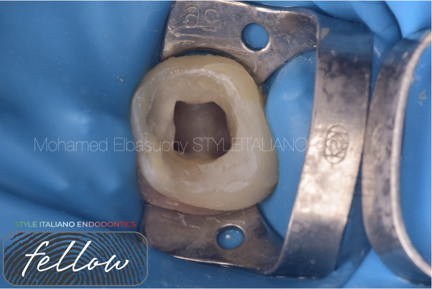 Radix Entomolaris: Case Report with Clinical Implication
Usually, first mandibular molars have one mesial and distal root but in some cases there are anatomical variations. Presence of an additional lingual root distally in mandibular molars is called radix entomolaris (RE). If present, an awareness and understanding of this unusual root and its root canal morphology can contribute to the successful outcome of root canal treatment. The article describes the endodontic management of mandibular molar with RE.
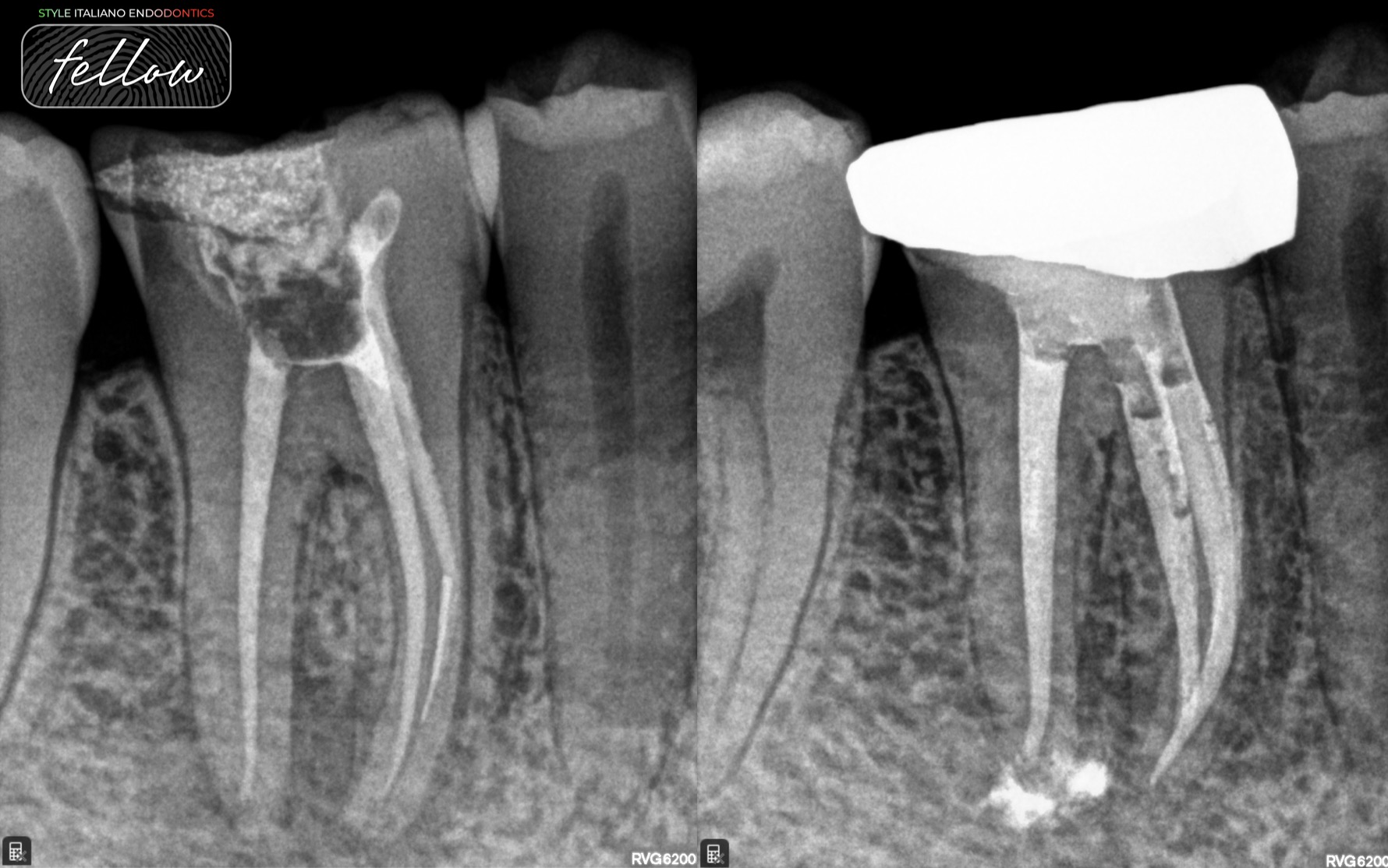 Retrieval and retreatment of mandibular molar
Retrieval along with Retreatment is one of the most challenging procedures in the endodontic therapy, This article showcases the management of a lower first molar with swparatedinstrumemnt and a periapical lesion. In […]
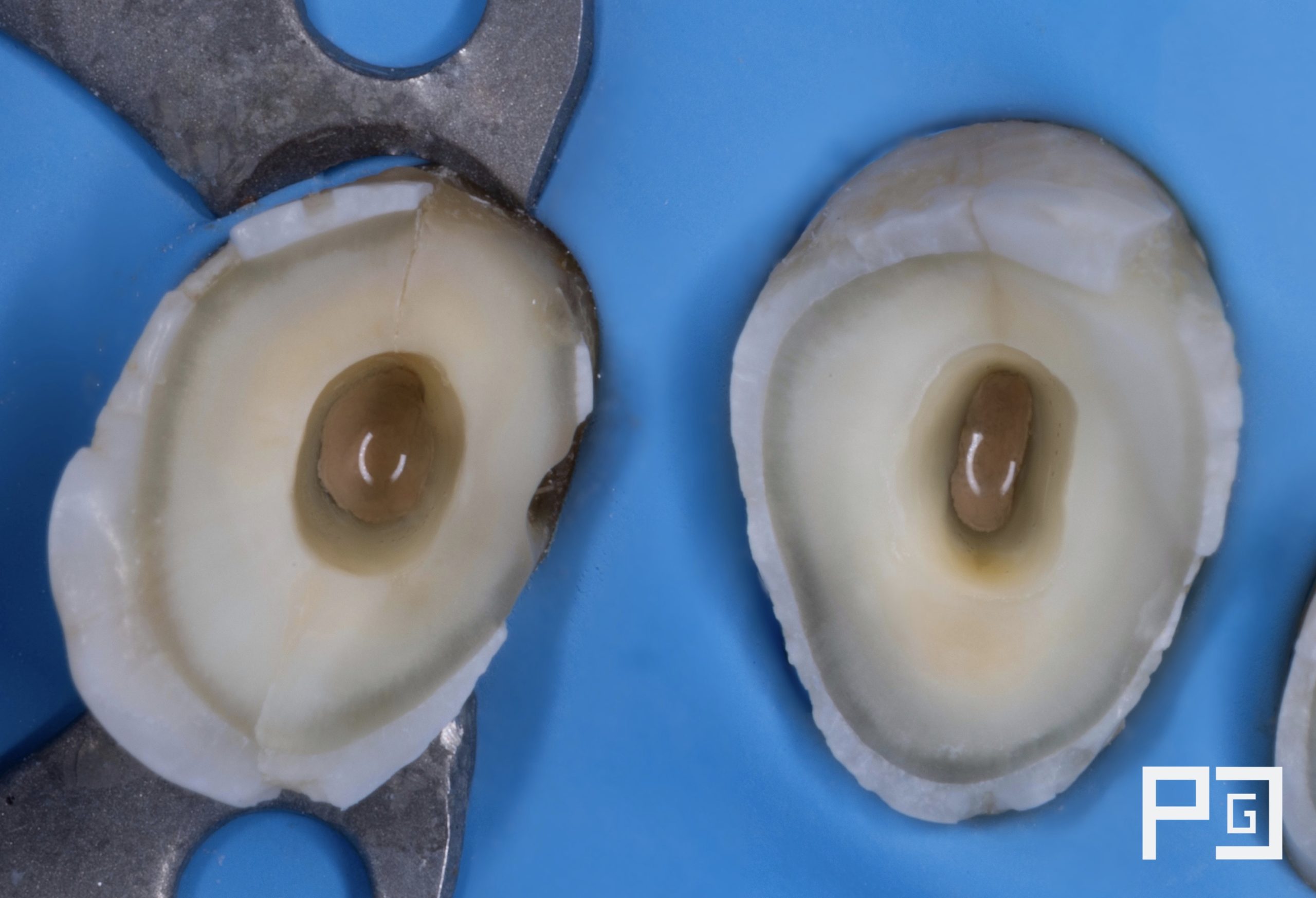 Management of complex anatomy in the first mandibular premolar, 2 cases in 1
We know that treating a tooth involves different phases or stages, but it is important to know the internal anatomy, in order to approach the case in the best possible […]