Bisphosphonate-related osteonecrosis of the jaws (BRONJ) is seen occasionally in bisphosphonate-receiving patients for several medical conditions such as osteoporosis, Paget’s disease or metastatic cancer. The term BRONJ was later changed […]

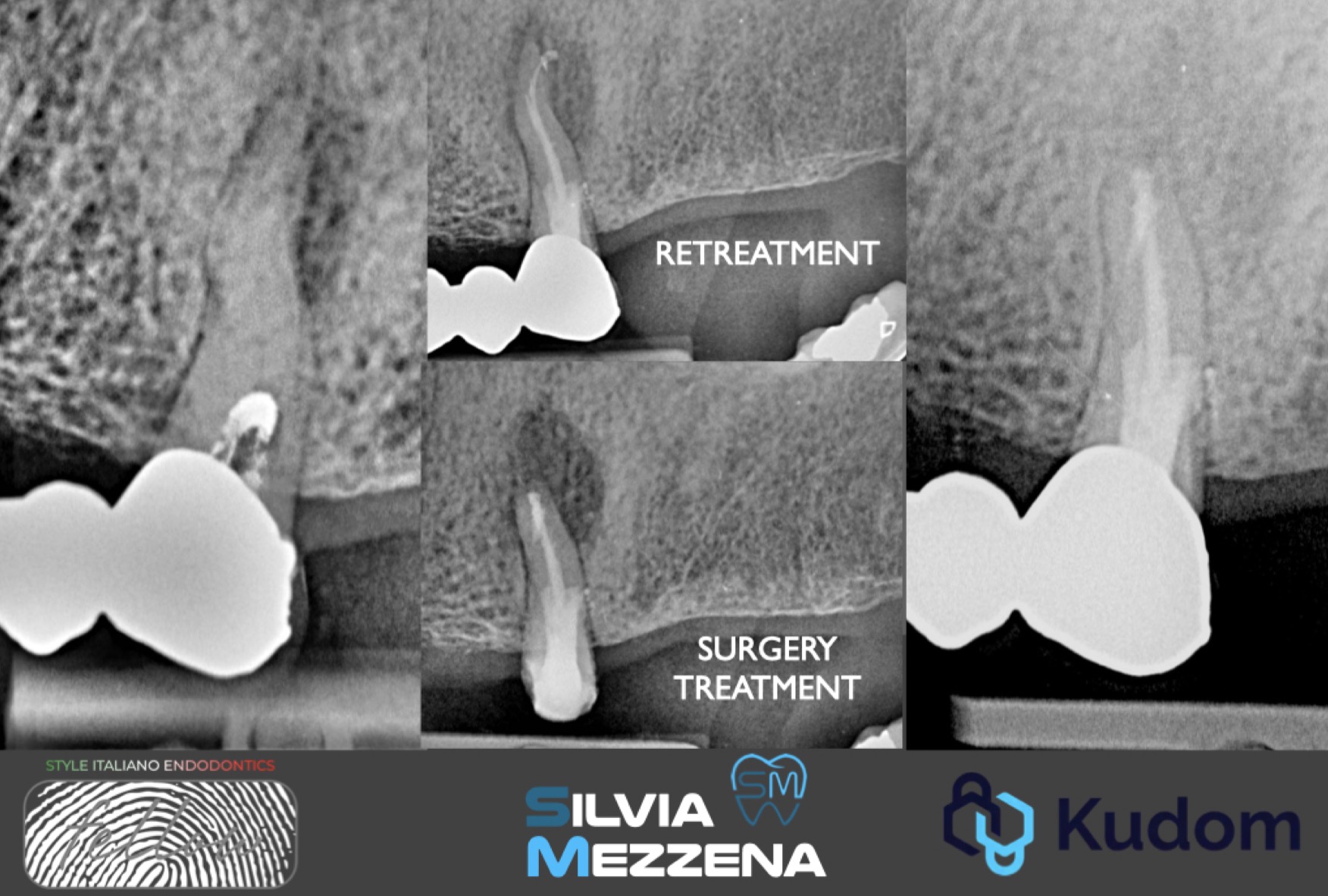 Endodontic management of tooth 2.5 in a patient taking bisphosphonates.
Bisphosphonate-related osteonecrosis of the jaws (BRONJ) is seen occasionally in bisphosphonate-receiving patients for several medical conditions such as osteoporosis, Paget’s disease or metastatic cancer. The term BRONJ was later changed […]
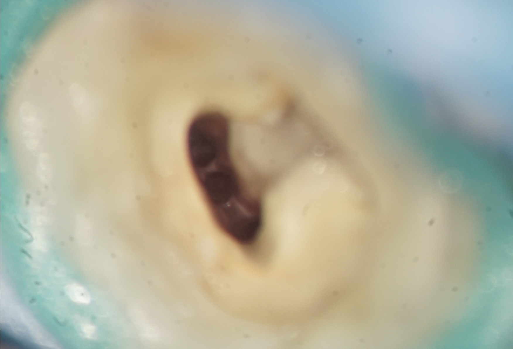 Clinical Insights into the Middle Distal Canal of Mandibular Molars: A Case Report.
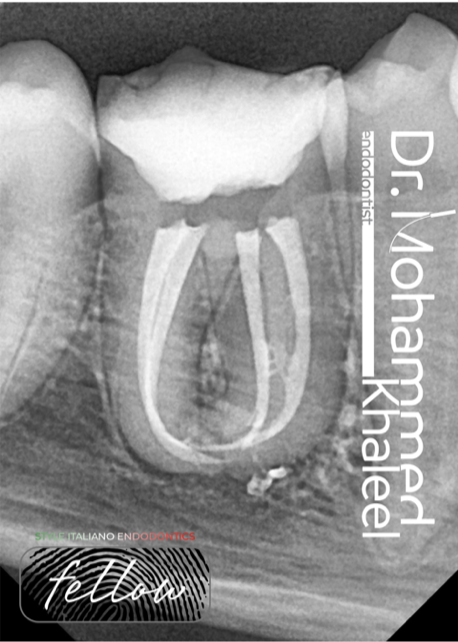
The middle distal canal in mandibular molars is a rare but clinically significant anatomical variation. Its prevalence varies from 0.26% to 9.6%, influenced by population differences and detection methods like […]
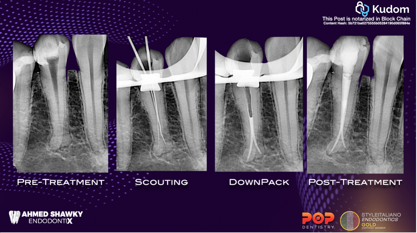
 Navigate the curve: the Rising protocol
Shaping narrow and curved canals remains one of the most complex challenges in endodontics, particularly when dealing with coronal and multiplanar curvatures.These anatomical variations increase the risk of procedural errors […]
 Deep challenges: splits remastered
Whenever root canal anatomy is discussed, variability is the rule!!
This requires careful attention of the clinician to anticipate such variation
 Endodontic Management of a Curved Upper Left Molar using the Rising Files
To perform an endodontic treatment and achieve a favorable outcome it is crucial that we eliminate the bacterial infection from the internal complex anatomy through proper shaping and cleaning.
In order to achieve this outcome we need a proper diagnosis, an appropriate treatment plan, a correct protocol approach and of course a set of files with great cutting efficiency and flexibility to deal with the challenges of the internal anatomy.
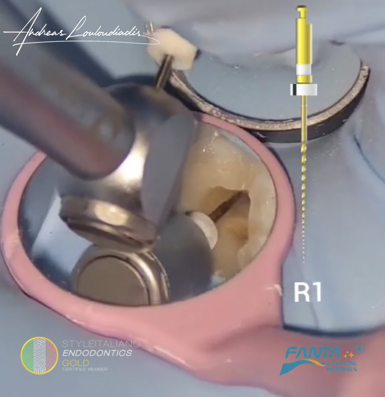
 R One Mini Case Study
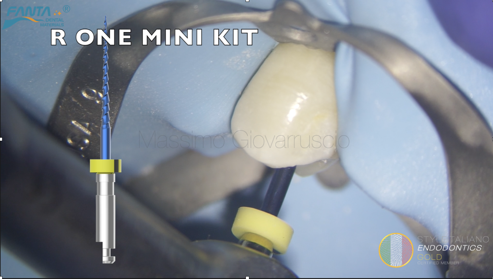
The R-One Mini System from Fanta Dental is an advanced reciprocating file system designed for efficient and conservative root canal shaping.It features a hybrid heat treatment short Sequence for enhanced […]
 External resorption Part II
Root resorption is the loss of hard dental tissue due to the action of osteoclastic cells. External resorptions present a challenging clinical situation; often, lesions are misdiagnosed and confused. Therefore, […]
 Tooth Autoreplantation of 1.6 with stripping
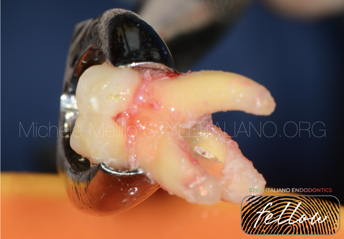
Tooth Replantation to approach the stripped Mb root on tooth 1.6 that was not possible to treat with a conventional conservative approach
 Dealing with intricate root canal system
In Endodontics, while managing curved canals , an exception to dealing with the intricate canal system could be when using advanced technology like rotary instruments or nickel-titanium files.
These tools can help simplify the process by efficiently cleaning and shaping the canals, making it easier to navigate through complex canal anatomy.
By utilizing such modern techniques, Endodontist can overcome the challenges posed by intricate canal systems more effectively.
 The curve you don’t expect
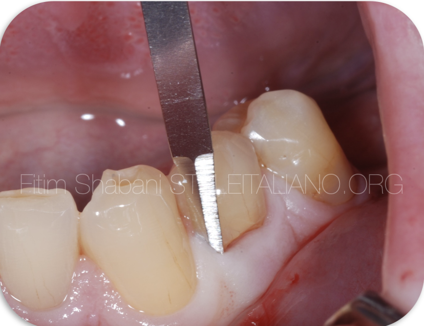
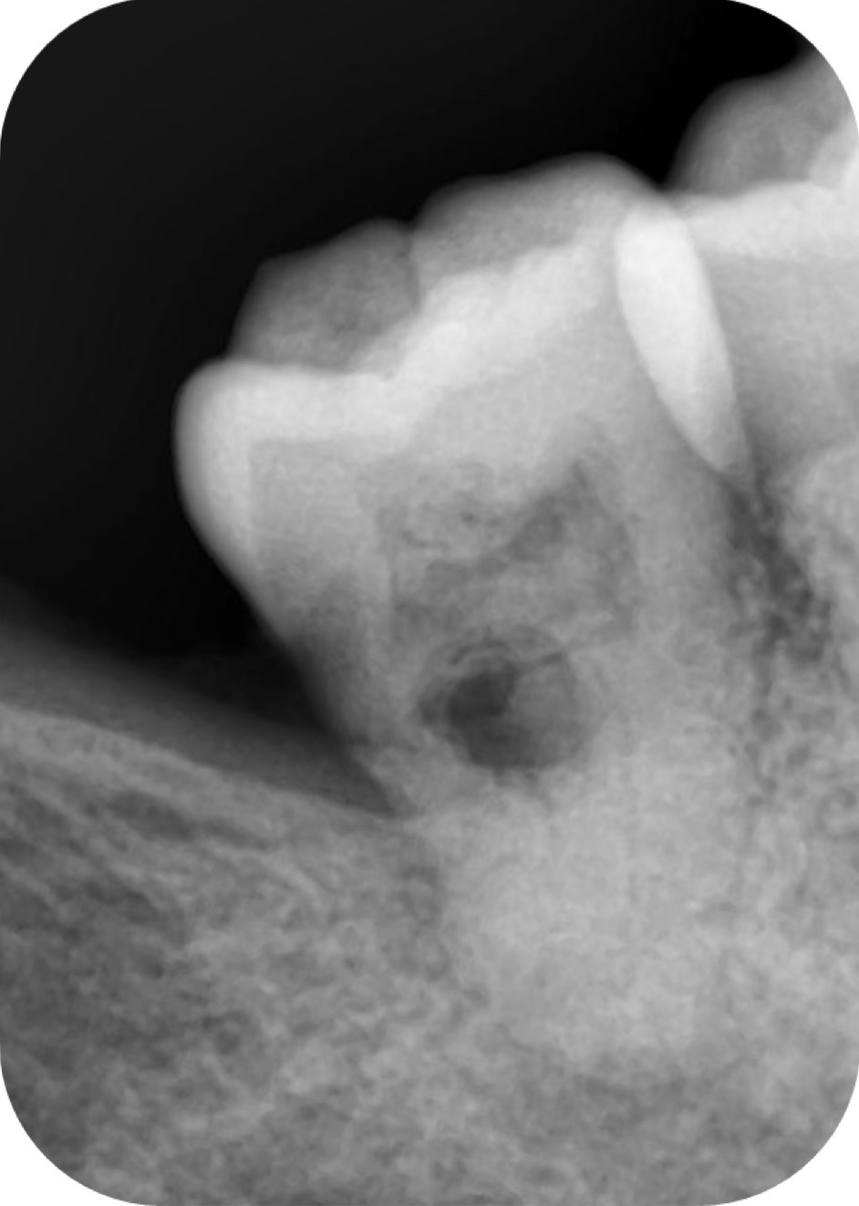
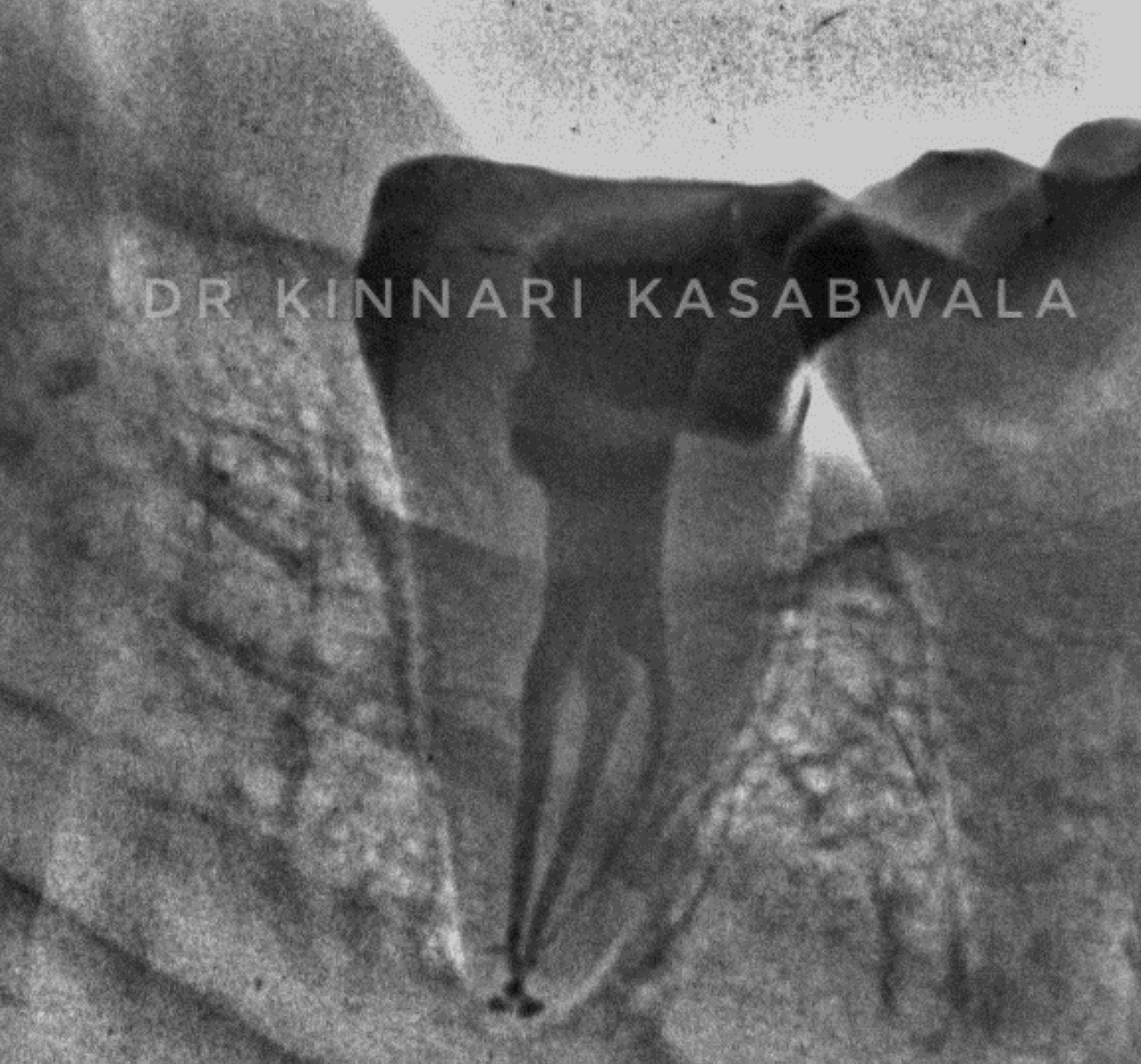
Patient has been referred due to symptoms on Upper Right Second Premolar. The tooth is tender to percussion and does not respond positively to vital test. Diagnosis is apical periodontitis […]
 Internal root resorption in permanent mandibular molar: A rare entity
Internal resorption is usually uncommon thing which starts from the root canal & destroy the tooth structure. It usually occurs as a result of a continuous chronic inflammatory process . […]
 Management of C shaped canal in mandibular second molar
C shaped root canal configuration is an aberrant canal anatomy, common in the mandibular second molars with prevalence ranging from 2.7% to 45.5% in different populations. Failure of fusion of Hertwig’s […]