A story of a dilacerated premolar
31/05/2024
Fellow
Warning: Undefined variable $post in /home/styleendo/htdocs/styleitaliano-endodontics.org/wp-content/plugins/oxygen/component-framework/components/classes/code-block.class.php(133) : eval()'d code on line 2
Warning: Attempt to read property "ID" on null in /home/styleendo/htdocs/styleitaliano-endodontics.org/wp-content/plugins/oxygen/component-framework/components/classes/code-block.class.php(133) : eval()'d code on line 2
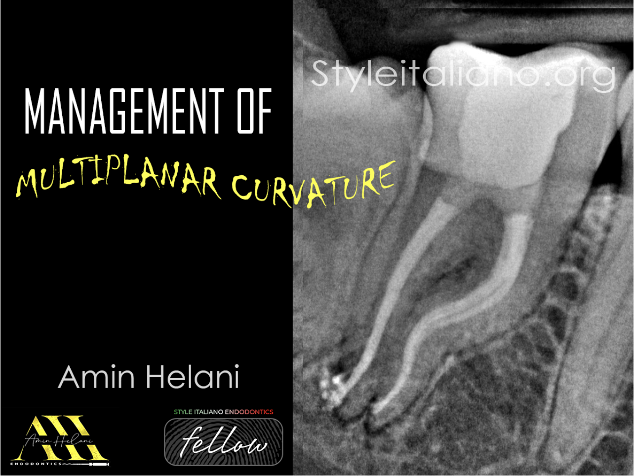
Premolars are the most versatile teeth in regards of anatomy. One of the most challenging anatomical variations we have to face is dilaceration. Abrupt curvatures complicate the RCT especially when we have more than one.
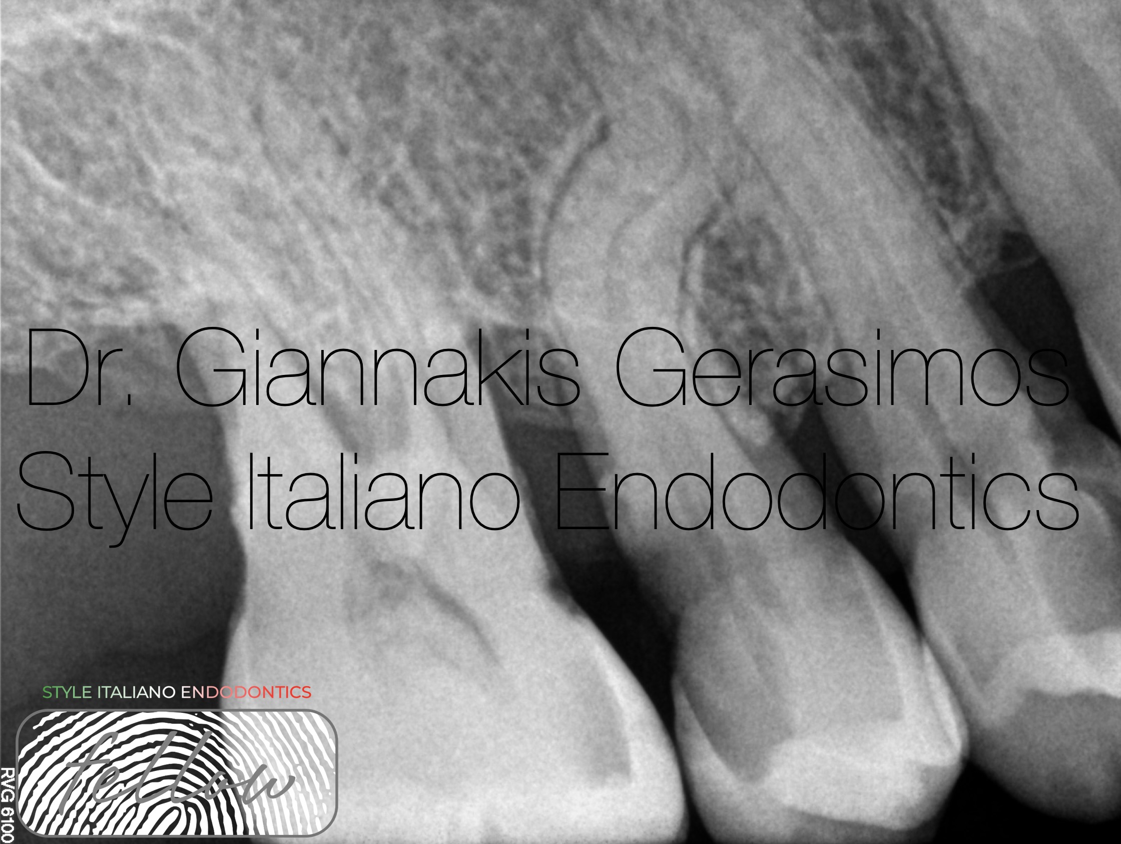
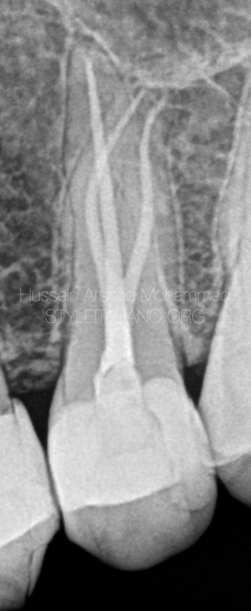
Fig. 1
Radiographic evaluation revealed deep distal decay and dental tissue loss. Also, we can observe that we have two canals. The canal anatomy seems intricate with abrupt dilaceration
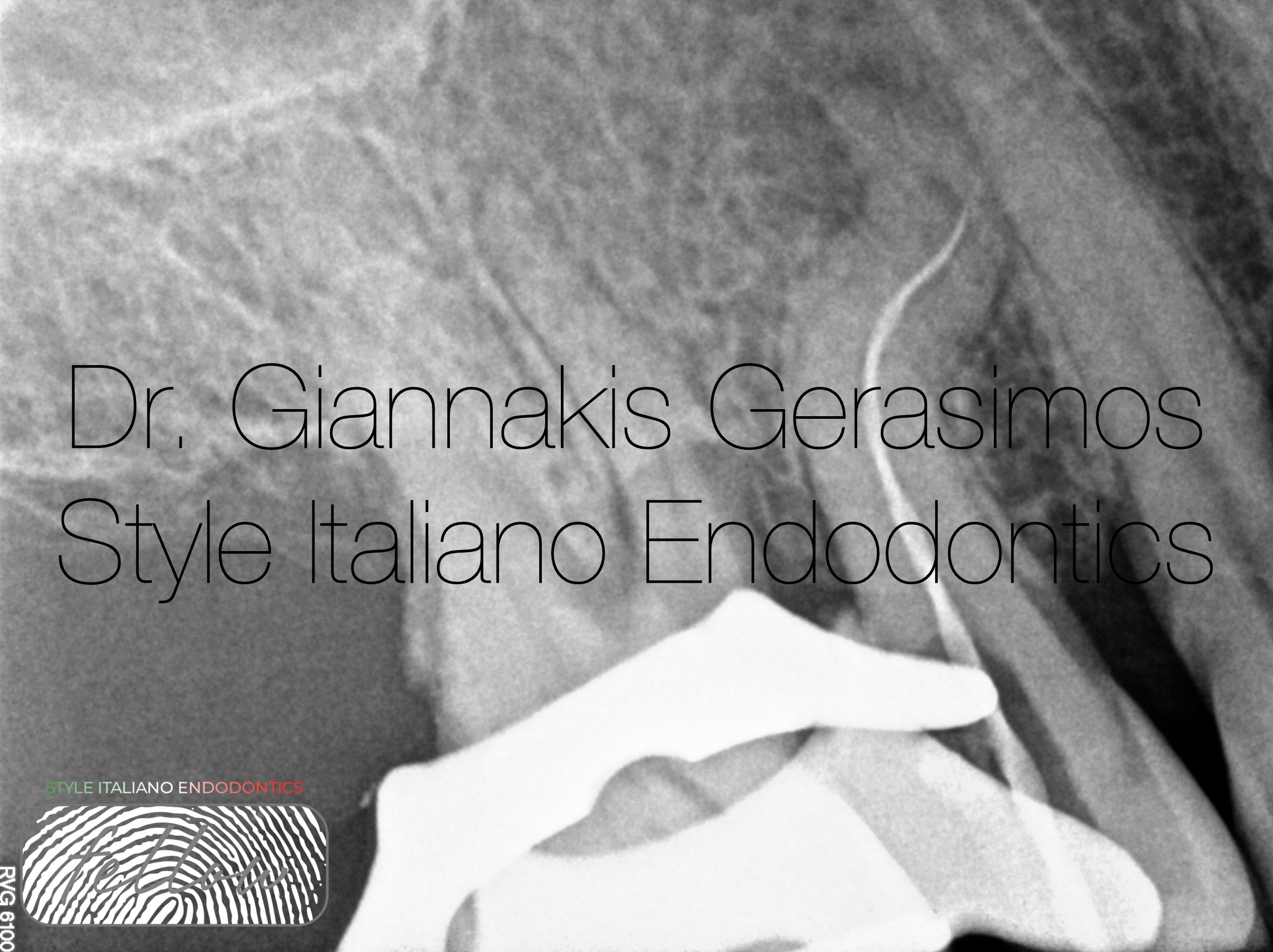
Fig. 2
After proper anesthesia and isolation, we removed decay. Then.
we did some preflaring in order to make sure that all files reach the point of the first curvature unobstructed. Next, with a hand file we determined the exact point of the second curvature but did not try to negotiate it at this point. Finally, we prepared with mechanical files up to his point. This is the zone technique. Of course, irrigation is mandatory between each file to avoid blocking the canal. The secret to this point is to completely forget the apical curvature and focus first to instrumenting up to that point.
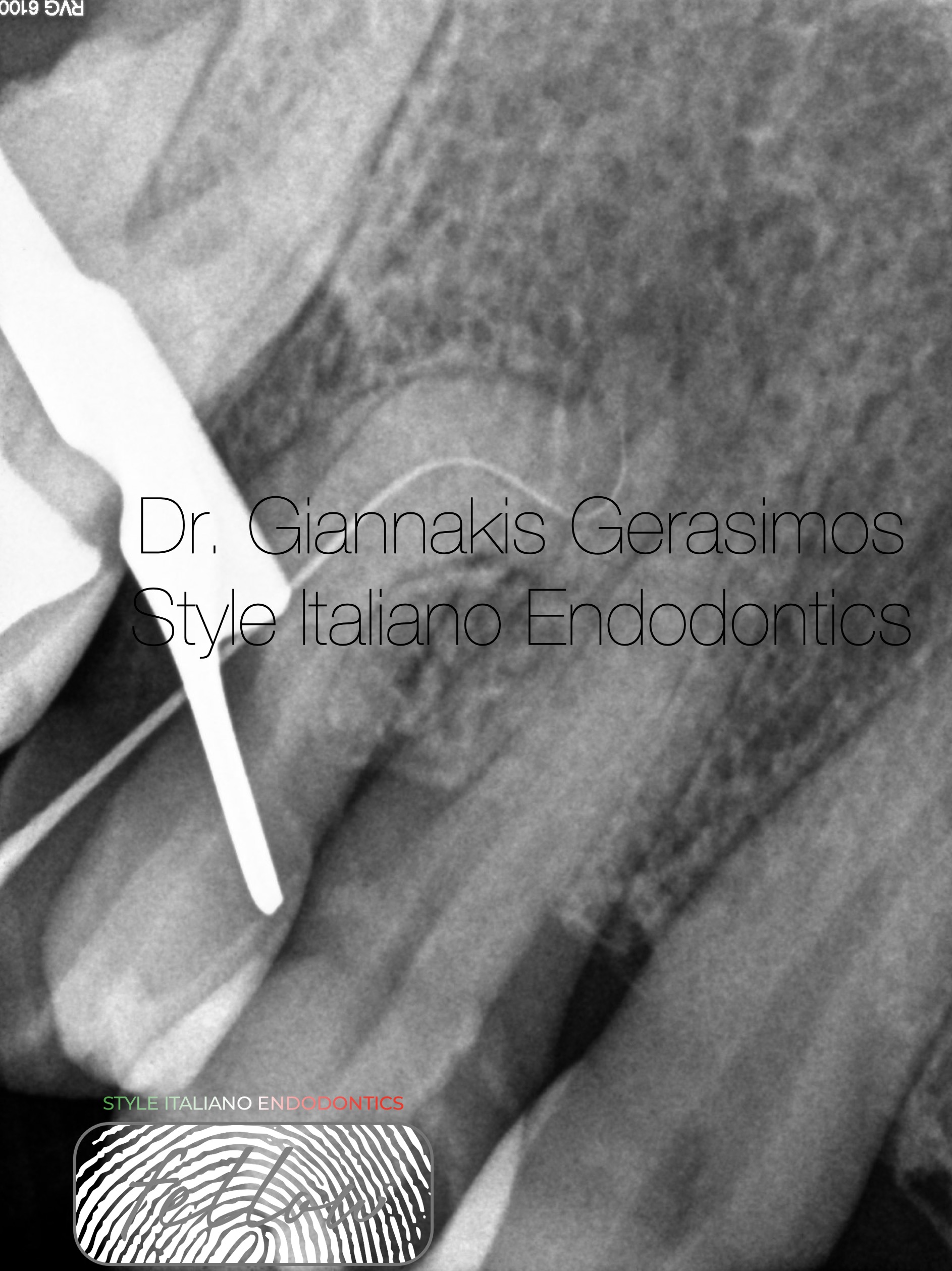
Fig. 3
Now is the time to precurve hand files ( c-pilot VDW ) No 8 & 10.
Crown – Down technique with balanced force allowed us to reach up to this point. After the second curvature I used only hand files because it was abrupt in a buccal distal direction and we had more control. However, as we can observe there is apical pathology so we dedicated a lot of time to irrigation and activation with internal heating method.
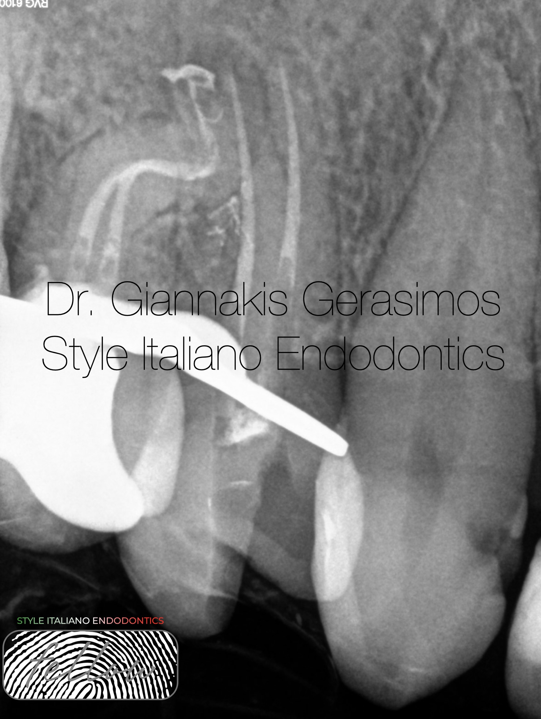
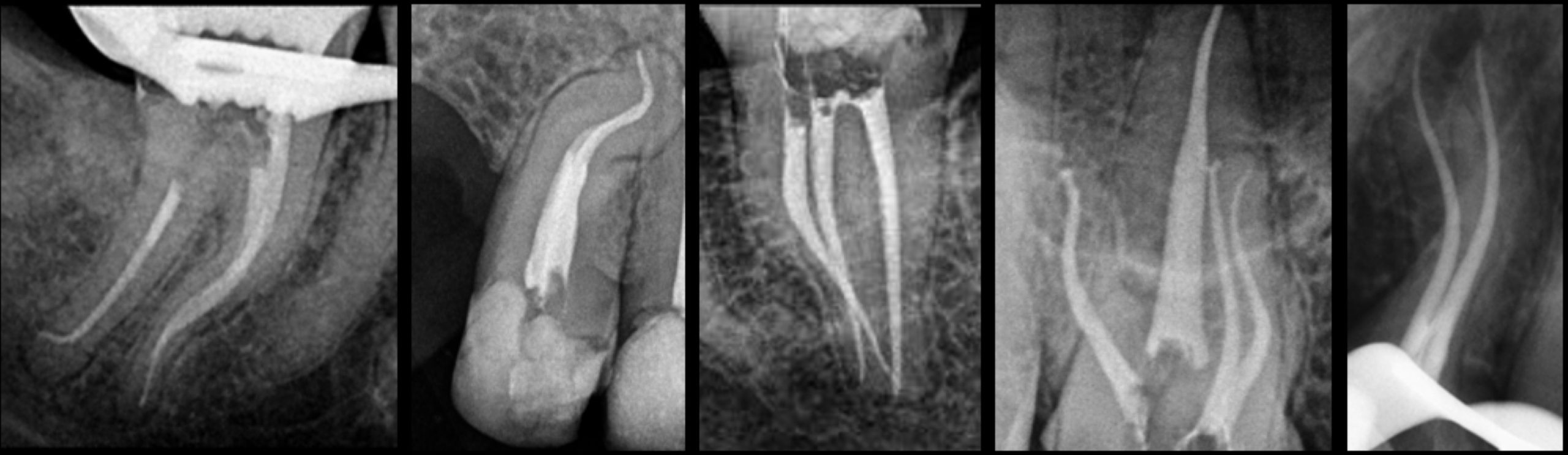
Fig. 4
This is the obturation radiograph of teeth # 14, 15
We can observe the abrupt curvature in the second premolar but also the apical ramifications and lateral canals.

Fig. 5
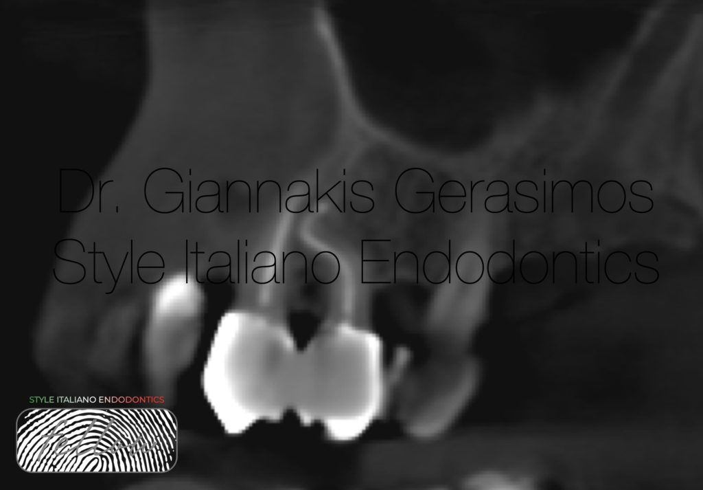
Now the most important : Follow-up
The patient did a CBCT to evaluate bone for an implant
So we can observe that 1,5 year later the area seems to have a good healing.

Fig. 6
Gerasimos Giannakis received the Doctor of Dental Surgery degree from the University of Athens in 2018.
He has special interest in the field of endodontics, restorative dentistry and microsurgical techniques.
Since then has completed
STYLE ITALIANO Daily menu 40th edition , Milan 2020
TOOTH WEAR MASTER COURSE, by Didier Dietschi 2021
EXPERT COURSE IN MASTERING SOFT TISSUE SURGERY,
In cooperation with the University of Cologne 2022
MICROSCOPE SIMULATION COURSE ON CANAL BLOCKAGE MANAGEMENT by Chaniotis, Sousa Dias, 2020
He is working as an endodontics associate in a clinic, responsible also for the restoration of endodontically treated teeth.
Working exclusively under the microscope and rubber dam, has the opportunity to document the cases.
In the Style Italiano Congress in Greece 2023, was part of the bootcamp and became fellow of the Style Italiano Endodontics community.
Conclusions
Dilacerated premolars are a big challenge in terms of negotiating and disinfecting this anatomy. We need a protocol, feasible-teachable-repeatable. So, preflaring. zone technique, precurving our files to follow the anatomy are essential tips to handle such cases. Also, patience and tactile sensation are very important.
Bibliography
Present status and future directions: Management of curved and calcified root canals
Antonis Chaniotis , Ronald Ordinola-Zapata
Root canal treatment of dilacerated second maxillary premolars: Planning the shaping procedure
Davide Mancino and Naji Kharouf
Intracanal heating of sodium hypochlorite: Scanning electron microscope evaluation of root canal walls
Alfredo Iandolo , Massimo Amato , Alberto Dagna, Claudio Poggio, Dina Abdellatif , Vittorio Franco , Giuseppe Pantaleo