Management of S-shaped root canals
31/07/2023
Fellow
Warning: Undefined variable $post in /home/styleendo/htdocs/styleitaliano-endodontics.org/wp-content/plugins/oxygen/component-framework/components/classes/code-block.class.php(133) : eval()'d code on line 2
Warning: Attempt to read property "ID" on null in /home/styleendo/htdocs/styleitaliano-endodontics.org/wp-content/plugins/oxygen/component-framework/components/classes/code-block.class.php(133) : eval()'d code on line 2
Straight simple root canals are rare in the human dentition; the majority present with complexities and curvatures in different planes, making the endodontic treatment a complex task that requires adaptation according to each specific case (1). One of these special anatomies is the S-shaped root canal.
The purpose of this paper is to present 2 cases with S-shaped root canals and to describe the methodology followed to treat such anatomies.
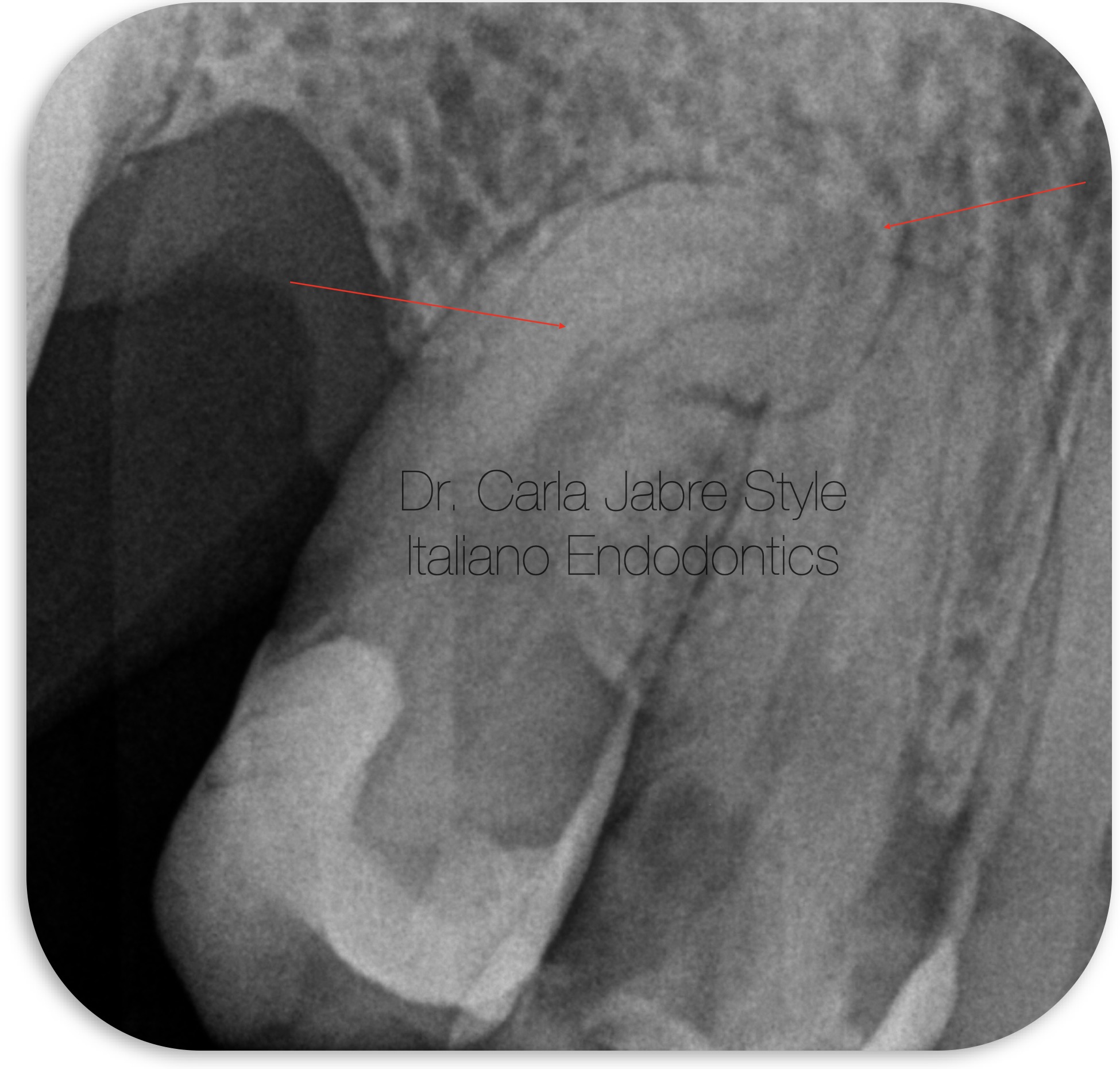
Fig. 1
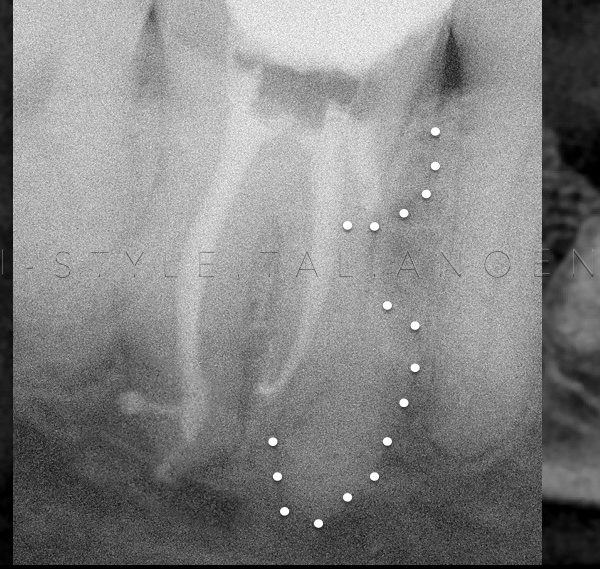
A 40 year old patient was referred for the endodontic treatment of teeth (14) and (15) for prosthetic reasons.
Examination of the initial radiograph reveals a sharp curvature in the middle third, followed by a second abrupt one more apically in tooth (15).
After anesthesia, rubber dam placement and access cavity, shaping began with an early coronal preflaring using a 25/.08 file.
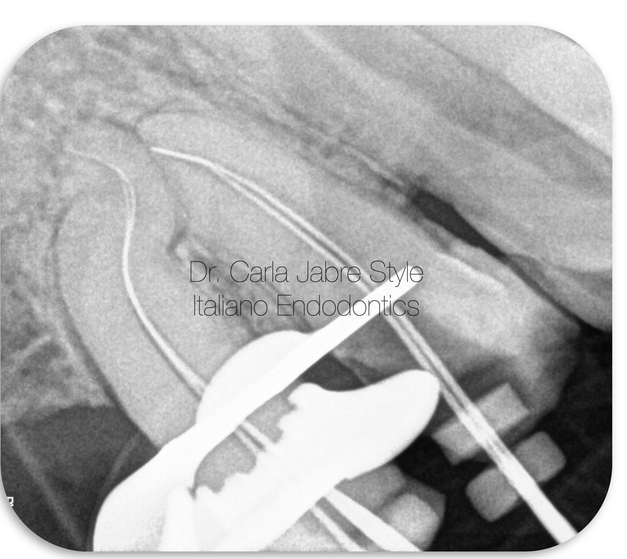
Fig. 2
A 08 K file was used for scouting. It stopped at the level of the first curvature. No extra pressure was applied to force the file to the apex. On the contrary, a glide path file was used coronal to the first curvature to remove the interferences that were blocking the progression of the file.
After glide path and irrigation with NaOCl, the 08 K file reached the electronic working length. It was not removed from the canal, but gentlly used in a pull push motion until it became loose in the canal. If the file is removed prematurely, it becomes extremely difficult to reach working length again. (2)
Then, a 10 K file was able to reach passively that same length and also used in the same filing motion.
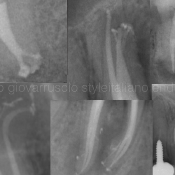
Working length radiograph was taken with a 10 K file.
A smooth glide path was obtained with a rotary instrument size 15/.04. The glide path is a mandatory step before shaping because it decreases the fracture rate of shaping files and it prevents inaccurate shaping (3).
Shaping the middle and apical thirds was done with controlled memory NiTi files up to size 25/.04.
Controlled Memory NiTi files undergo a special thermomechanical process that makes them stay in their martensitic phase during clinical use (4). The result is an extremely flexible and bendable instrument but without the shape memory of other NiTi files. This minimizes the risk of iatrogenic mishaps and the file follows the anatomy of the canal very closely because it does not rebound to its original shape (2,5,6). They also have increased resistance to cyclic fatigue because of their unique manufacturing process (5,7).
In addition to that, files with a bigger taper were not used apically in order to avoid straightening the curvature (3,8).
Canal patency was maintained by using a 08 K file at 0.5 mm beyond the canal terminus and copiously irrigating with NaOCl along with sonic activation of the solution.
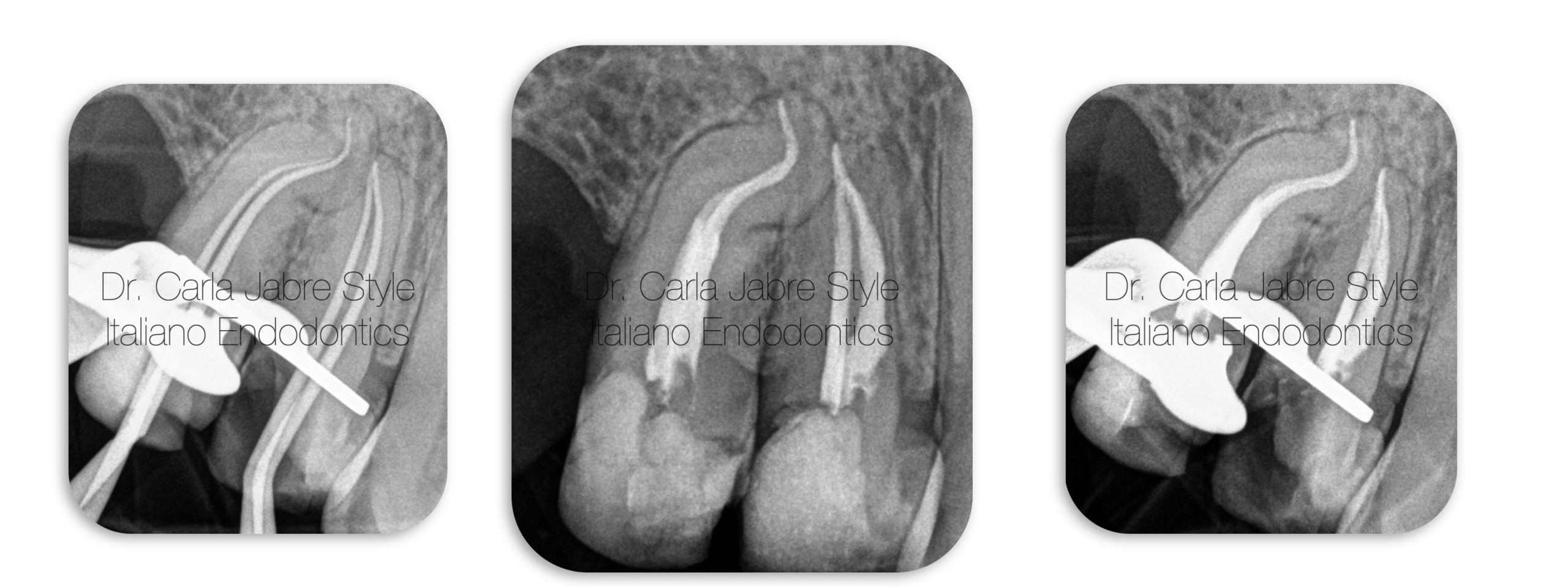
Fig. 3
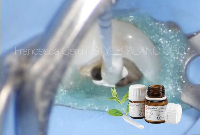
For obturation, pluggers could not reach the recommended length for the continuous wave of condensation technique. Therefore, single cone technique with bioceramic sealer was chosen.
The sealer was gently injected until the middle third, and the gutta-percha cones were coated with sealer in their apical part and inserted at working length, then cut and packed at the level of the orifice.
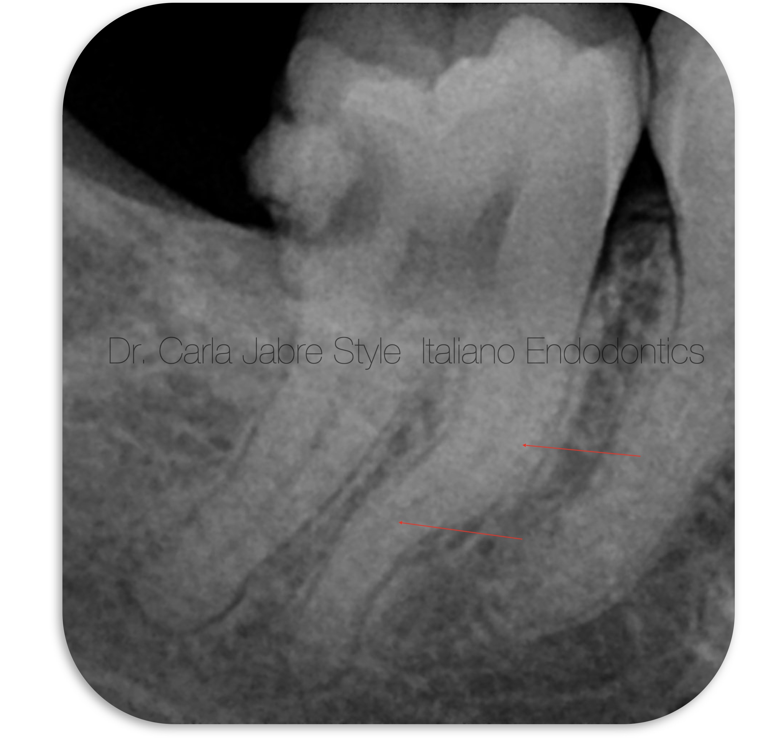
Fig. 4
A 35 year old patient presented to the clinic with irreversible pulpitis on tooth 47.
The initial radiograph shows a double curvature in the middle third of the mesial root.
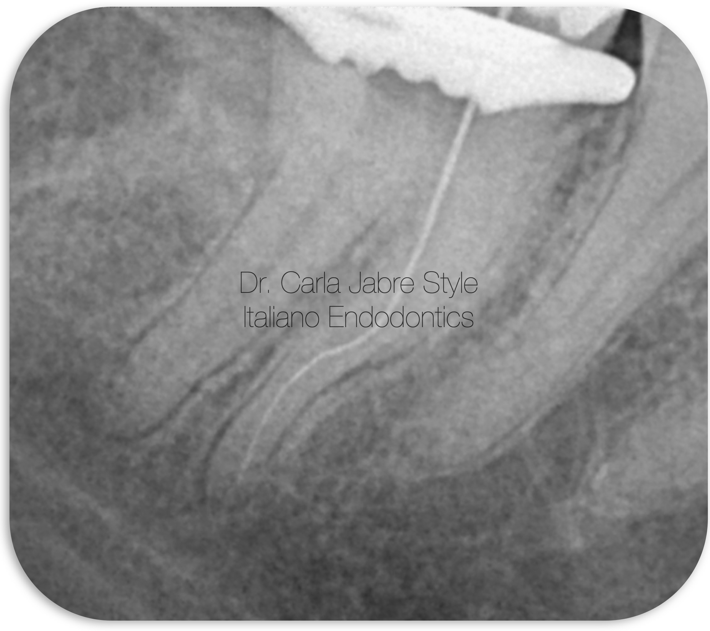
Fig. 5
After anesthesia, rubber dam placement and access cavity, shaping began with an early coronal preflaring using a 25/.08 file.
As the previously described case, the 08 K file did not reach the apical terminus directly. Coronal interferences were progressively removed using glide path mechanical files.
When the scouting 08 K file reached WL, it was used in a pull push motion until it became loose in the canal. Same was done with the 10 K file.
Glide path was done mechanically until size 15/.04.
Then for shaping, controlled memory instruments in continuous rotation were used until size 19/.05.
Canal patency was maintained by using a 08 K file 0.5 mm beyond the canal terminus and copiously irrigating with NaOCl, along with sonic activation of the solution.
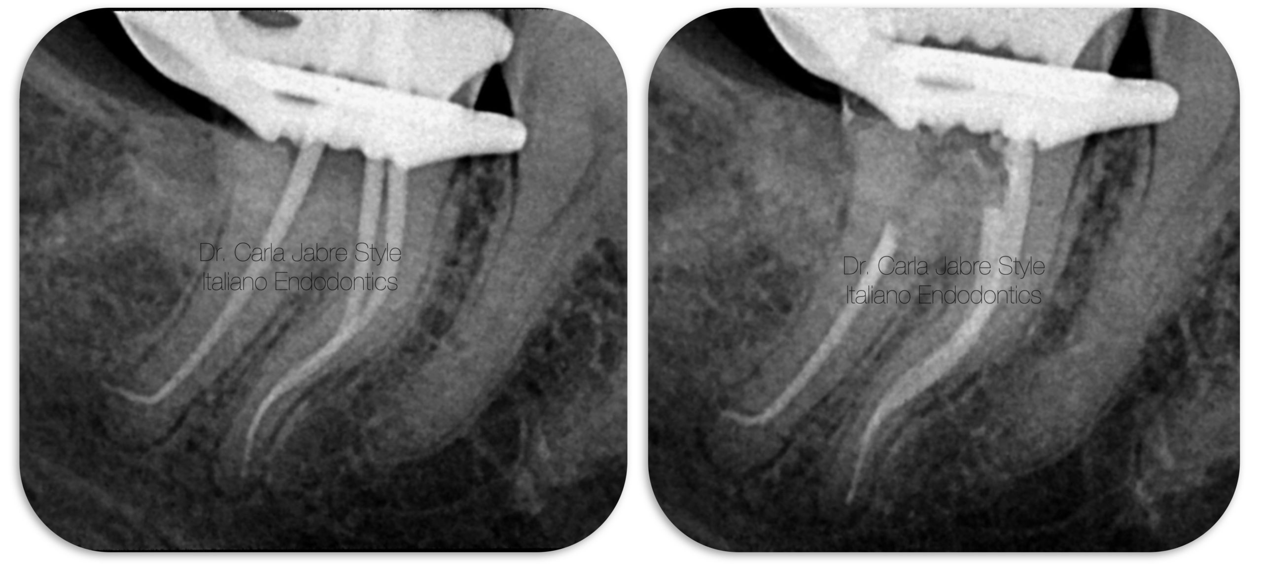
Fig. 6
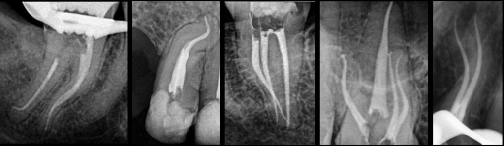
For obturation, single cone technique with bioceramic sealer was chosen and applied as previously described.

Fig. 7
Dr Carla Jabre
2008-2013: Undergrad studies at the faculty of Dentistry at Saint Joseph University, Beirut, Lebanon.
2013-2016: Masters in Endodontics at the faculty of Dentistry at Saint Joseph University, Beirut, Lebanon.
2018-2019: Masters in Biomaterials at Saint Joseph University, Beirut, Lebanon.
Dr Jabre has been a clinical instructor, guiding undergraduate students, in the department of Endodontics at Saint Joseph University, since 2016.
She is a member of the Lebanese Society of Endodontics, and a fellow in the Style Italiano Endodontics group.
Her private practice is limited to endodontics.
Conclusions
In summary, following simple steps can help clinicians manage S-shaped canals in a reproducible way. For that, a careful analysis of the initial and working length radiographs will help detect and understand this particular anatomy and adapt the armamentarium accordingly.
Bibliography
1: Machado R, Chaniottis A, Vera J, Saucedo C, Vansan LP, Silva E. S-Shaped Canals: A Series of Cases Performed by Four Specialists around the World, Case Reports in Dentistry, vol. 2014, Article ID 359438, 6 pages, 2014. https://doi.org/10.1155/2014/359438.
2: Deshmukh SN, Shenoy VU, Margasahayam SV, et al. Endodontic management of S - shaped root canal in a maxillary premolar using controlled memory nickel titanium rotary files – a case report. J Evolution Med Dent Sci 2020; 9(51):3894-3897, DOI: 10.14260/jemds/2020/853
3: Biasillo V, Castagnola R, Colangeli M, Panzetta C, Minciacchi I, Plotino G, Staffoli S, Marigo L, Grande NM. Comparison of shaping ability of the Reciproc Blue and One Curve with or without glide path in simulated S-shaped root canals. Restor Dent Endod. 2022 Feb;47(1):e3. https://doi.org/10.5395/rde.2022.47.e3
4: Shen Y, Zhou HM, Zheng YF, Peng B, Haapasalo M. Current challenges and concepts of the thermomechanical treatment of nickel-titanium instruments. J Endod. 2013 Feb;39(2):163-72. doi: 10.1016/j.joen.2012.11.005. PMID: 23321225.
5: Tabassum S, Zafar K, Umer F. NiTi Rotary Systems: What’s New? Eur Endod J 2019; 3: 111-7
6: Liang, Y., Yue, L. Evolution and development: engine-driven endodontic rotary nickel-titanium instruments. Int J Oral Sci 14, 12 (2022). https://doi.org/10.1038/s41368-021-00154-0
7: Dablanca-Blanco A-B, Castelo-Baz P, Miguéns-Vila R, Álvarez-Novoa P, Martín-Biedma B. Endodontic Rotary Files, What Should an Endodontist Know? Medicina. 2022; 58(6):719. https://doi.org/10.3390/medicina58060719
8: Donnermeyer, D., Viedenz, A., Schäfer, E. et al. Impact of new cross-sectional designs on the shaping ability of rotary NiTi instruments in S-shaped canals. Odontology 108, 174–179 (2020). https://doi.org/10.1007/s10266-019-00450-6