A Comprehensive Clinical Protocol From Diagnosis of Failure to Aesthetic Composite Rehabilitation.
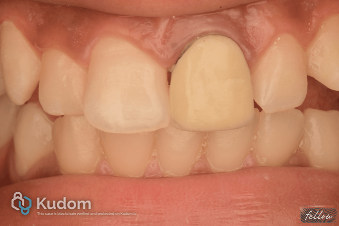
Endodontic failure in immature permanent teeth presenting with open apices poses a significant clinical challenge, compounded further when gross over-obturation, coronal leakage, and intrinsic discoloration coexist. This article describes the non-surgical retreatment of a maxillary left central incisor in a 14-year-old female patient who presented with a chronically failing, PFM-crowned tooth exhibiting a clinically detectable open margin, a draining sinus tract, a history of recurrent swelling, periapical pathology, an incompletely formed apex (open apex), and gross over-obturation of approximately 5–6 mm beyond the radiographic apex.