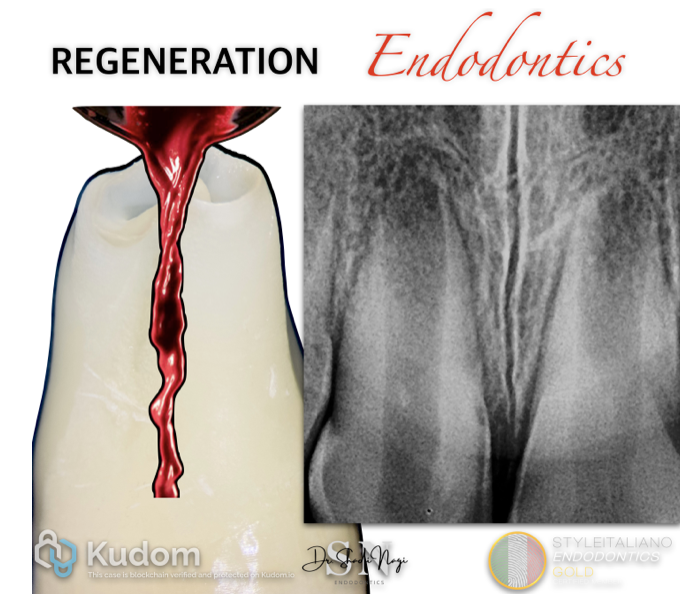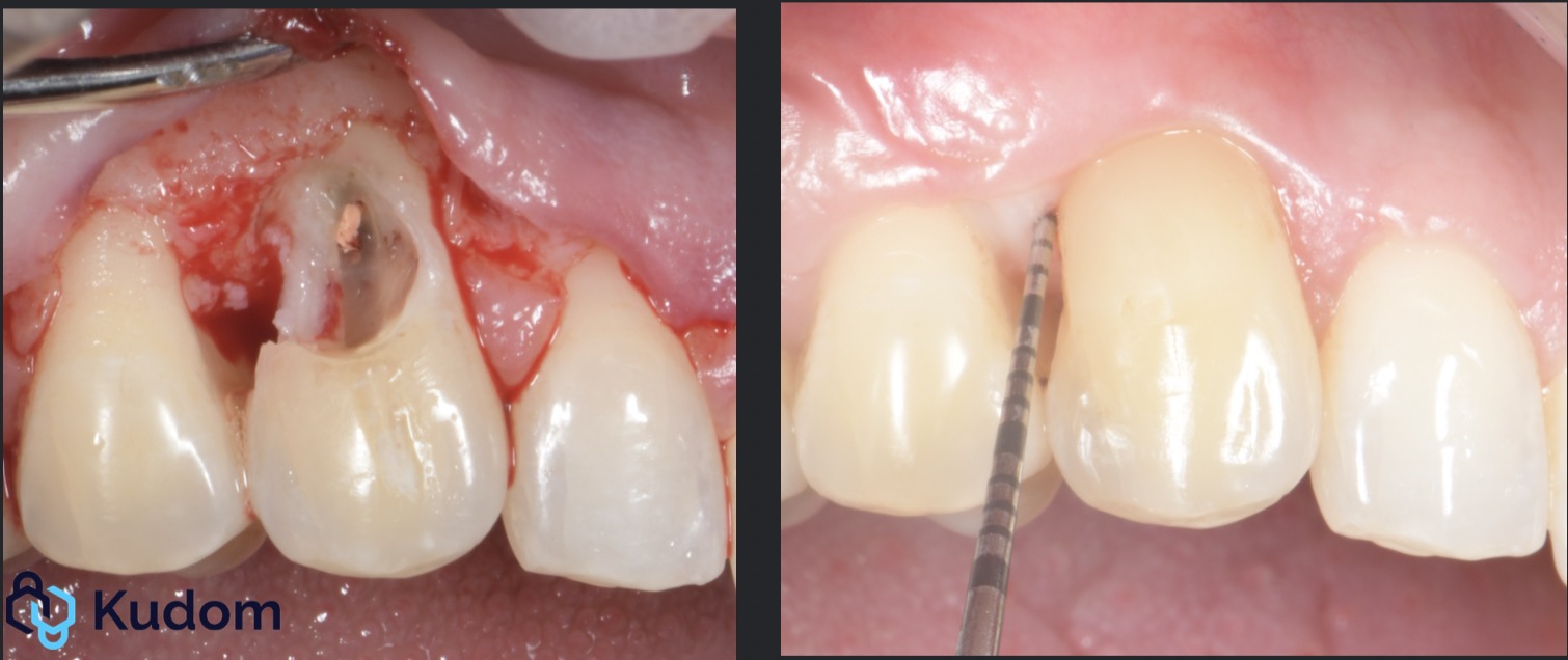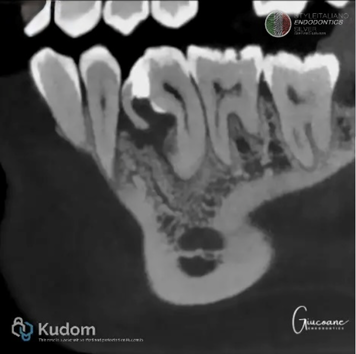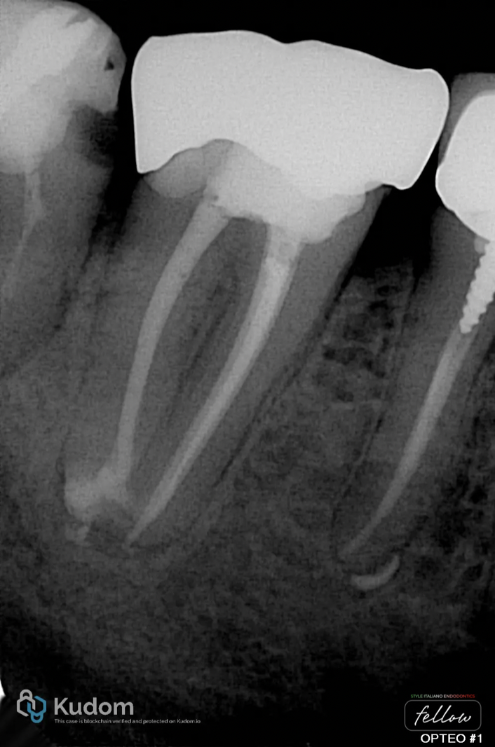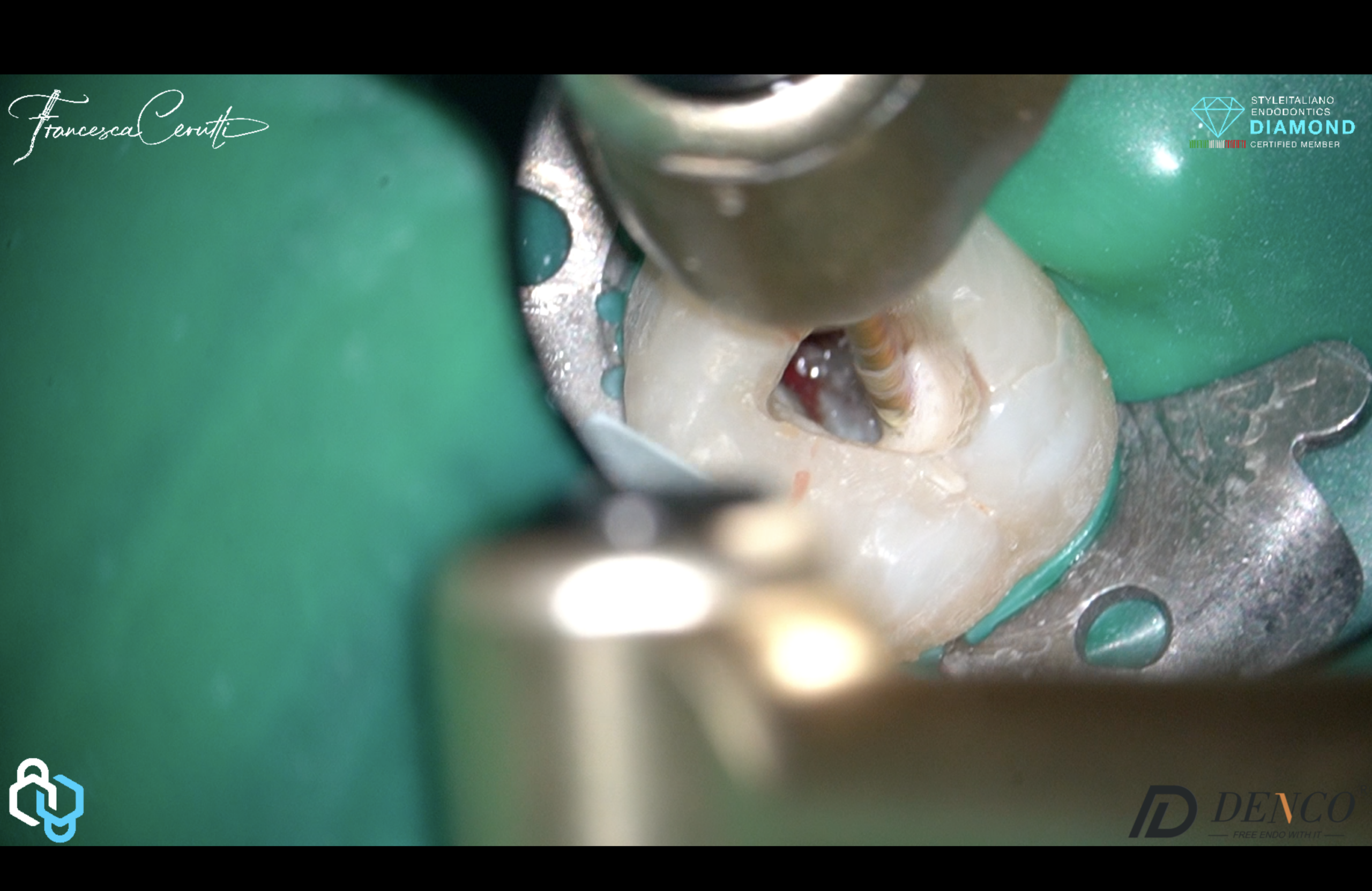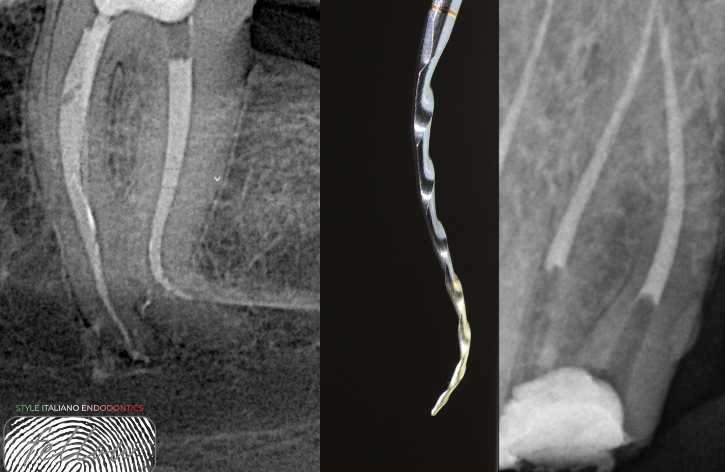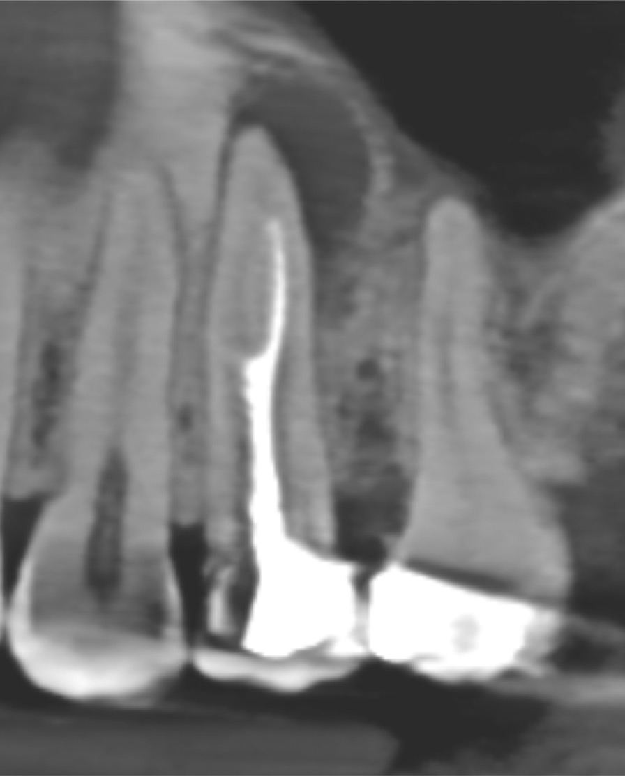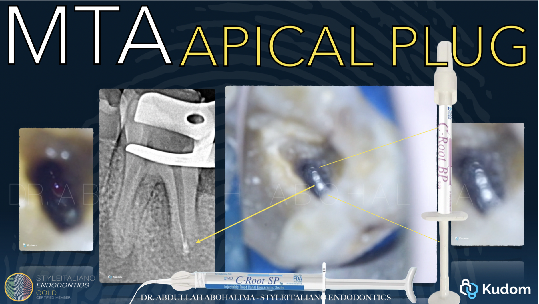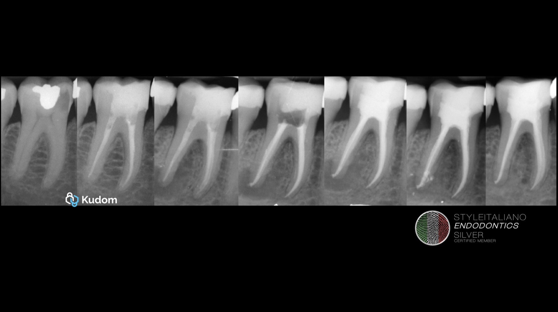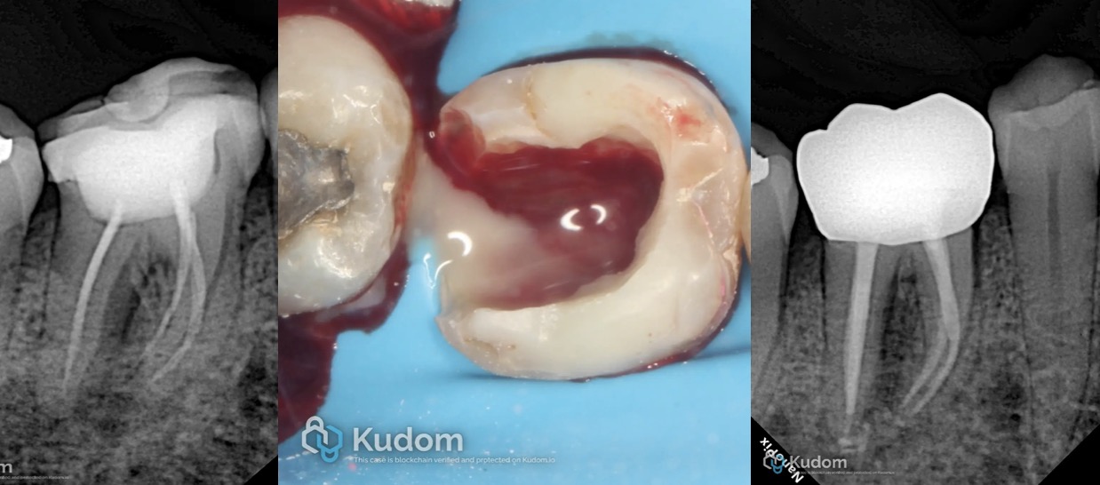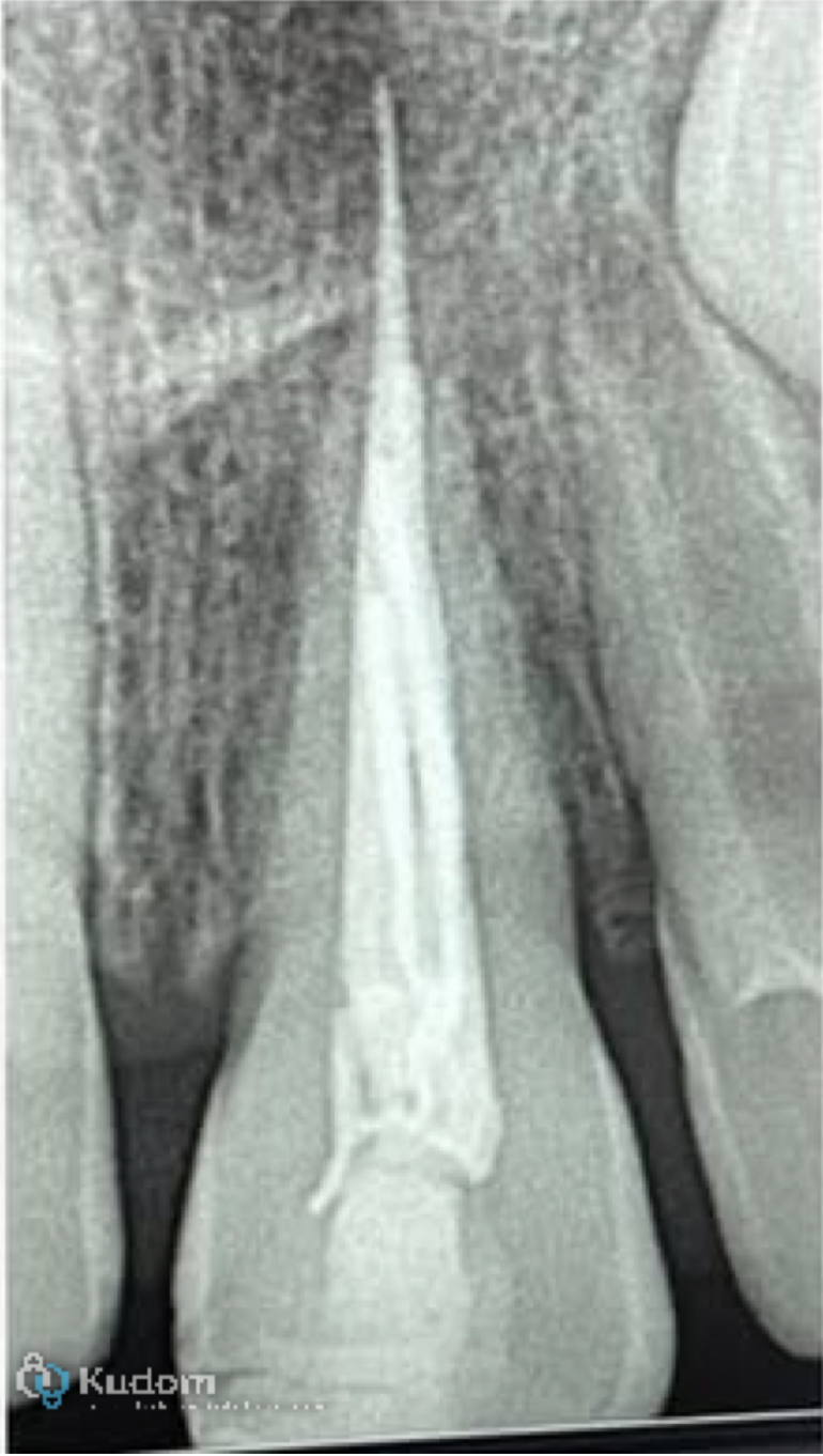
Overextended root canal fillings beyond the apical foramen can lead to persistent symptoms and periapical inflammation. This article discusses a retreatment case involving a maxillary anterior tooth, where the previous gutta-percha obturation was extended beyond the apex. The case was managed using H-file mechanical removal and obturation with bioceramic putty. Radiographic images demonstrate pre- and post-treatment conditions with clear clinical resolution.