Three-rooted maxillary premolars represent an uncommon but clinically important anatomical variation. The maxillary first premolar most frequently presents with two roots, while the occurrence of three separate roots is considerably […]

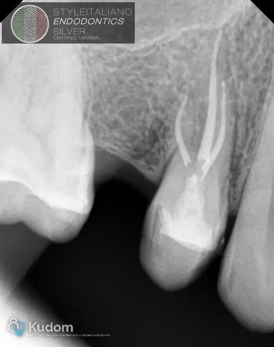 Three-rooted maxillary premolars: an uncommon anatomical variation.
Three-rooted maxillary premolars represent an uncommon but clinically important anatomical variation. The maxillary first premolar most frequently presents with two roots, while the occurrence of three separate roots is considerably […]
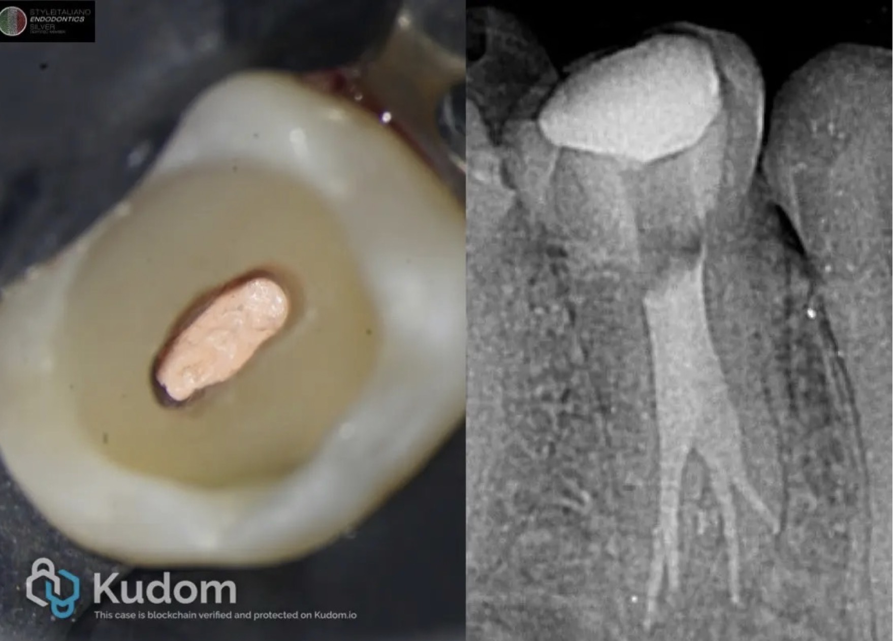 Expect the unexpected : Four canals in mandubular premolar
According to Vertucci, mandibular premolars have one single canal in approximately 74% of the cases. On occasion, mandibular premolars have two canals, in around 25.5%, but higher numbers are very […]
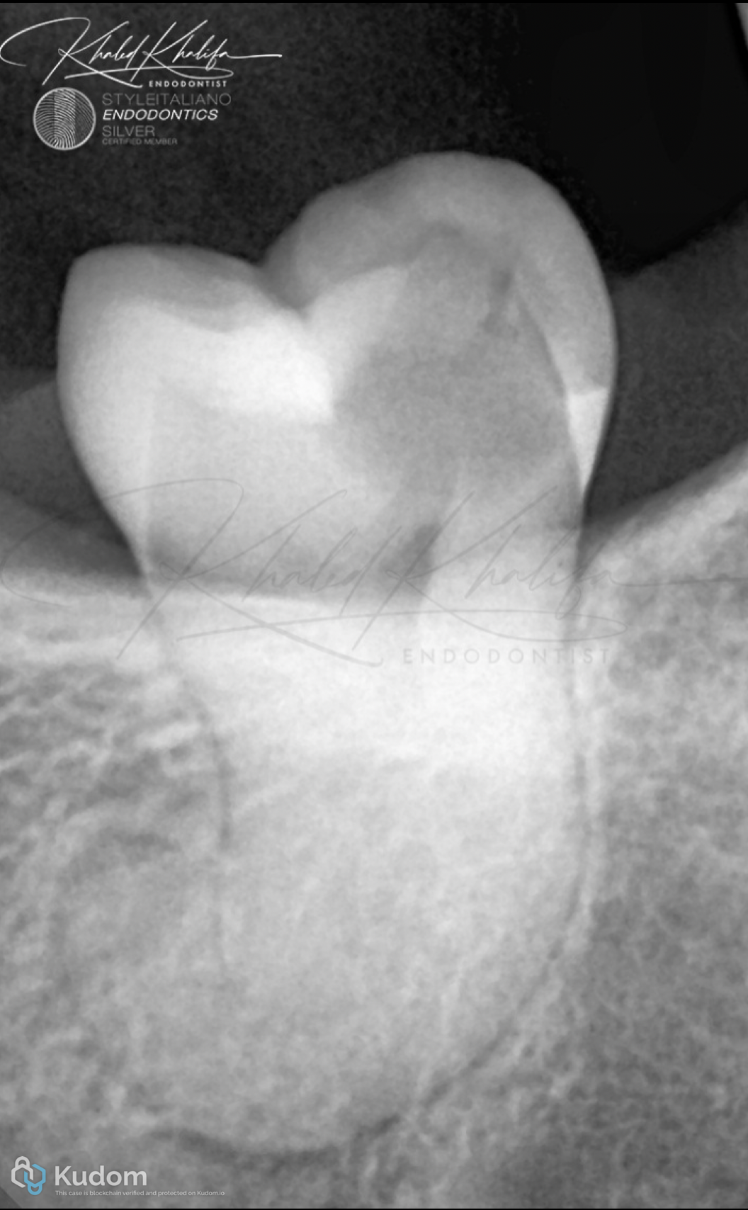 Impossible is possible with the right protocol
Impossible cases might be possible and worth your time.
But you should always approach cases like these with the right protocols to avoid making iatrogenic errors.
Dilacerated roots are one of the hardest anatomies any clinician can ever face.
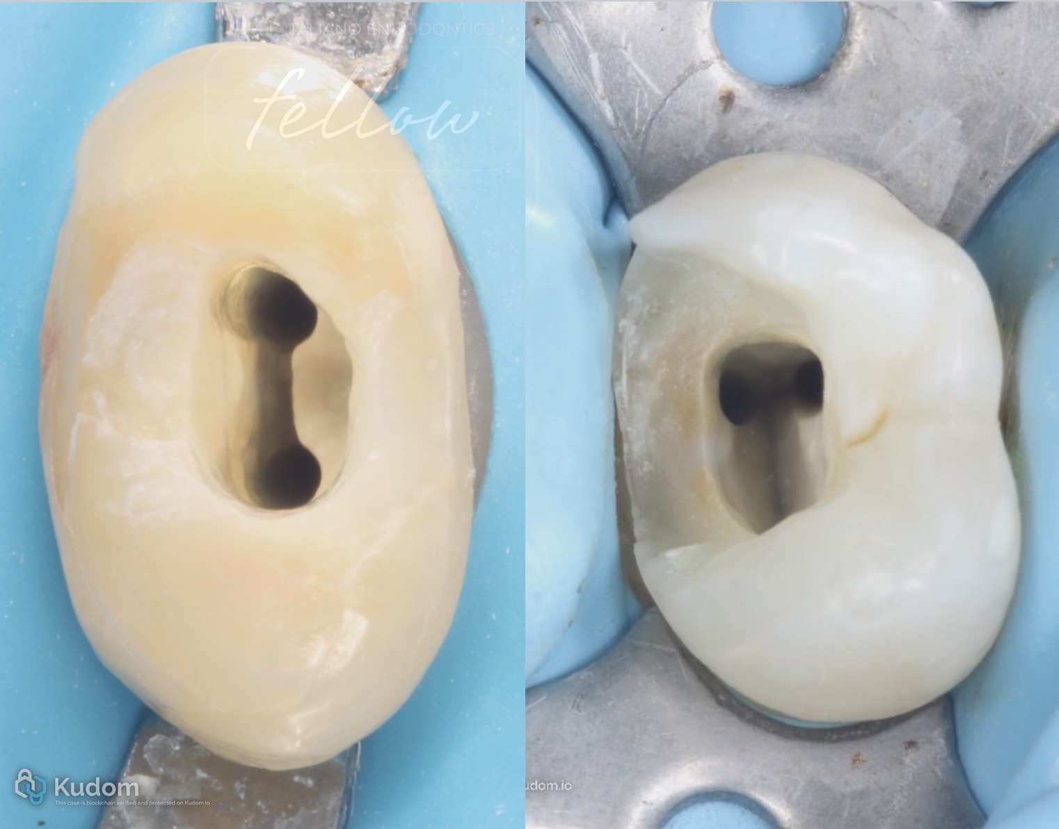 Beyond Canal Number: Anatomical and Clinical Challenges in Maxillary Premolars – A Case Series
Maxillary premolar cases highlight how anatomy and technical factors can turn expected root canal treatment into a clinical challenge.
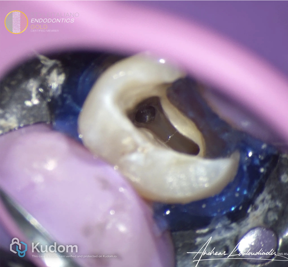 Expect the Unexpected: Anatomy Twist in Maxillary Second Premolar
The maxillary second premolars are generally known to have a single root and one or two canals, with the most common canal configuration being one canal (Type I) or two canals merging into one (Type II or III).
However, a rare and noteworthy anatomical variation exists: the presence of three roots.
While this variation is more frequently seen in maxillary first premolars, three-rooted second premolars are uncommon and present unique clinical challenges. This article explores the prevalence, clinical implications, and endodontic management considerations for three-rooted upper second maxillary premolars.
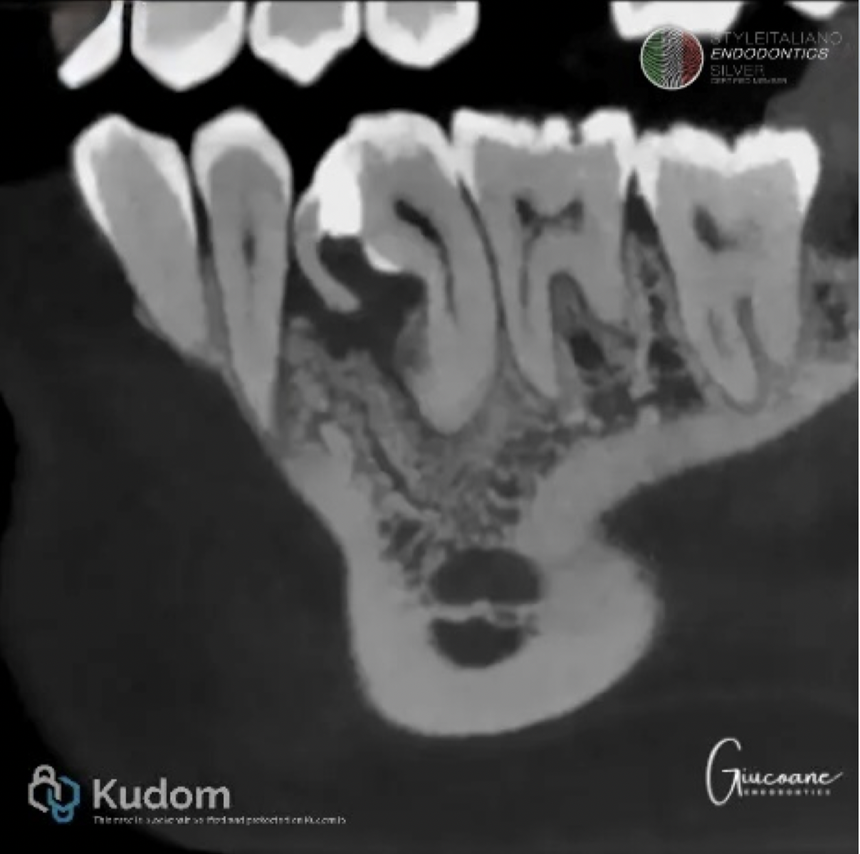 Premolar gemination: a case report
Gemination in lower premolars is a very rare condition. This aspect makes it difficult to treat and it should be approached by an endodontist using a microscope to ensure proper debridement, disinfection and obturation.
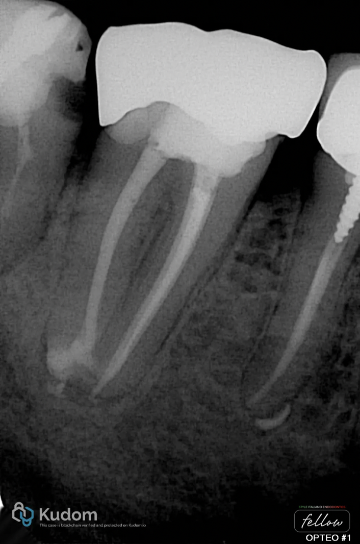 Endodontic Retreatment of a Molar with Complex Anatomy
How a good analysis and a efficient protocol can turn and complex case into success.
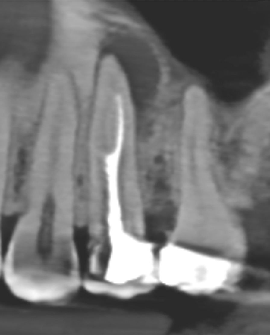 Case report of retreatment of molarized premolar
This article showing the retreatment of molarized premolare upper 5.
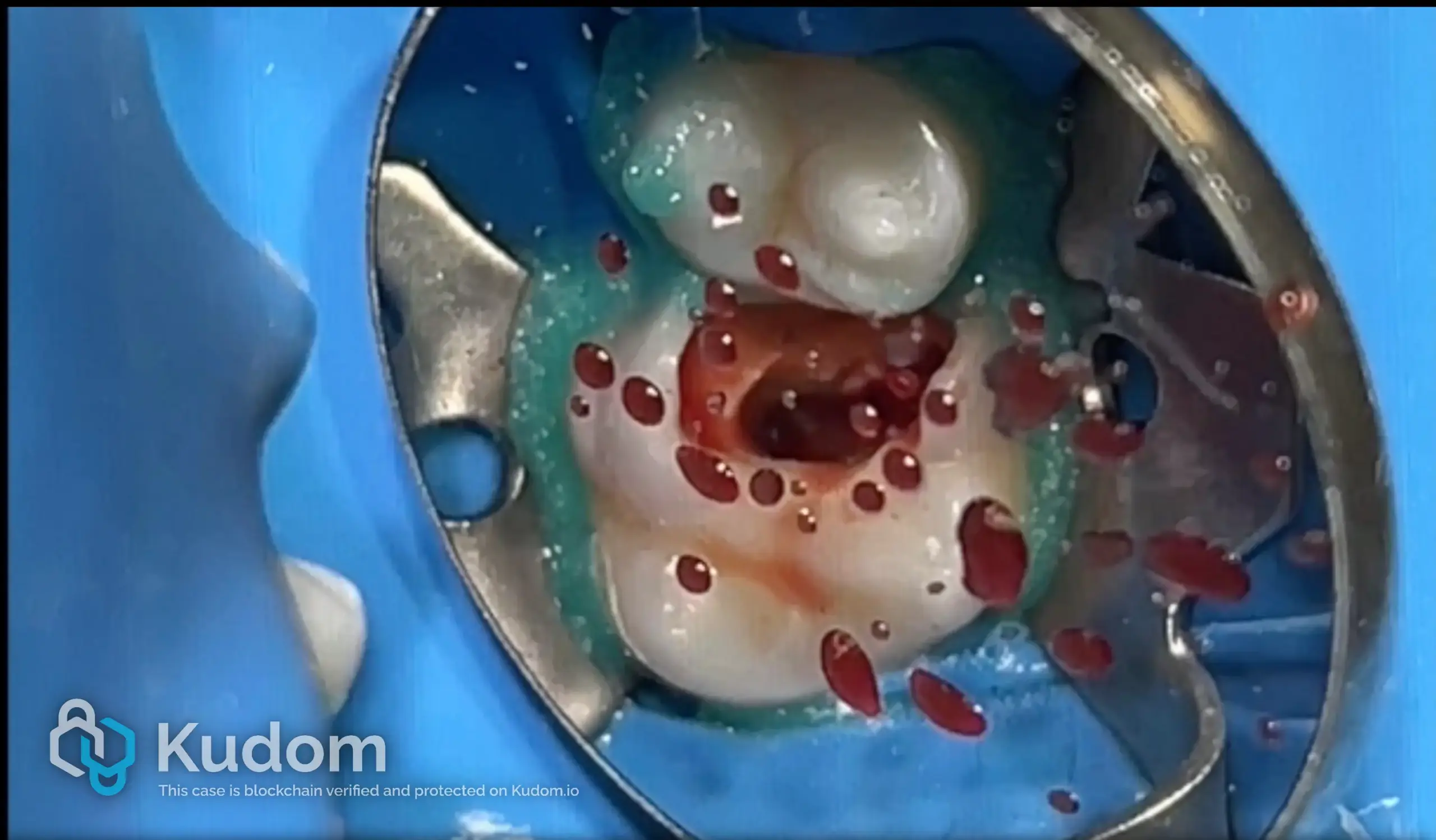 Endodontic-maxillary sinus comunications.
The anatomical relationship between maxillary posterior teeth and the maxillary sinus represents a clinically relevant factor during endodontic treatment. Variations in sinus pneumatization may lead to close proximity or even […]
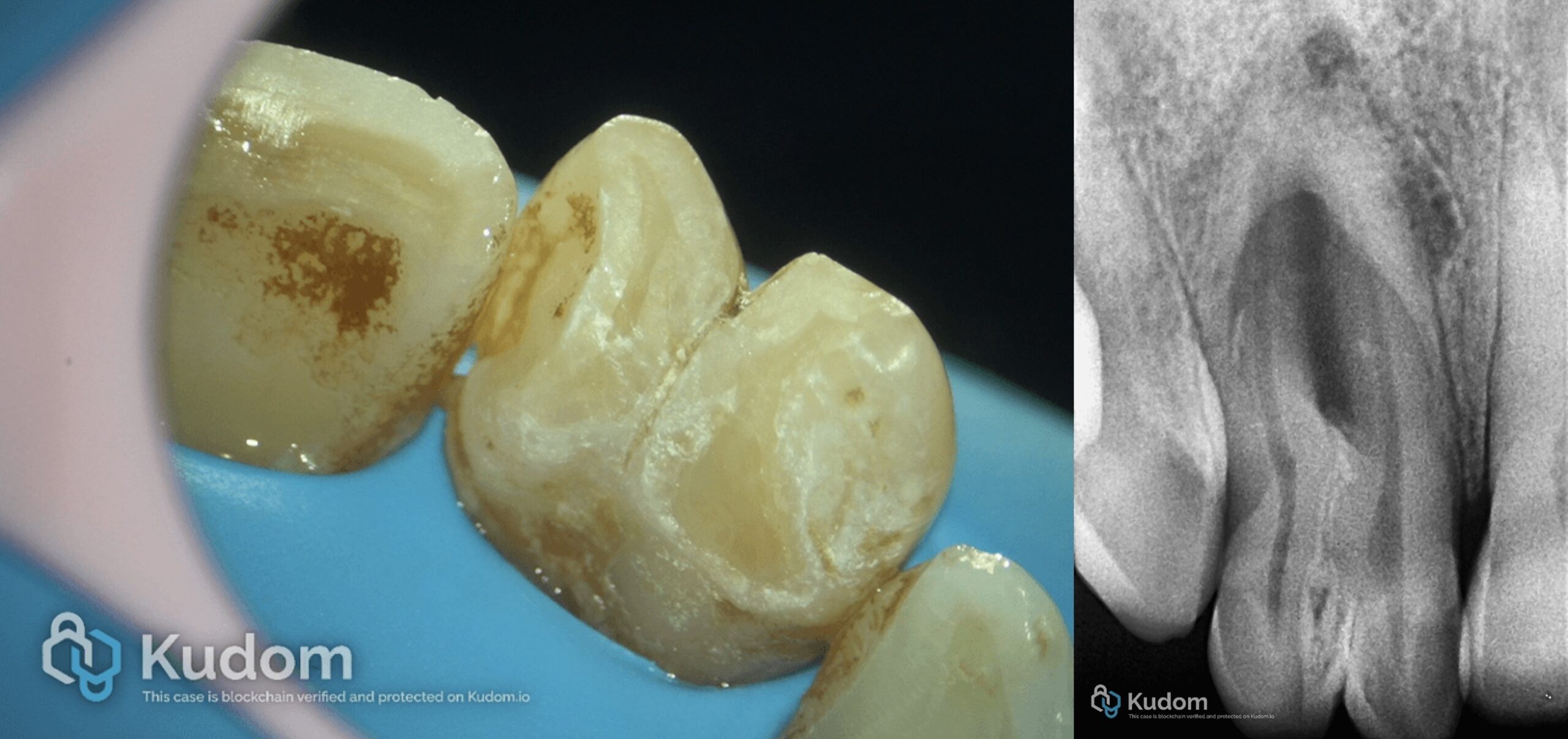 Rare anatomy Fusion+ Dens in dent Type ااا in upper lateral incisor.
The management of a necrotic maxillary lateral incisor presenting with fusion and Type III dens invaginatus associated with a periapical lesion represents one of the most biologically and technically demanding […]
 Clinical and Anatomical Challenges in Mandibular Molars: A Comparative Analysis of Two-Canal versus Six-Canal Systems with Apical Pathosis
Mandibular molars exhibit one of the highest degrees of anatomical variability in the human dentition. While the classical configuration involves two or three canals, extreme variations exist, ranging from simplified […]
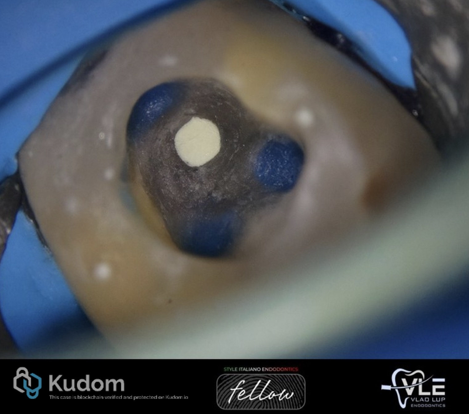 Endodontic Management of a Mandibular Molar with Iatrogenic Perforation, Ledges and Missed Canals
Successful endodontic therapy begins with a properly designed access cavity, which is the foundation for locating, cleaning, and shaping the root canal system. Inadequate or misdirected access can lead to […]