Treatment of an extensive radicular fracture: RCT and restoration of a structurally compromised tooth. Part. 2
25/06/2022
Davide Guglielmi
Warning: Undefined variable $post in /var/www/vhosts/styleitaliano-endodontics.org/endodontics.styleitaliano.org/wp-content/plugins/oxygen/component-framework/components/classes/code-block.class.php(133) : eval()'d code on line 2
Warning: Attempt to read property "ID" on null in /var/www/vhosts/styleitaliano-endodontics.org/endodontics.styleitaliano.org/wp-content/plugins/oxygen/component-framework/components/classes/code-block.class.php(133) : eval()'d code on line 2
Trauma related fractures are one of the most common types of dental injury in the permanent dentition.
The purpose of this case report is to present a multidisciplinary management of a sub-gingival crown- root fracture in a patient. Management of traumatic dental injuries (TDI) should involve a multidisciplinary approach with the aim of optimizing the healing while maintaining function and aesthetics.
The challenge is to identify the most suitable treatment for a given patient combining evidence-based guidelines withclinical experience.
The prognosis of ETT depends not only on the quality of endodontic treatment, but also on the subsequent restorative techniques. Reported reasons for extraction of teeth after endodontic treatment include endodontic failures, prosthodontic complications, coronal and root fracture, decay, or periodontal disease.
Due to all of these reasons, special care should be taken while restoring endodontically treated molar and premolar teeth. The conventional approach to restore fractured and retreated posterior teeth would be cast post and crown, the evolution of bonding material along with similar dentine mechanical properties of glass fiber post allows nowadays clinicians to core the build up with a fiber post instead of using an aggressive stiff metal cast post. The purpose of this clinical article is to highlight the possible management of core build up with ITENA CLINICAL's Post-and-Core Restoration Kit.
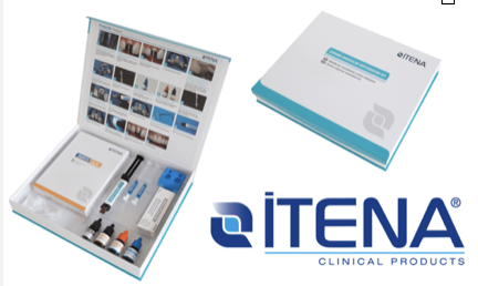
Fig. 1
Post-and-core restoration kit ITENA CLINICAL (6)
The case reported is the continuation of a previous paper: https://endodontics.styleitaliano.org/treatment-of-an-extensive-radicular-fracture-the-pre-endodontic-restoration-of-a-structurally-compromised-tooth-part-1/
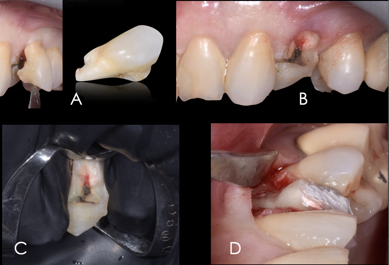
Fig. 2
Schematic illustration of the previous article. (A to D)
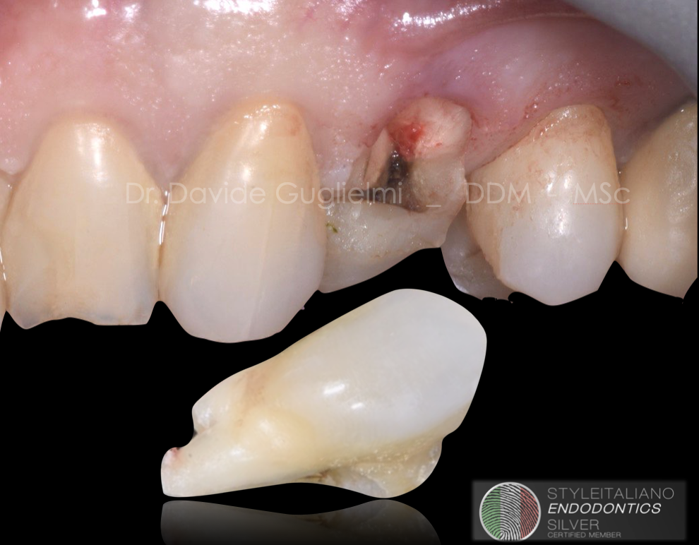
Case of a 66 years old patient referring pain in the left upper jaw when chewing.
Both sensibility testing and bleeding on probing were negative, pocket probing depth of the element 2.4 was ≤ 5 mm circumferentially.
Higher mobility and pain during the percussion test were recorded.
The tooth was isolated without suturing the underlying flap and than a pre-endodontic restoration was applied.
The case reported is the continuation of a previous paper:
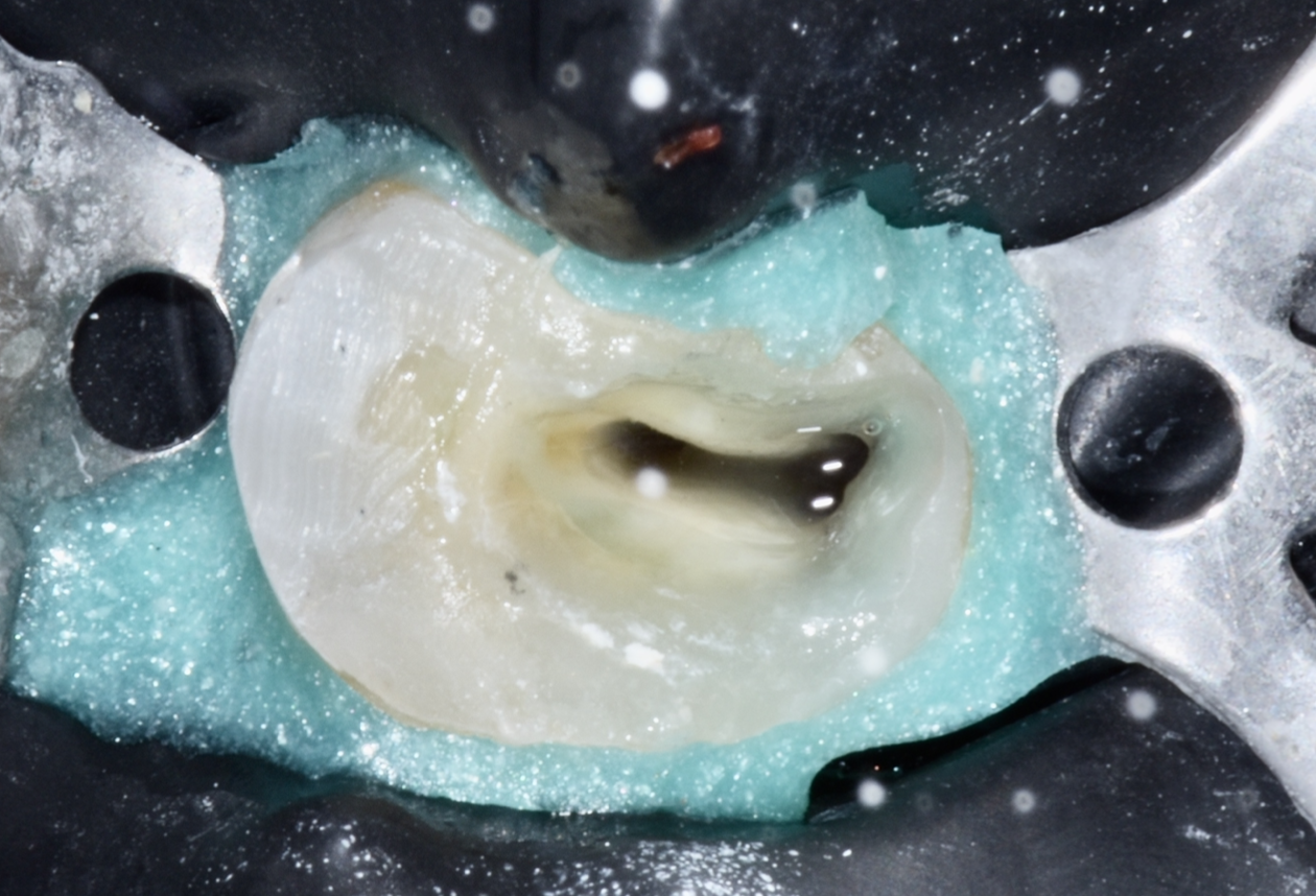
Fig. 3
Two weeks after the surgical therapy, a full isolated field was obtained by placing both the rubber dam in the classic manner plus a liquid dam in the most cervical portion of the tooth.
In this case, the pre-endodontic restoration was successful for various reasons, such as improving the isolation quality for the subsequent endodontics treatment and creating space for prolonged function of irrigation solutions (5).
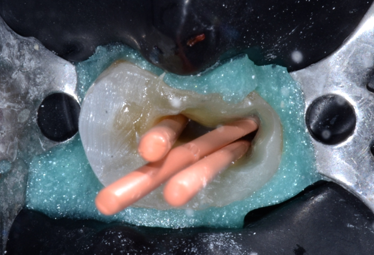
Fig. 4
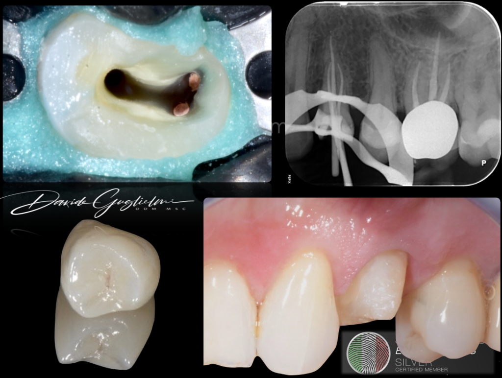
Cone fitting before the 3D canal obturation with warm vertical condensation (WVC) technique.
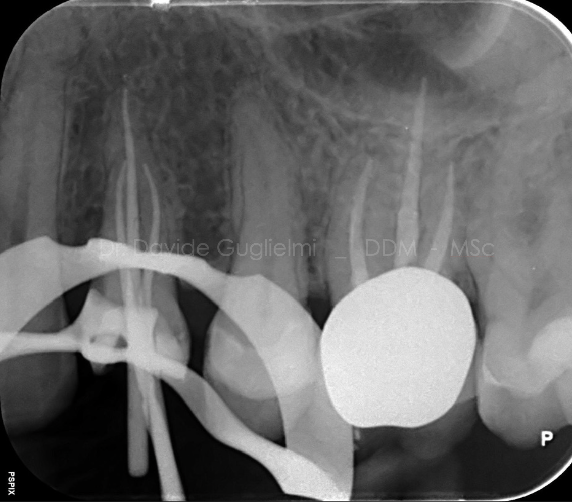
Fig. 5
Master cones X-ray taken during root canal treatment (RCT)
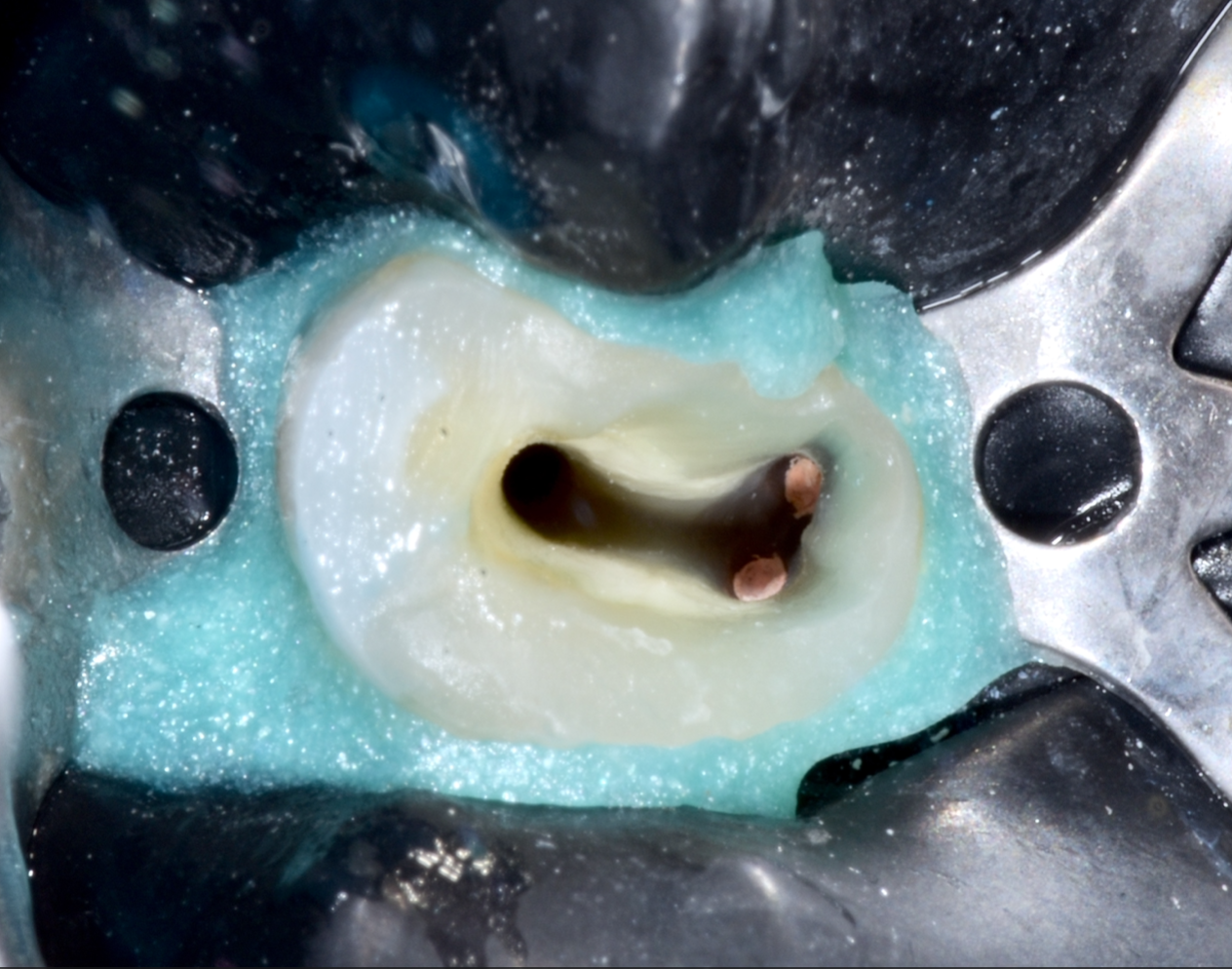
Fig. 6
Access cavity view after 3D obturation with focus on buccal canals.
The palatal canal has been prepared for the fiber post reconstruction.
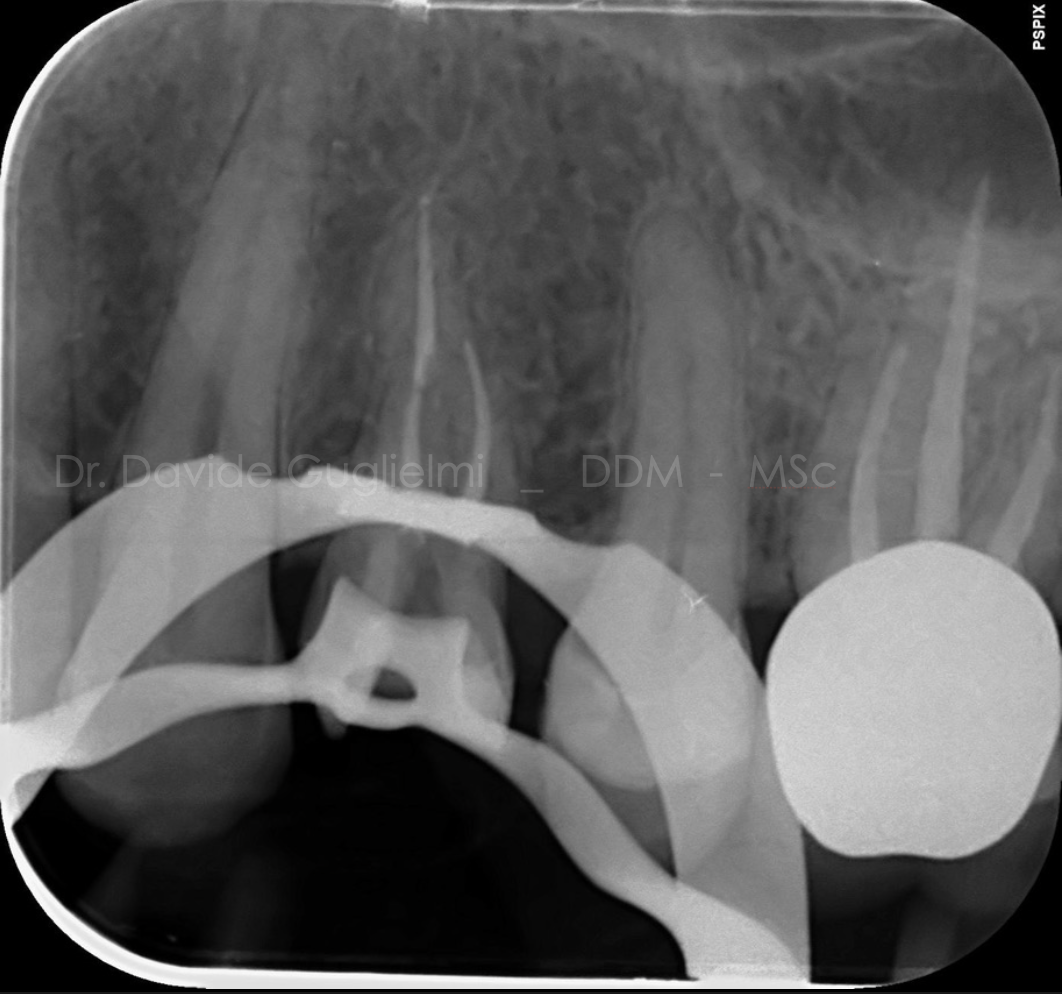
Fig. 7
Post Operative X-ray taken on the day of the RCT after warm vertical obturation.
Video of the endodontic treatment
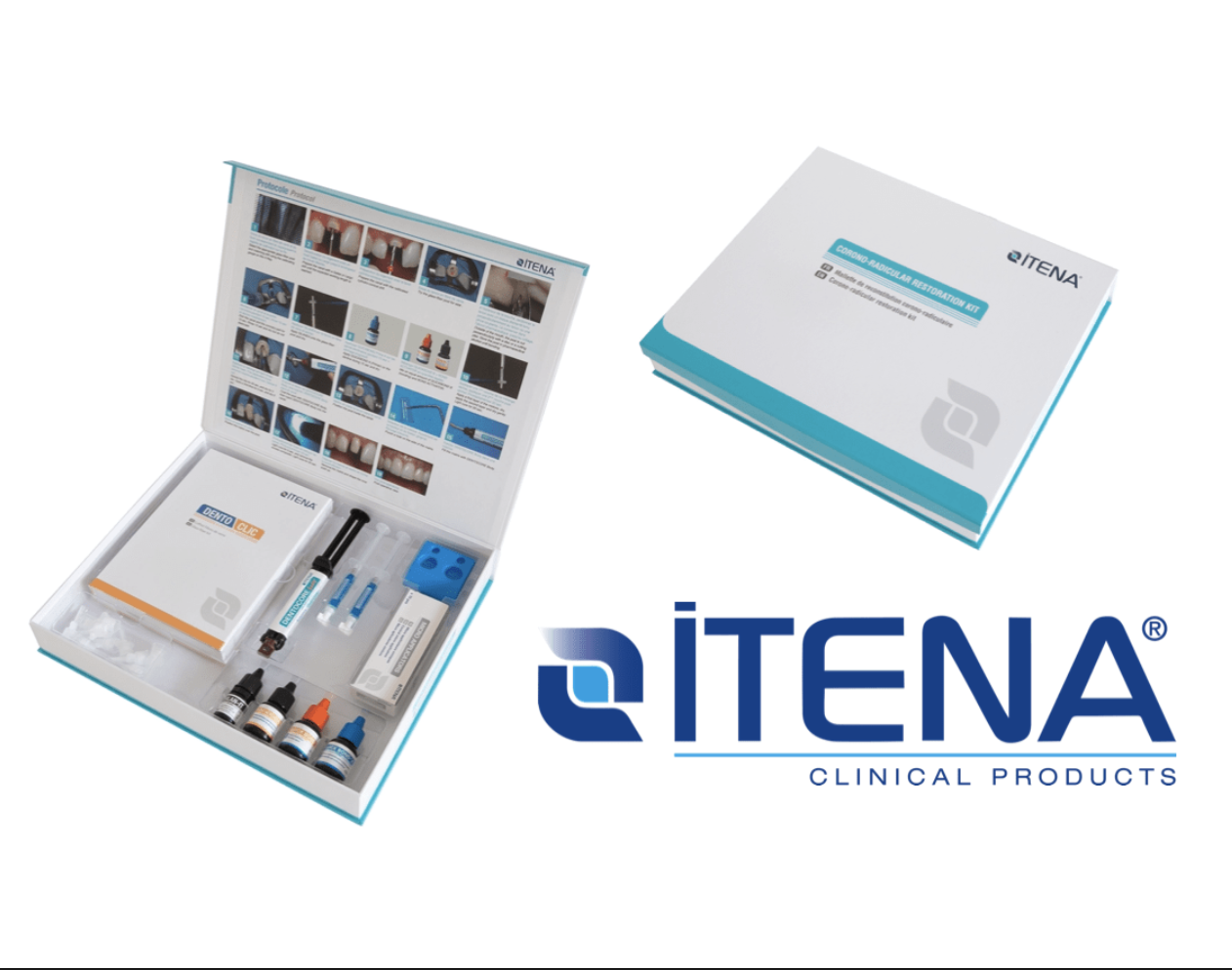
Fig. 8
Post-and-core restoration kit ITENA CLINICAL (6).
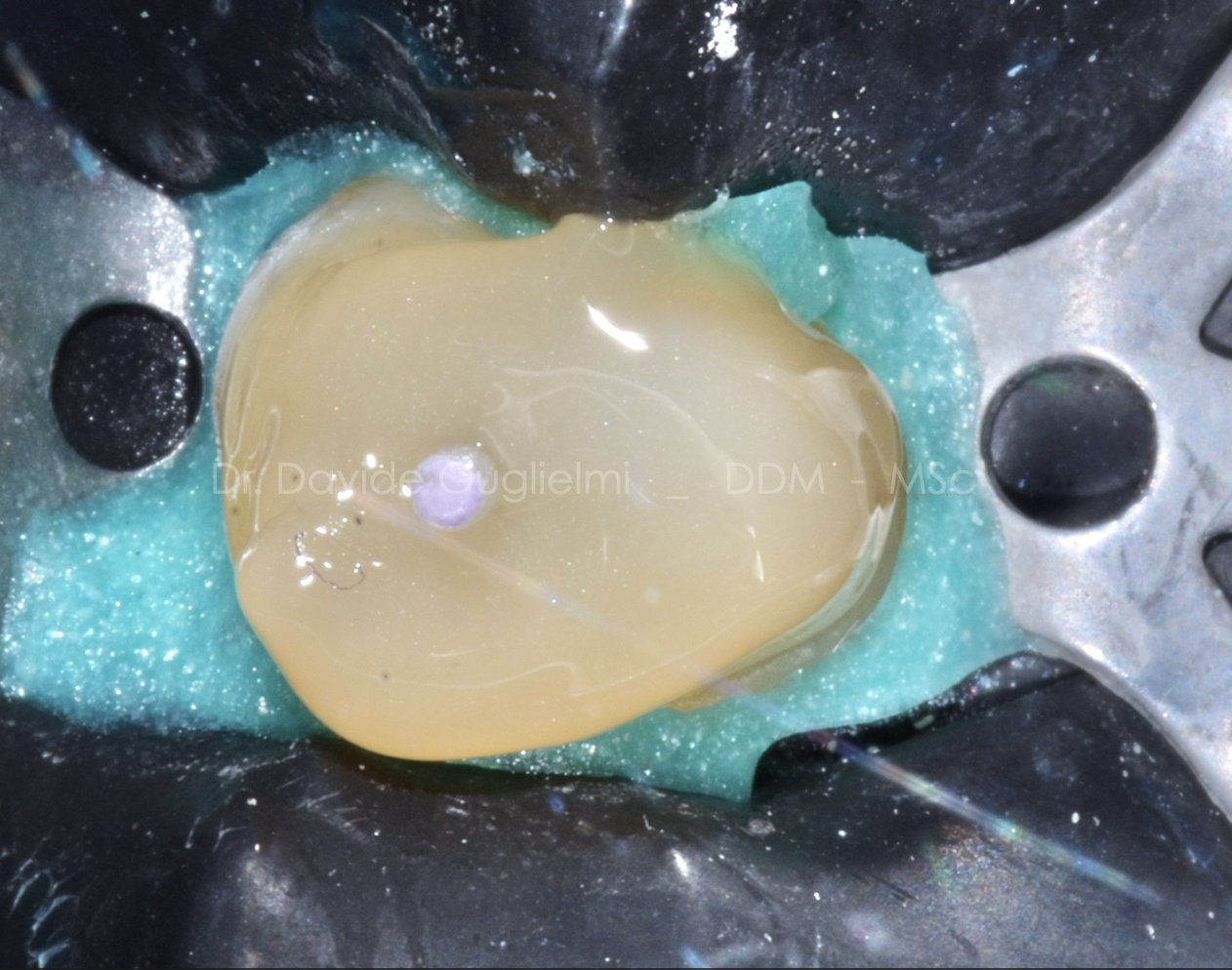
Fig. 9
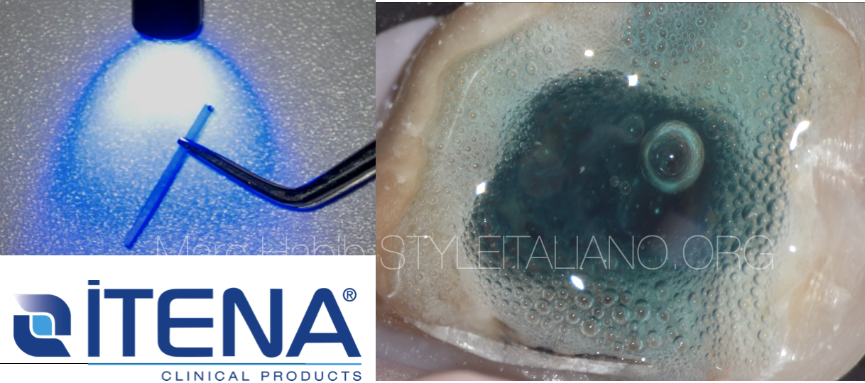
Nano - filled composite dual cure material was placed (Dentocore - ITENA CLINICAL) .
A recent study has demonstrated that the hardness of this material is close to the dentine one.
Furthermore, it has a low shrinkage rate, despite being a very fluid material useful to avoid bubbles in the post cementation.
The Dentocore (ITENA CLINICAL) is a dual curing Material.
Nevertheless it is suggested to cure this resin composite material for at least 40 seconds.
Due to properties of the posts composition, excellent distribution of the light in the whole material is guaranteed.
Furthermore it allows polymerization of the core build-up material on all of the post surfaces.
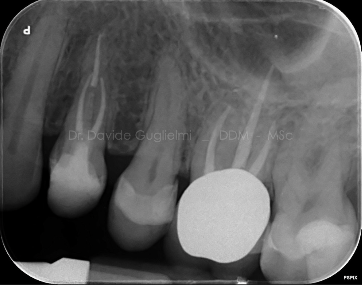
Fig. 10
Post core build up X-Ray with Dentoclic Purple Fiber Post and DentoCore Body (ITENA CLINICAL).
Continuity between root canal obturation and core build up without gaps or voids should always be reached for optimum results.
Due to fracture history and carious infiltration indirect full coverage crown was planned for tooth 2.5
Post endodontic restoration with fiber post luting
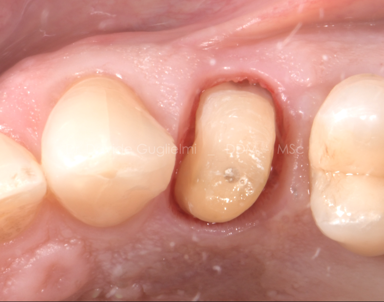
Fig. 11
After 8 weeks, the gingival tissue was stabilized and it was possible to take the impression to finalize the restoration. The dental preparation was applied without finishing lines, described as feather edge (1).
The absence of any finish line will make the procedure faster and simpler. The use of two retraction cords is strongly suggested in order to have a good reading of the sulcus and to help the technician during the laboratory procedures (2).
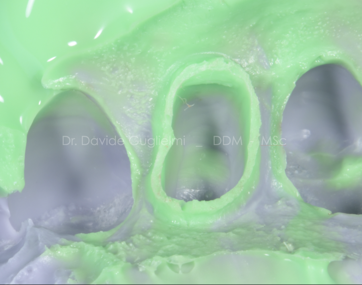
Fig. 12
Light and Heavy body polyvinylsiloxane impression for FDP fabrication.
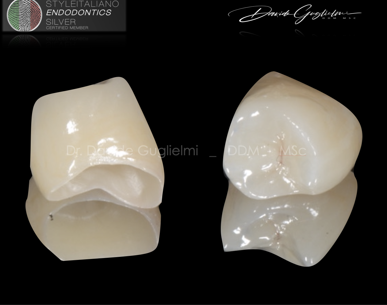
Fig. 13
For vertical preparations, the margin is positioned by the laboratory technician on the base of the gingival tissue informations.
The material chosen for the restoration of this element was a monolithic zirconia (7).
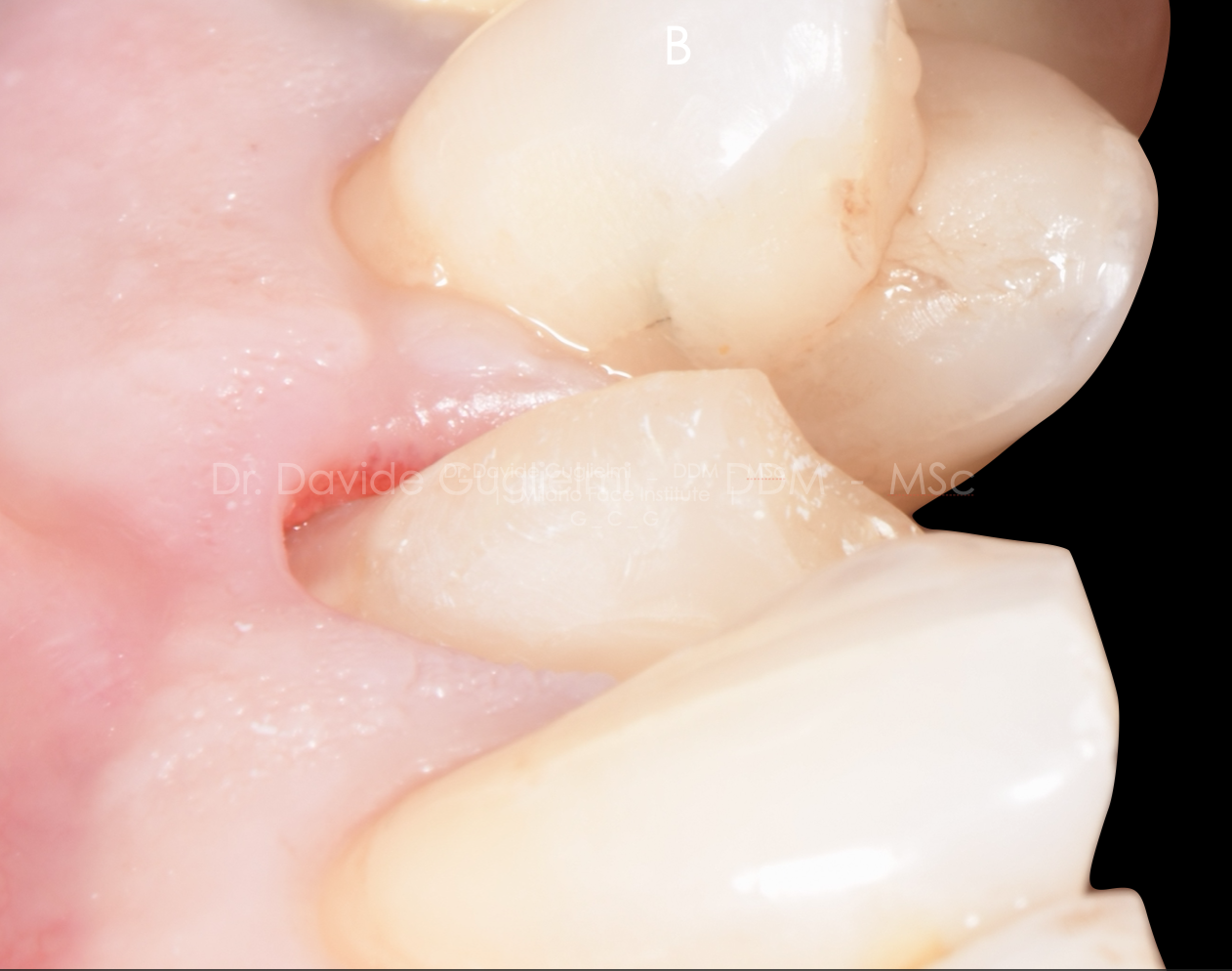
Fig. 14
Clinical image before cementation of the fixed dental prosthesis (FDP) focused on the periodontal tissues healing (2).
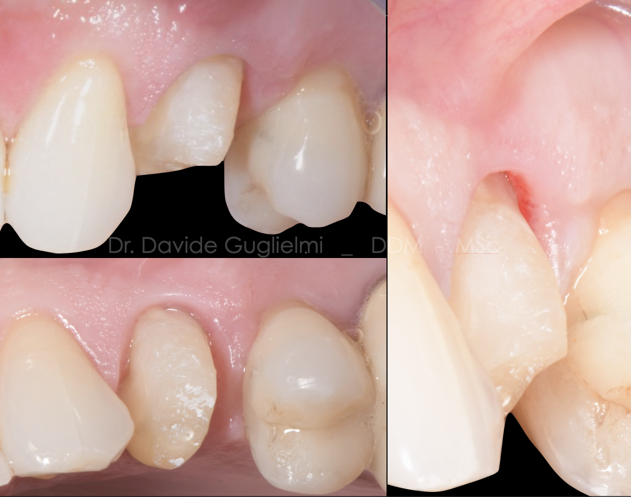
Fig. 15
Side and occlusal views of the clinical situation before cementation of the fixed dental prosthesis (FDP) focused on the periodontal tissues healing.
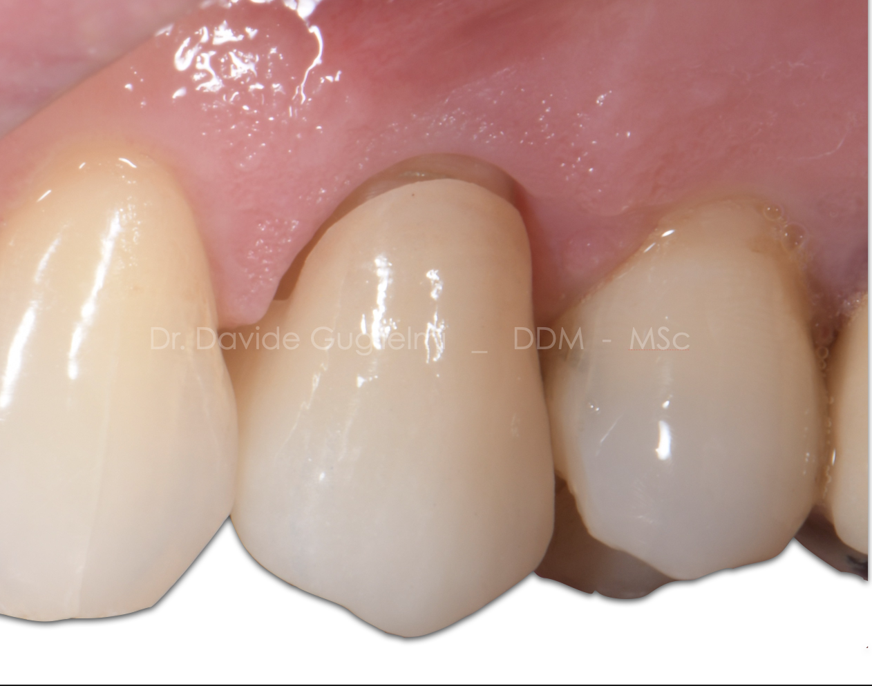
Fig. 16
Clinical image of the fitting of FDP
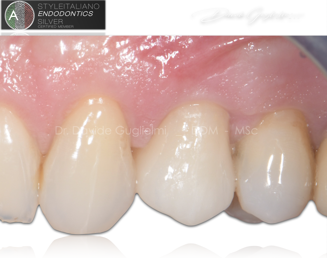
Fig. 17
Before cementation the dentine and build up were prepared by using a micro-etcher sandblaster (aluminium and silicon oxide powder_particle size 20-80 micron) with the aim of increasing the cleanliness and improving the adhesion of the bonding agents.
The image was taken one week after cementation.
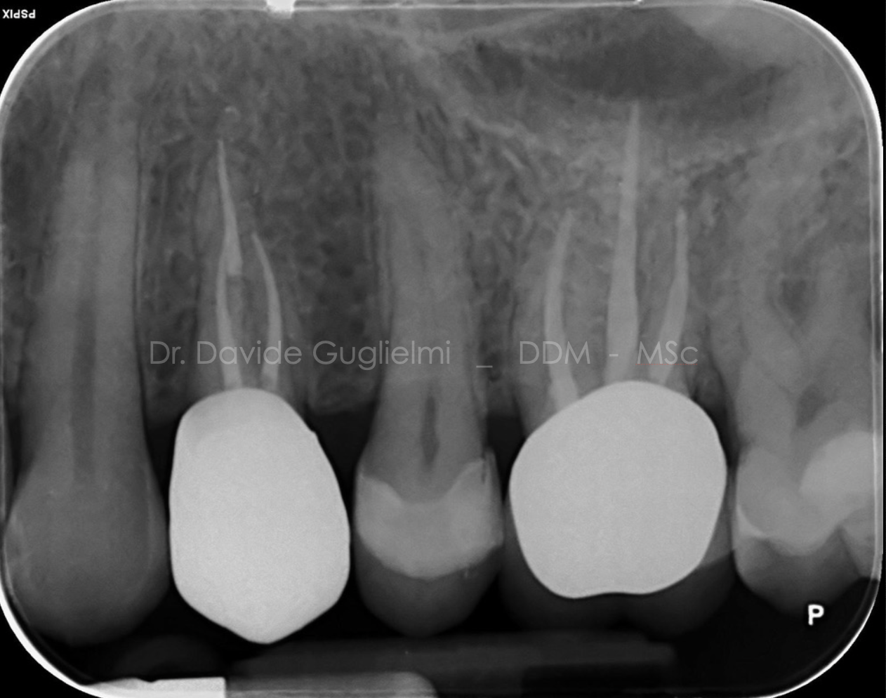
Fig. 18
Post cementation x-ray.
Conclusions
Based on author knowledge and experience, the post-and-core restoration kit ITENA CLINICAL contains all the products and accessories needed for a reconstruction with fiberglass posts.
The DENTOLIC products seems to be safe and effective for core build-up reconstruction and post cementation.
Nevertheless, due to the high fluidity of the DENTOCORE Body, it is not practical to create a reconstruction in a single stroke of material when the tooth to be reconstructed has no retaining walls. In fact, in these cases the material must be placed in small increments.
Bibliography
- Loi, I., & Di Felice, A. (2013). Biologically oriented preparation technique (BOPT): a new approach for prosthetic restoration of periodontically healthy teeth. The European Journal of Esthetic Dentistry, 8(1), 1–14.
- Ercoli, C., & Caton, J. G. (2018). Dental prostheses and tooth-related factors. Journal of Clinical Periodontology, 45(5S), S207–S218. http://doi.org/10.1111/jcpe.12950
- Guglielmi, D., & F. Gorni. (2019). Immediate Crown Replacement: a case report of extensive radicular fracture with intra-canal anchorage. Giornale Italiano Di Endodonzia, (33), 57–64. Gavriil, D., Kakka, A., Myers, P., & O Connor, C. J. (2021).
- De Sanctis M, Clementini M. Flap approaches in plastic periodontal and implant surgery: crit- ical elements in design and execution. (2014). Journal of Clinical Periodontology. 2014. 41(s15), S108–S122.
- Bhuva B, Giovarruscio M, Rahim N, Bitter K, Mannocci F. The restoration of root filled teeth: a review of theclinical literature. Int Endod J. 2021 Apr;54(4):509-535. doi: 10.1111/iej.13438. Epub 2021 Jan 5. PMID:33128279.
- Figueiredo FE, Martins-Filho PR, Faria-E-Silva AL. Do metal post-retained restorations result in more root fractures than fiber post-retained restorations? A systematic review and meta-analysis. J Endod. 2015 Mar;41(3):309-16. doi: 10.1016/j.joen.2014.10.006. Epub 2014 Nov 11. PMID: 25459568.
- Al-Bermani ASA, Quigley NP, Ha WN. Do zirconia single-retainer resin-bonded fixed dental prostheses present a viable treatment option for the replacement of missing anterior teeth? A systematic review and meta-analysis. J Prosthet Dent. 2021 Dec 7:S0022-3913(21)00588-6.