Endo-resto treatment. Management of confluent canals - Part II
15/04/2021
Fitim Shabani
Warning: Undefined variable $post in /var/www/vhosts/styleitaliano-endodontics.org/endodontics.styleitaliano.org/wp-content/plugins/oxygen/component-framework/components/classes/code-block.class.php(133) : eval()'d code on line 2
Warning: Attempt to read property "ID" on null in /var/www/vhosts/styleitaliano-endodontics.org/endodontics.styleitaliano.org/wp-content/plugins/oxygen/component-framework/components/classes/code-block.class.php(133) : eval()'d code on line 2
Case evaluation before treatment
In endodontic treatments of teeth with more than one canal, we must allways think about the presence of possible confluences between the canals, there are several methods for their determination.
In this clinical case I will present only one among others, which is performed by inserting a gutta percha and a manual instrument at the same time into the canals where we suspect there are confluences.
With this method we are safer and avoid possible mistakes during the obturation phase.
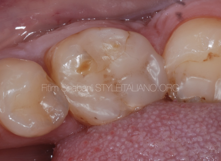
Fig. 1
Clinical data
Tooth 36
Dg: Pulpitis acute irreversible
Treatment plan :
Root canal treatment
Crown
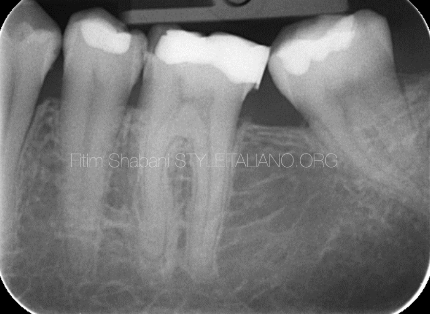
Fig. 2
Pre operative X-ray
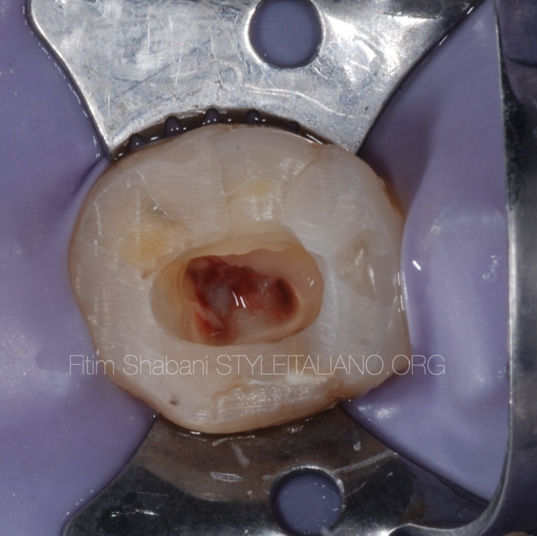
Fig. 3
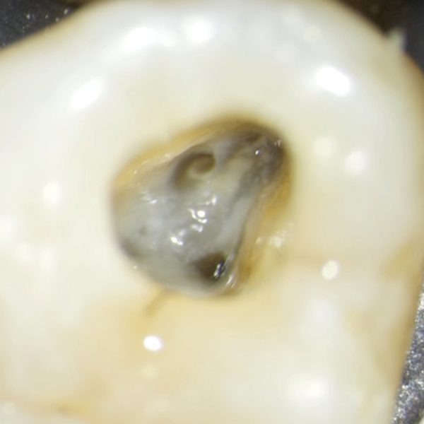
Isolation, initial preparation of endodontic access cavity
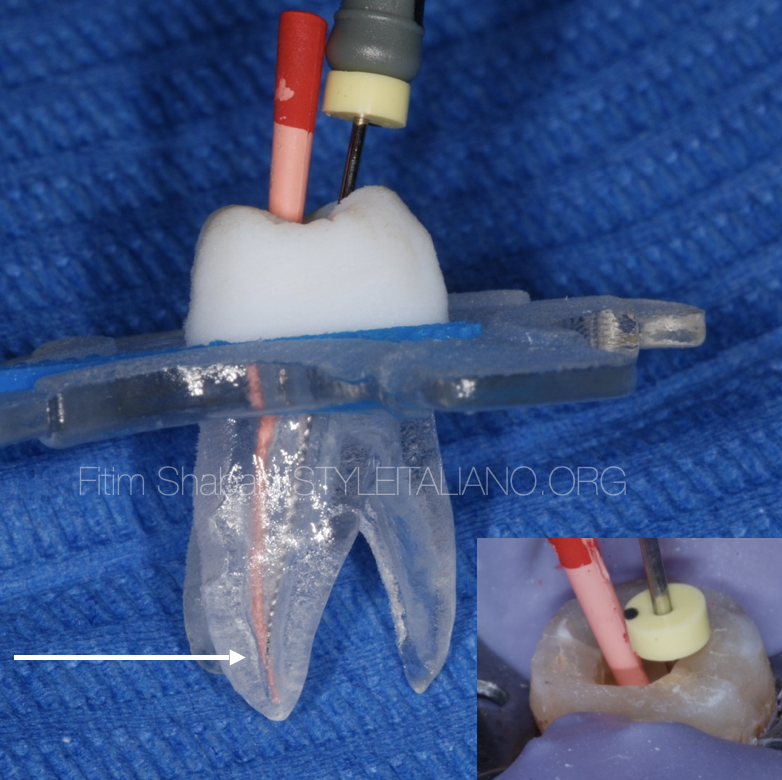
Fig. 4
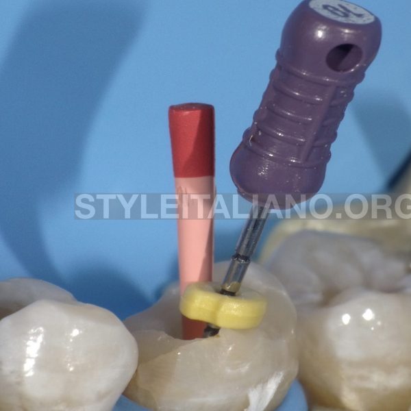
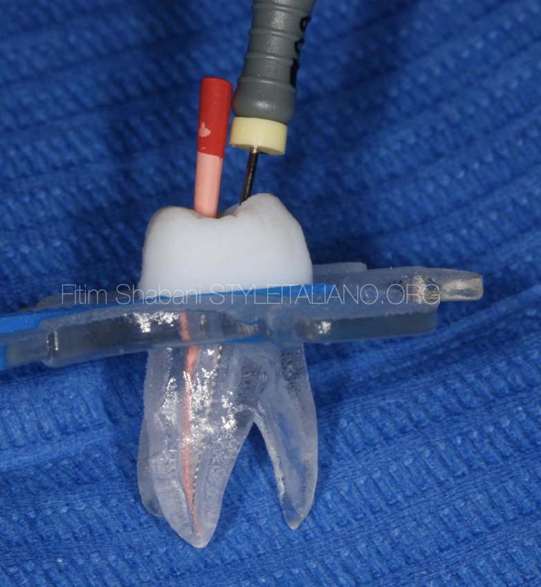
Detection of confluence in artificial tooth to illustrate the procedure
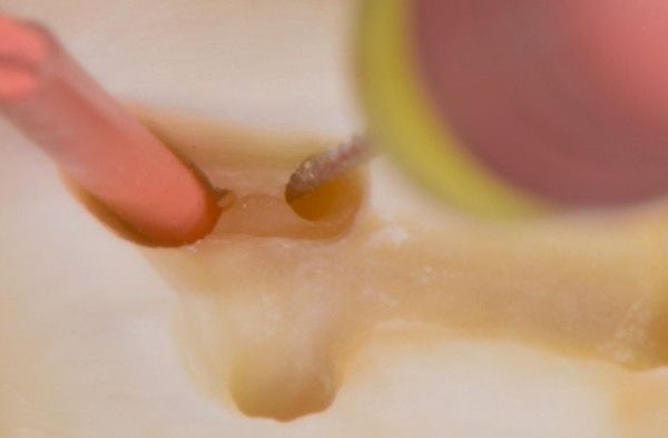
First we need to put the gutta-percha in the main canal, then with manual instrument ISO 08 or 10 we go into the other canal, in order to detect the level of confluences.
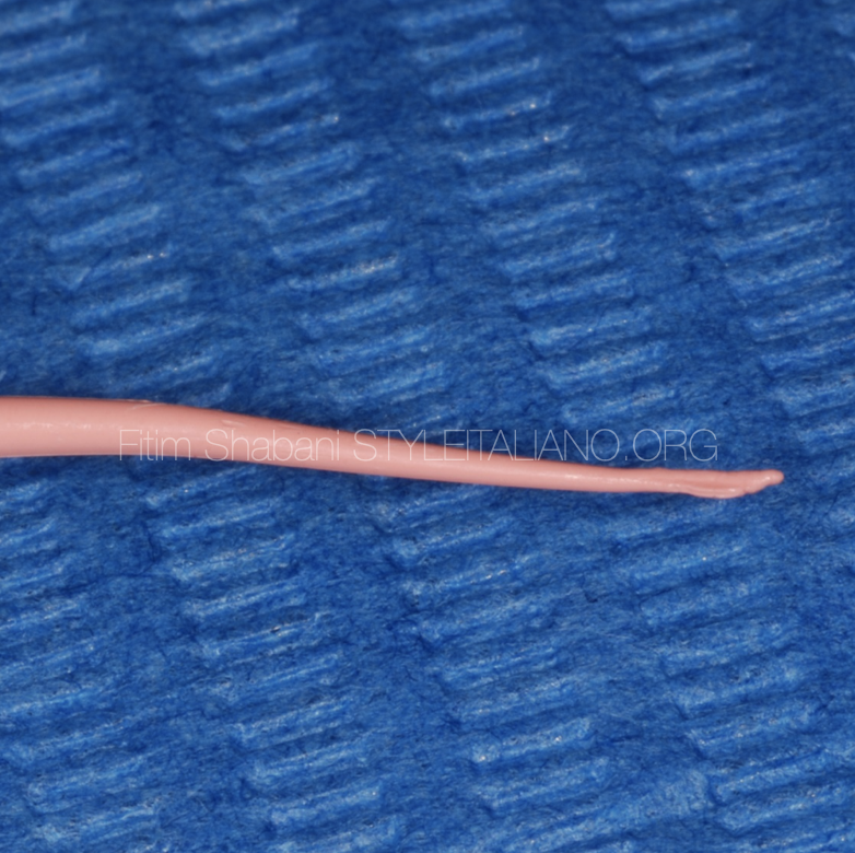
Fig. 5
In the gutta-percha will remain some scratches from the instrument, and this confirms the presence of confluences and determines their level
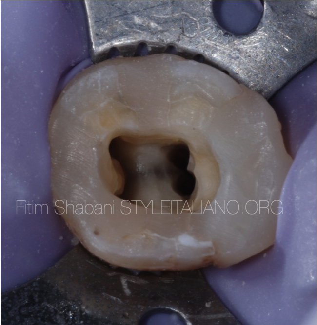
Fig. 6
The tooth is ready for obturation after all endodontic procedures
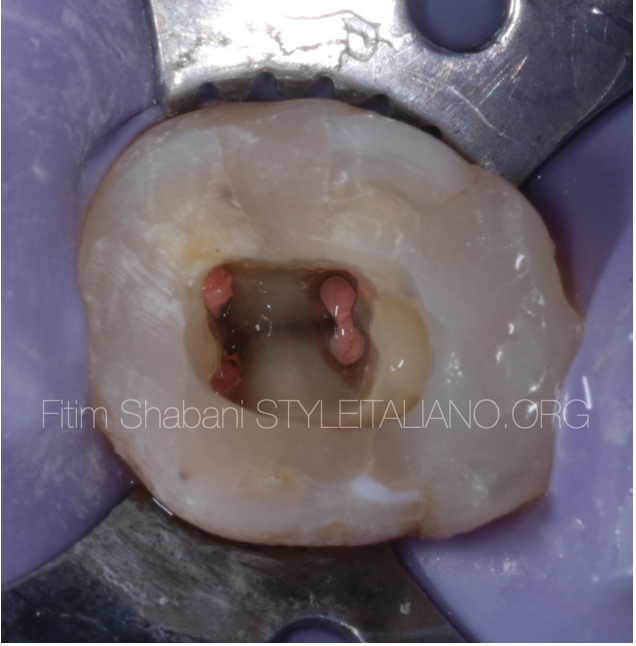
Fig. 7
Obturation
Warm vertical technique
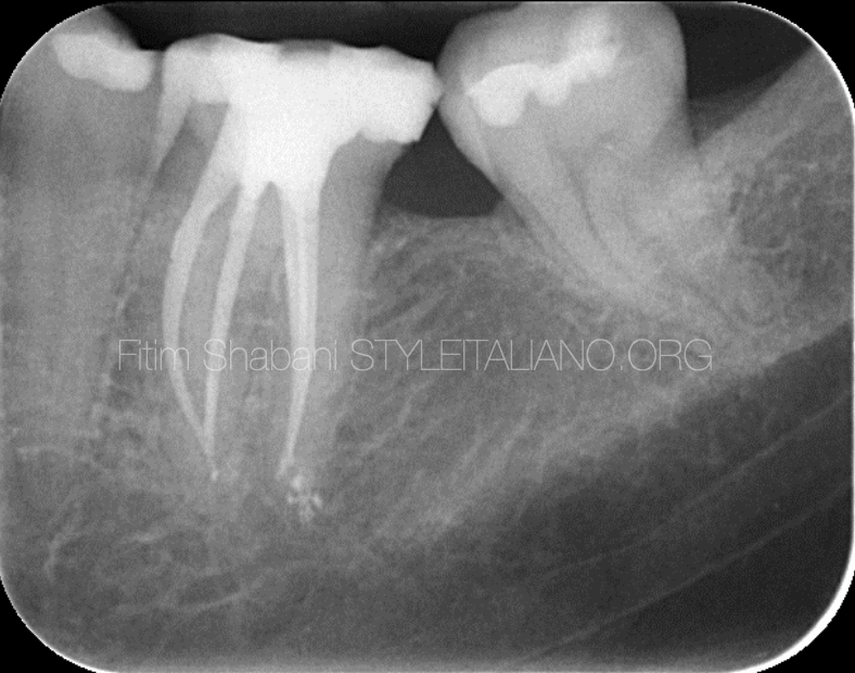
Fig. 8
X-ray after obturation
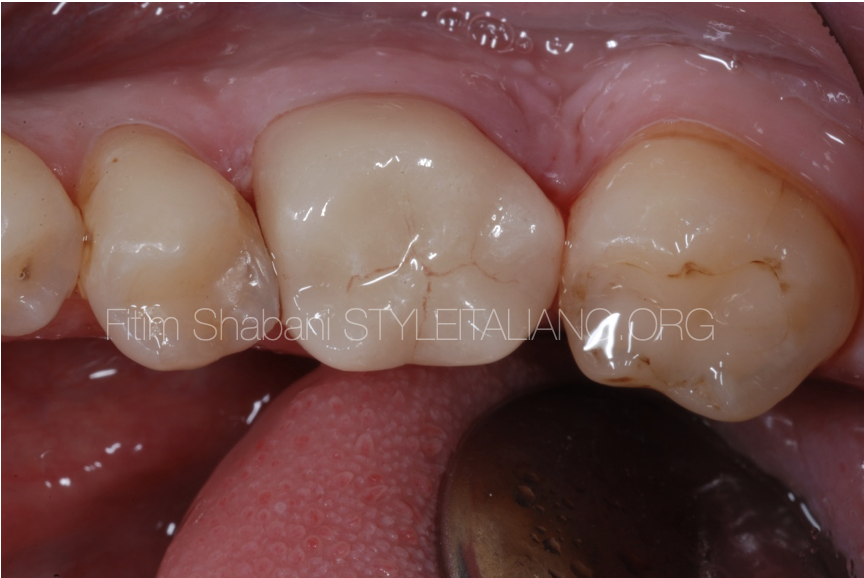
Fig. 9
Crown cementation
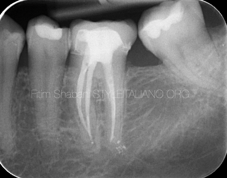
Fig. 10
X-ray after Crown cementation
Conclusions
Pre-treatment case evaluation greatly facilitates our work and avoids any errors that may occur during treatment.
Applying methods for detecting the confluences in this cases are inevitable.
If we have a pre-treatment plane, it will reduce the possibility of mistakes and surprises that may occur during the treatment.
Bibliography
Vertucci Fj. Root canal morphology and its relationship to endodontic procedures, Endodontic topics,2005
Arnaldo Castellucci , Two canals in a single root: clinical and practical considerations, 2003
Furri,M , Differences in the confluence of mesial canals in mandibular molar with three or four root canals, International endodontic journal, 2008
Gianluca Gambarini et AL. Cone-beam computed tomographic analysis on root and canal morphology of mandibular first permanent molar among multiracial population in Western European population, European Journal of Dentistry, 2018