Endo-resto treatment Management of confluent canals
30/05/2020
Fitim Shabani
Warning: Undefined variable $post in /home/styleendo/htdocs/styleitaliano-endodontics.org/wp-content/plugins/oxygen/component-framework/components/classes/code-block.class.php(133) : eval()'d code on line 2
Warning: Attempt to read property "ID" on null in /home/styleendo/htdocs/styleitaliano-endodontics.org/wp-content/plugins/oxygen/component-framework/components/classes/code-block.class.php(133) : eval()'d code on line 2
A 27 years old patient came to our clinic with pain on the right upper side. After examination a root canal treatment was needed. On the pre-operative X-ray we understood that we had to deal with a non routine, very interesting and challenging case.
But just a simple root canal treatment would not solve the problem entirely without resolving the restorative part. Therefore we set the pre-treatment plan based on clinical and radiograph data.
The endodontic treatment was done in one visit.
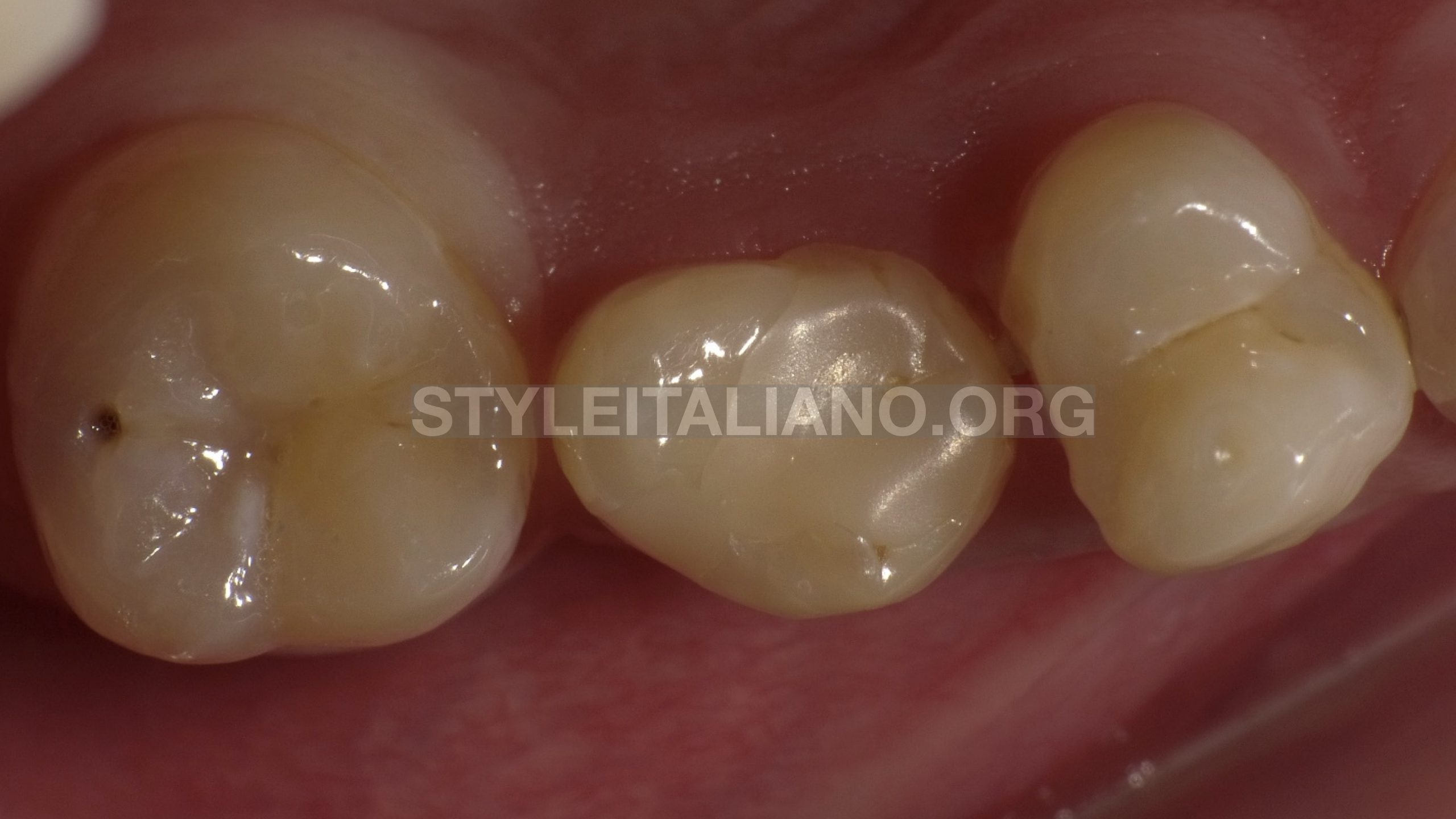
Fig. 1
Clinical data
Tooth 15, upper right second premolar
Dg: Pulpitis acute irreversible
Rotation of tooth due to early loss of tooth 16.
Treatment plan :
Root canal treatment
Crown
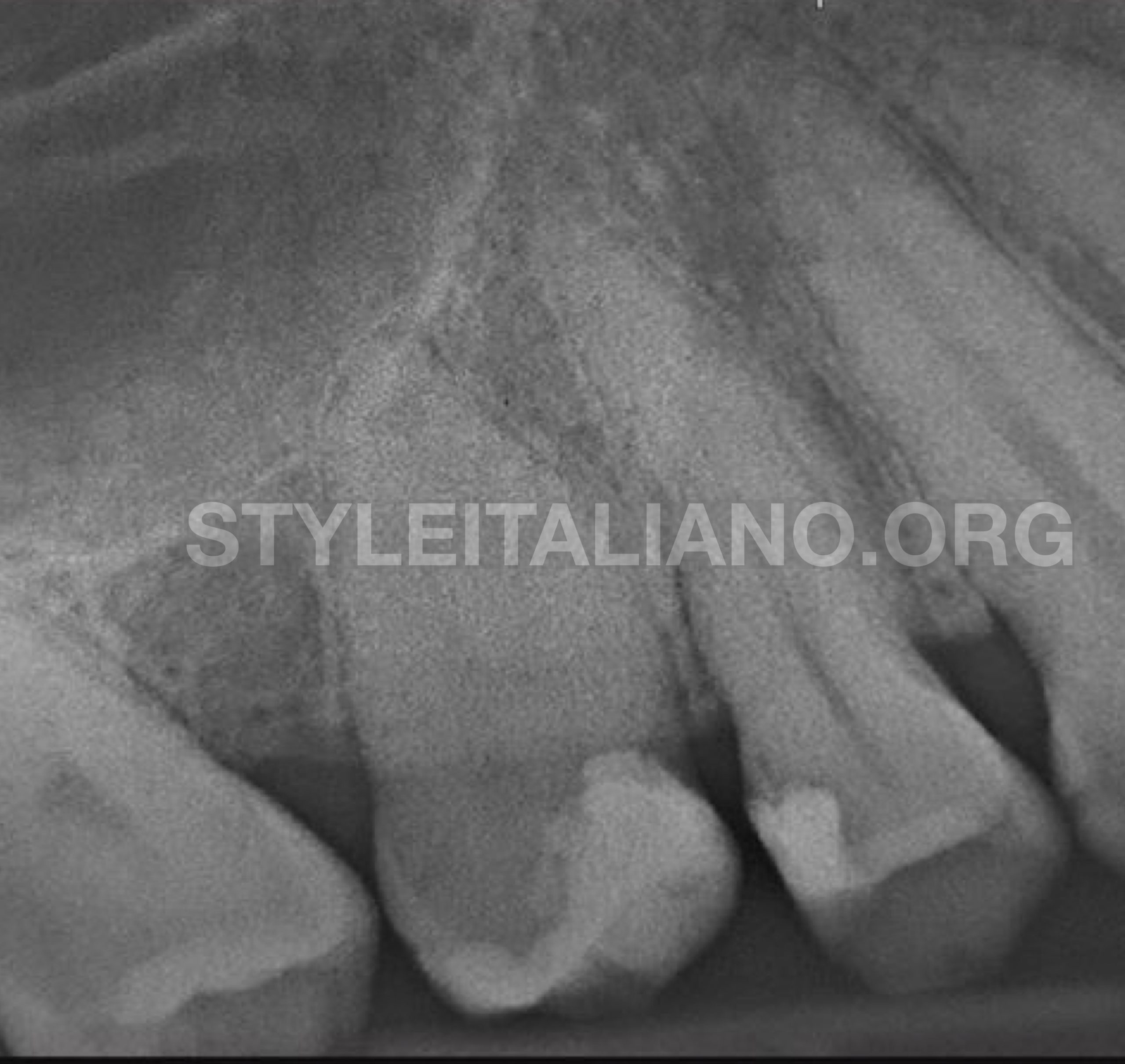
Fig. 2
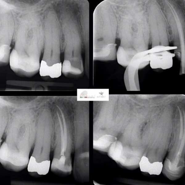
Pre operative X-ray :No pathological changes in the apical area Secondary caries under the deep filling.
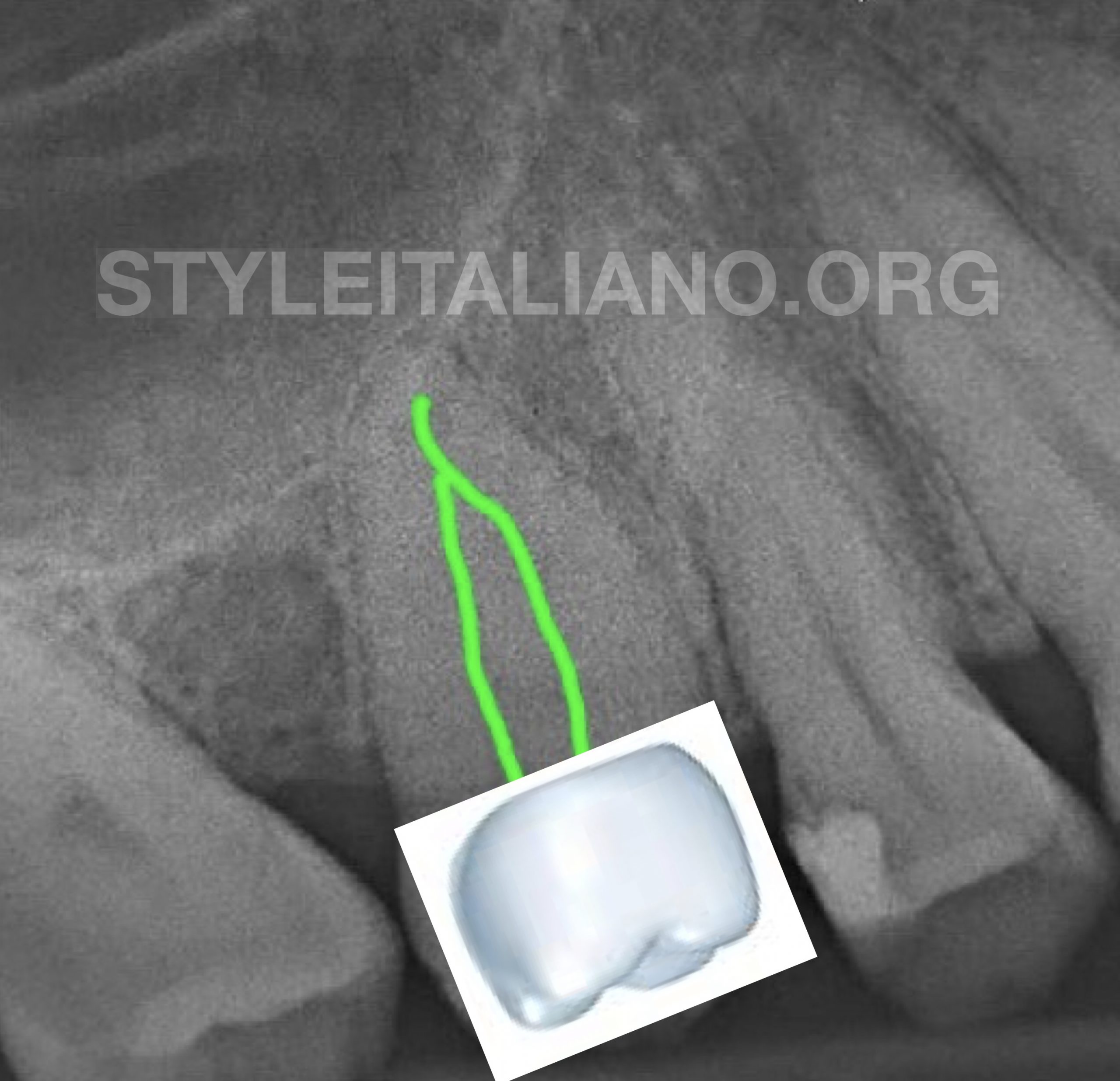
Fig. 3
Pre operative X-ray evaluation:
The only advantage of the tooth rotation was that the evaluation of the anatomy of the canals was easier to make.
Here we had to deal with the confluent canals that join in the apical third.
How the tooth was rotated, we decided to make a molar crown on the premolar.
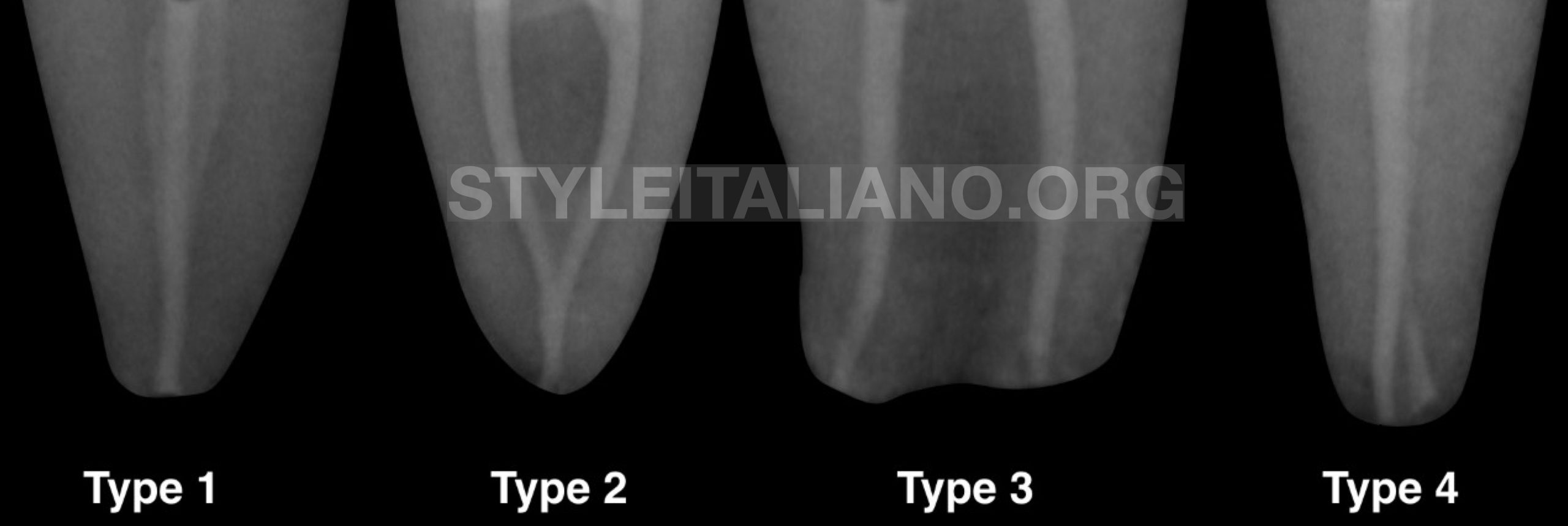
Fig. 4
According to Wayne`s classification, in this case we were dealing with Type 2 of confluent canals classification
image ( courtesy of Dr. Calogero Bugea)
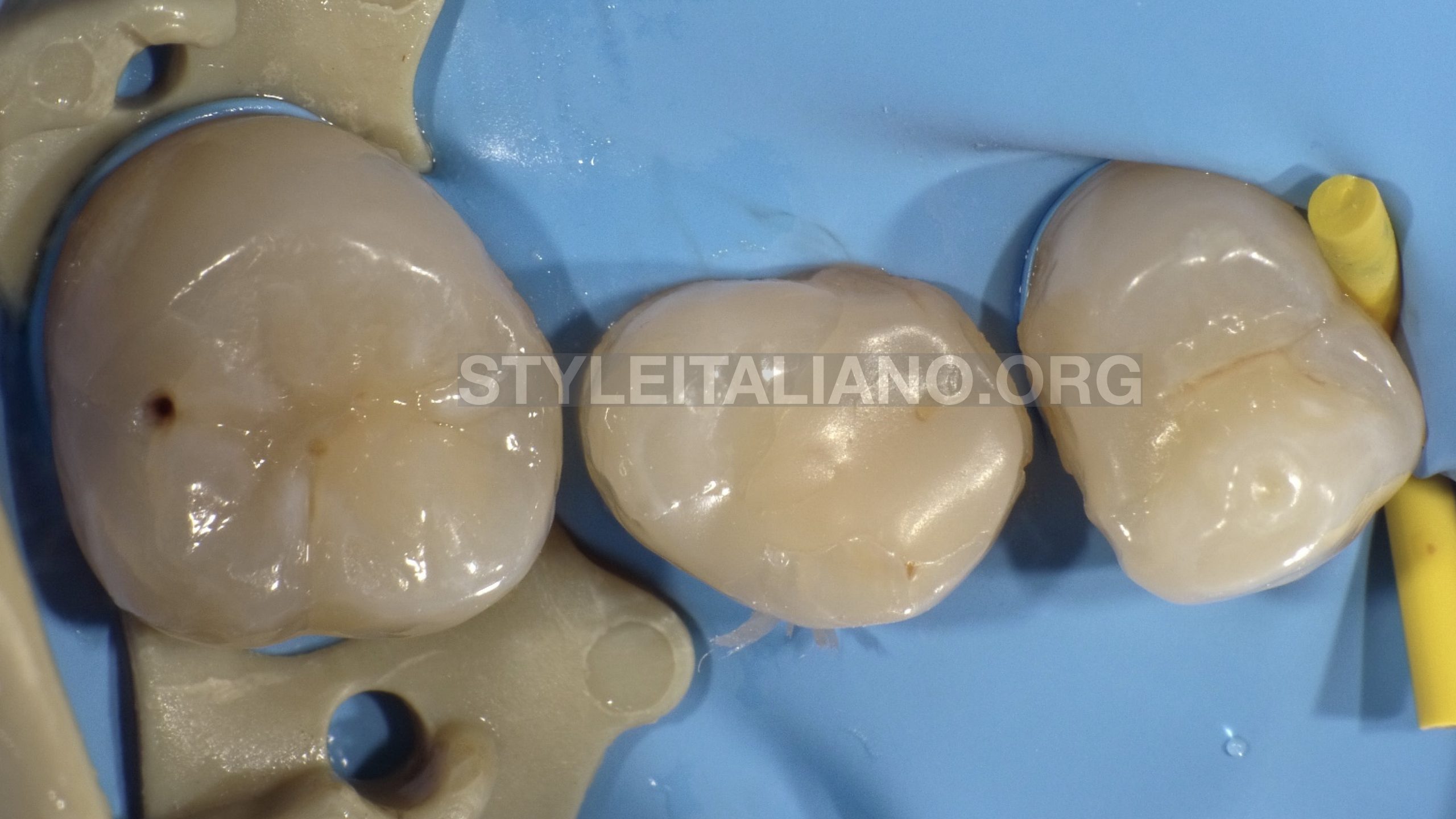
Fig. 5
Multiple tooth isolation
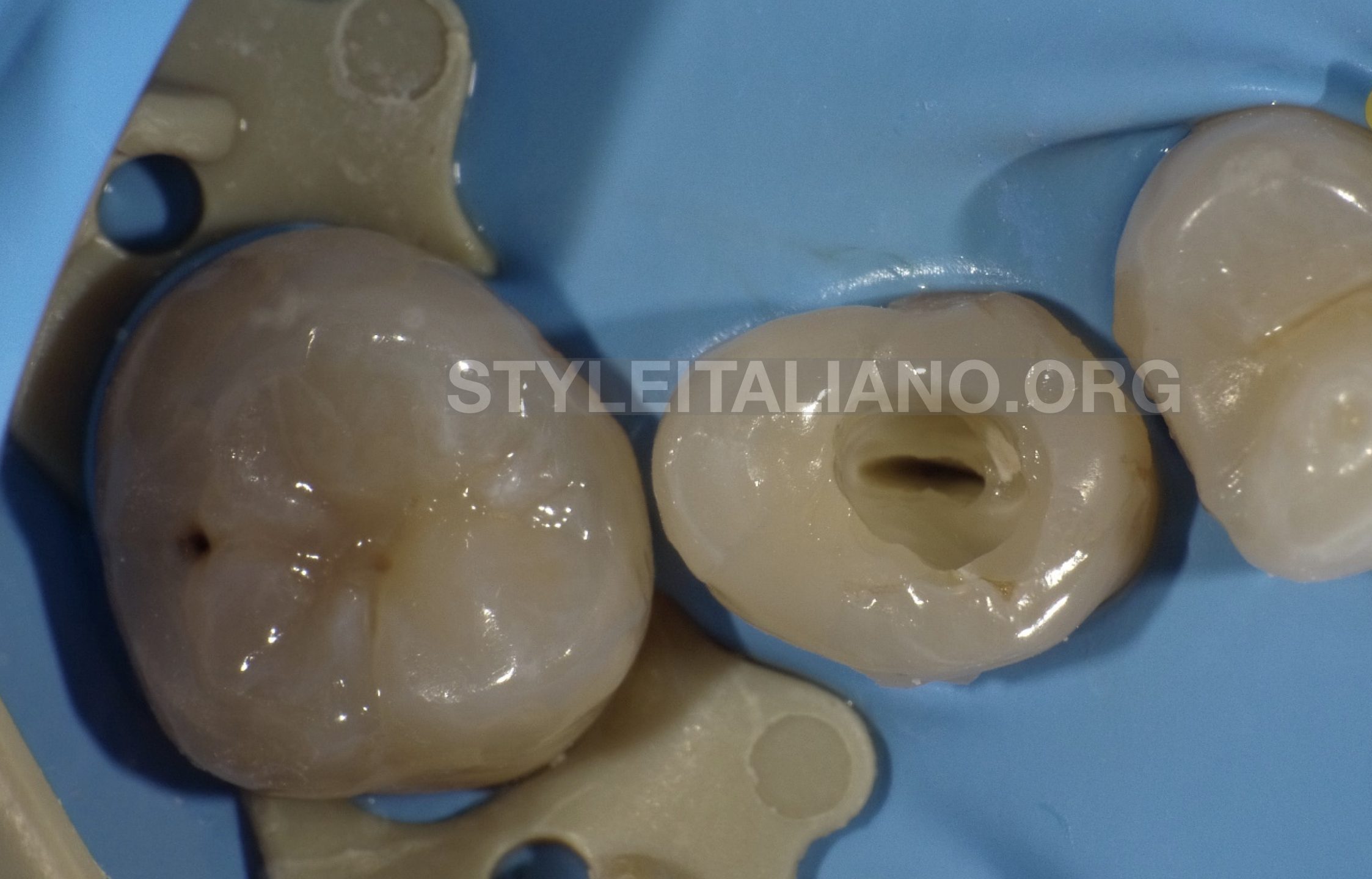
Fig. 6
Access cavity

Fig. 7
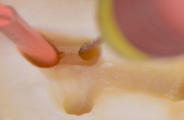
There are some methods to detect the confluences. In my opinion this method is simpler and faster compared to other methods.
First we need to put the gutta-percha in the main canal, then with manual instrument ISO 08 or 10 we go into the other canal, in order to detect the level of confluences.
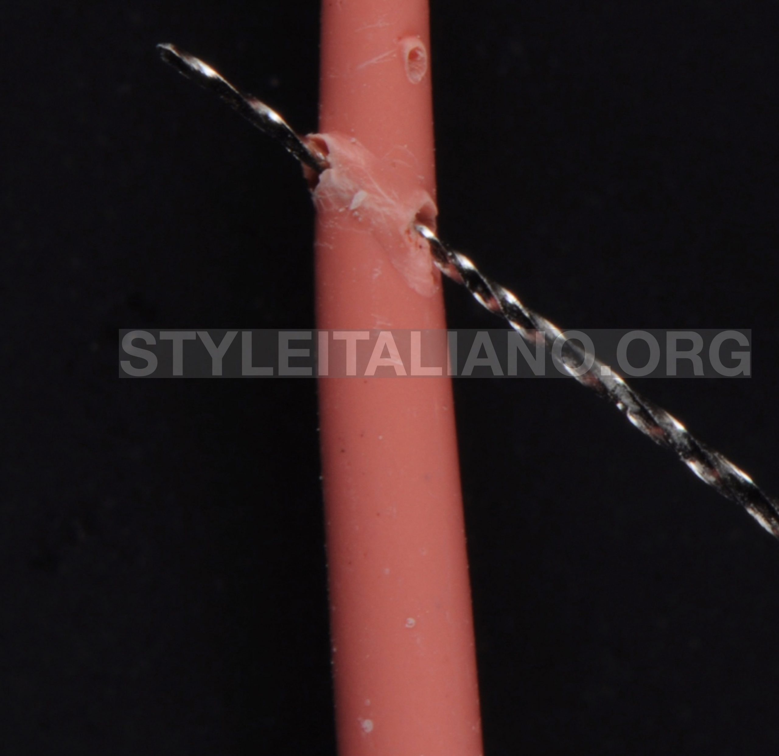
Fig. 8
The manual instrument ISO 08 or 10 will scratched the gutta-percha ( courtesy of Dr. Calogero Bugea)

Fig. 9
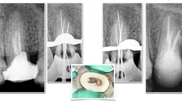
After removing the gutta-percha and the manual instrument ISO, we need to check traces or scratches left on the gutta-percha from ISO instrument. ( courtesy of Dr. Calogero Bugea)
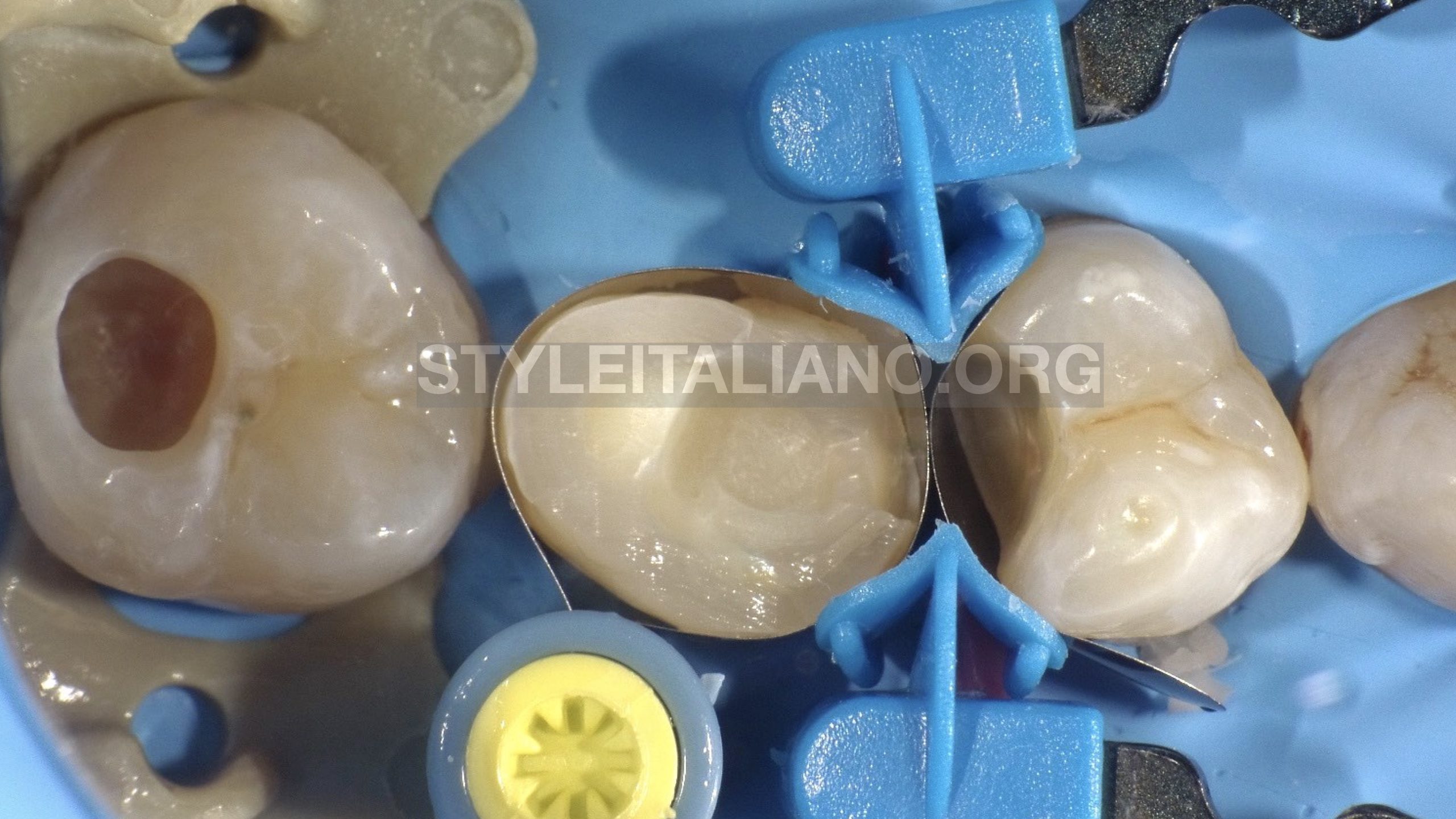
Fig. 10
Restoration of teeth 14 and 17 with composit, and build up of the tooth 15
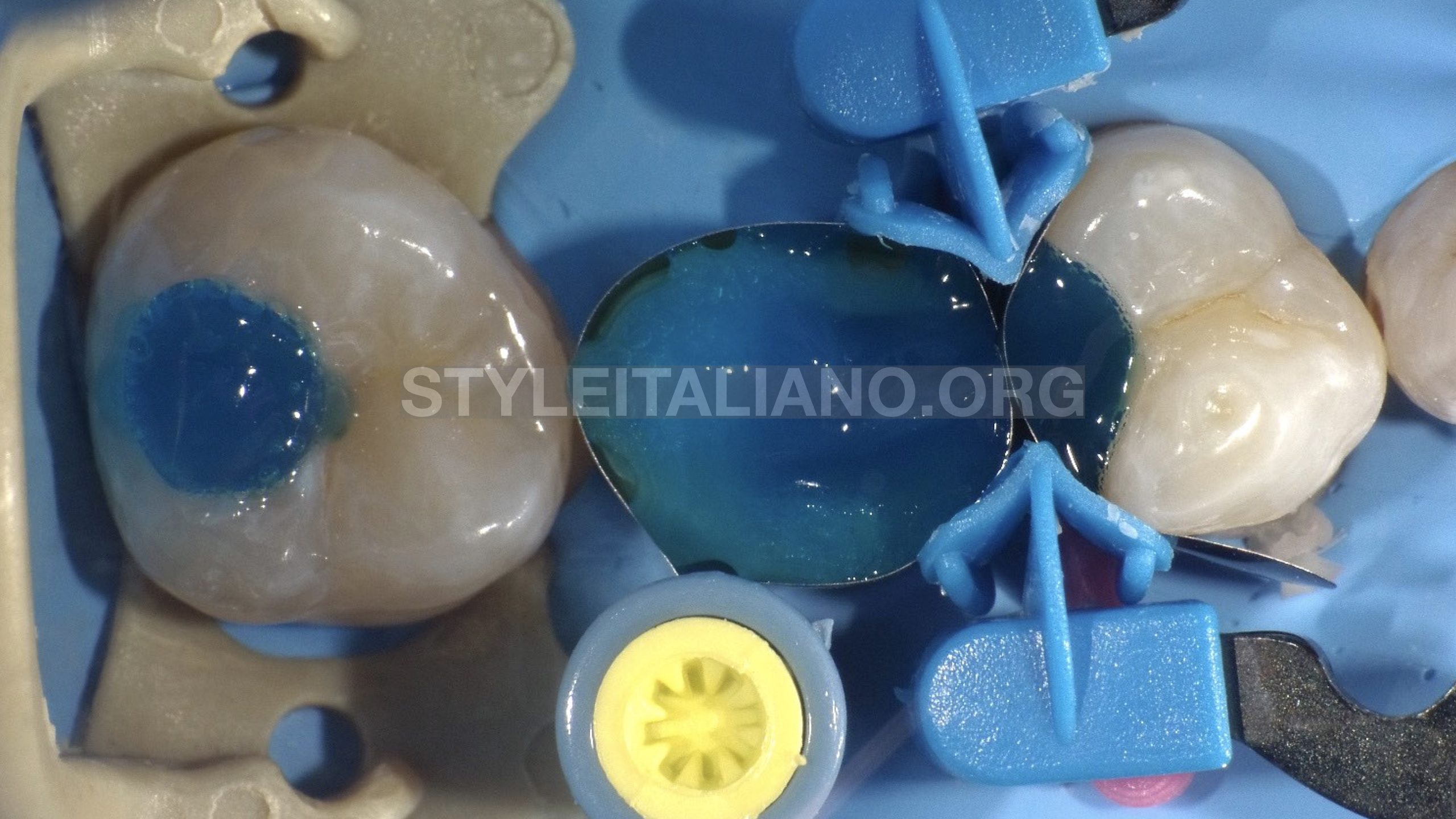
Fig. 11
Etching
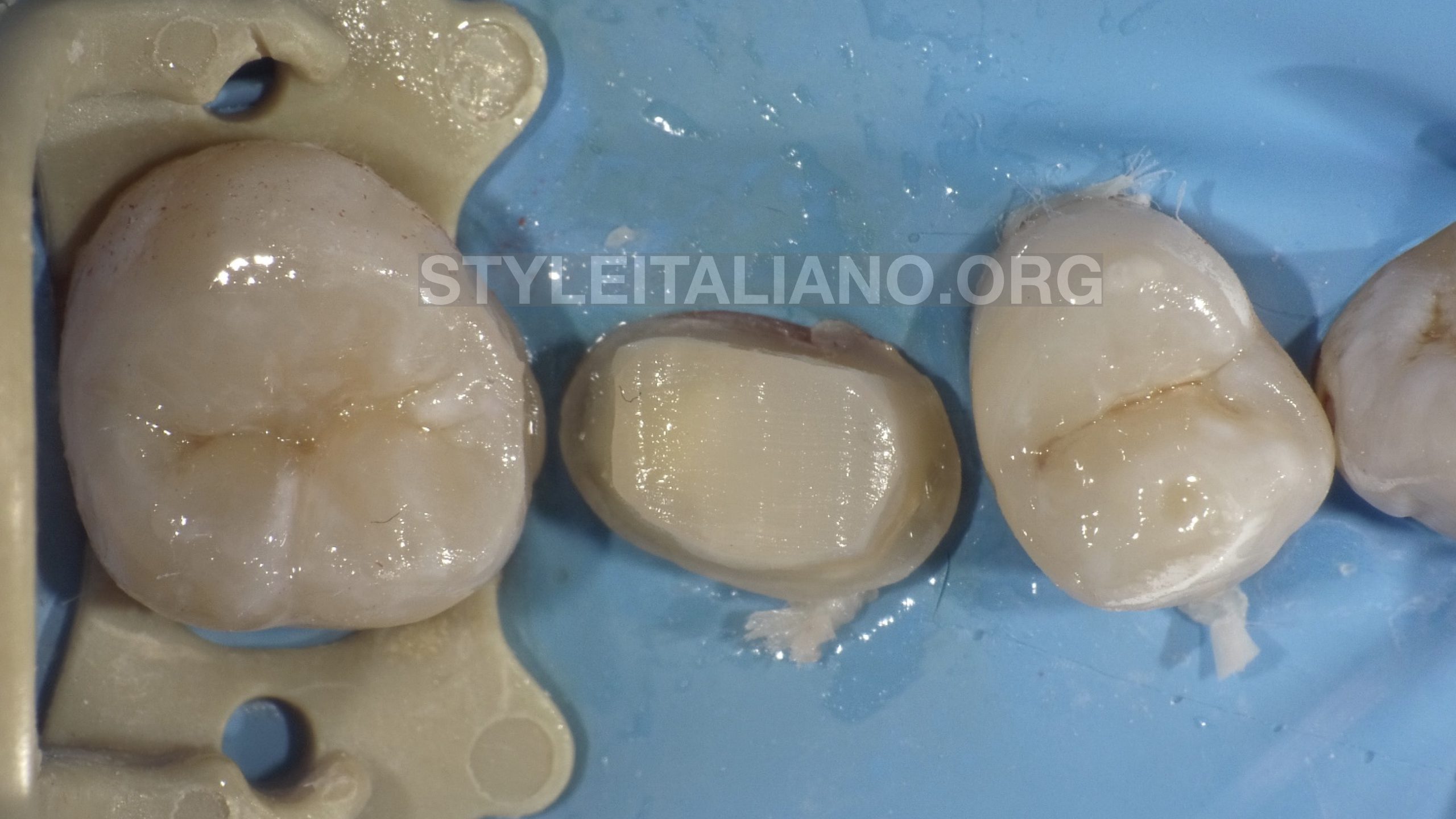
Fig. 12
Final preparation
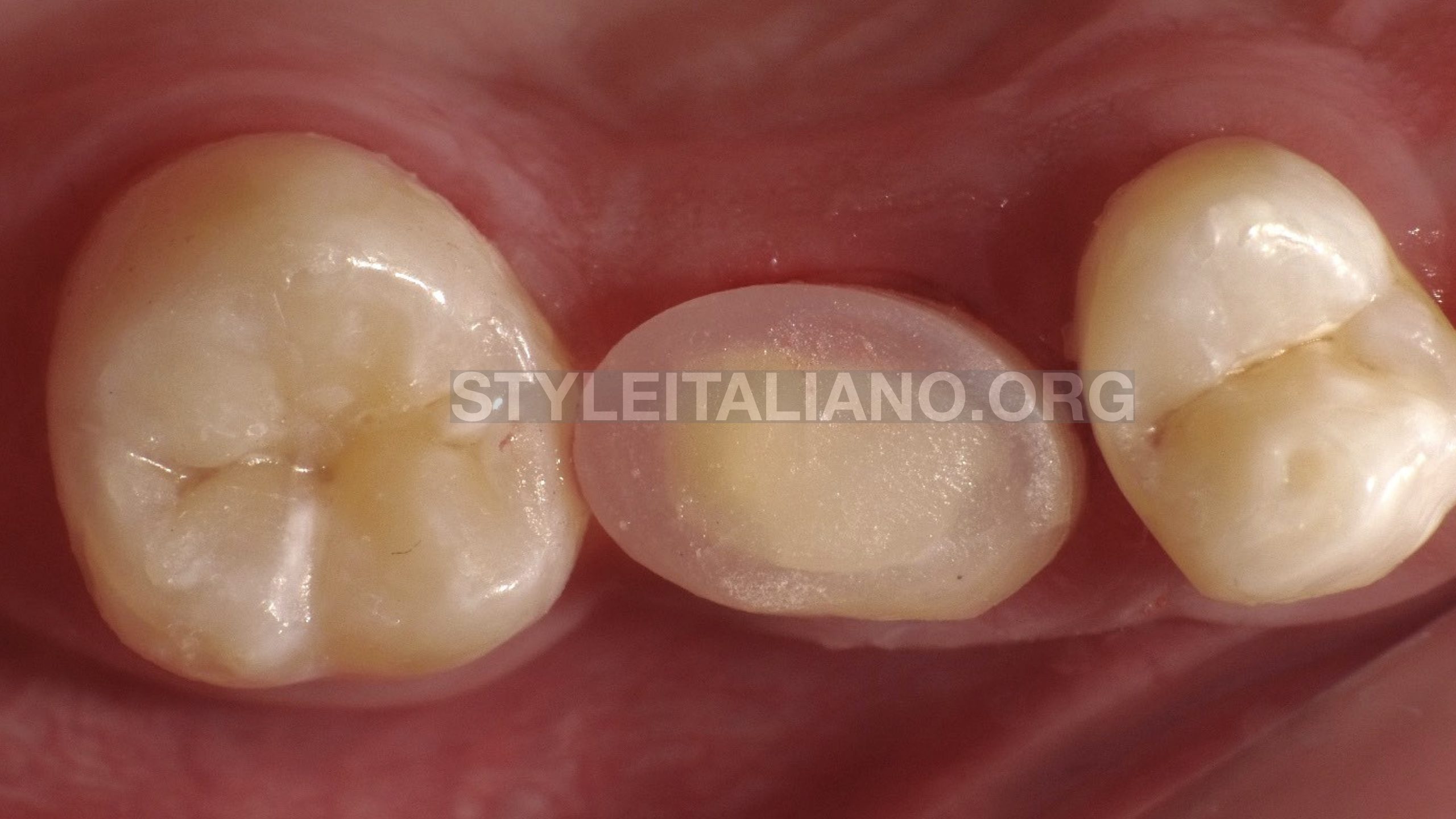
Fig. 13
Temporary filling
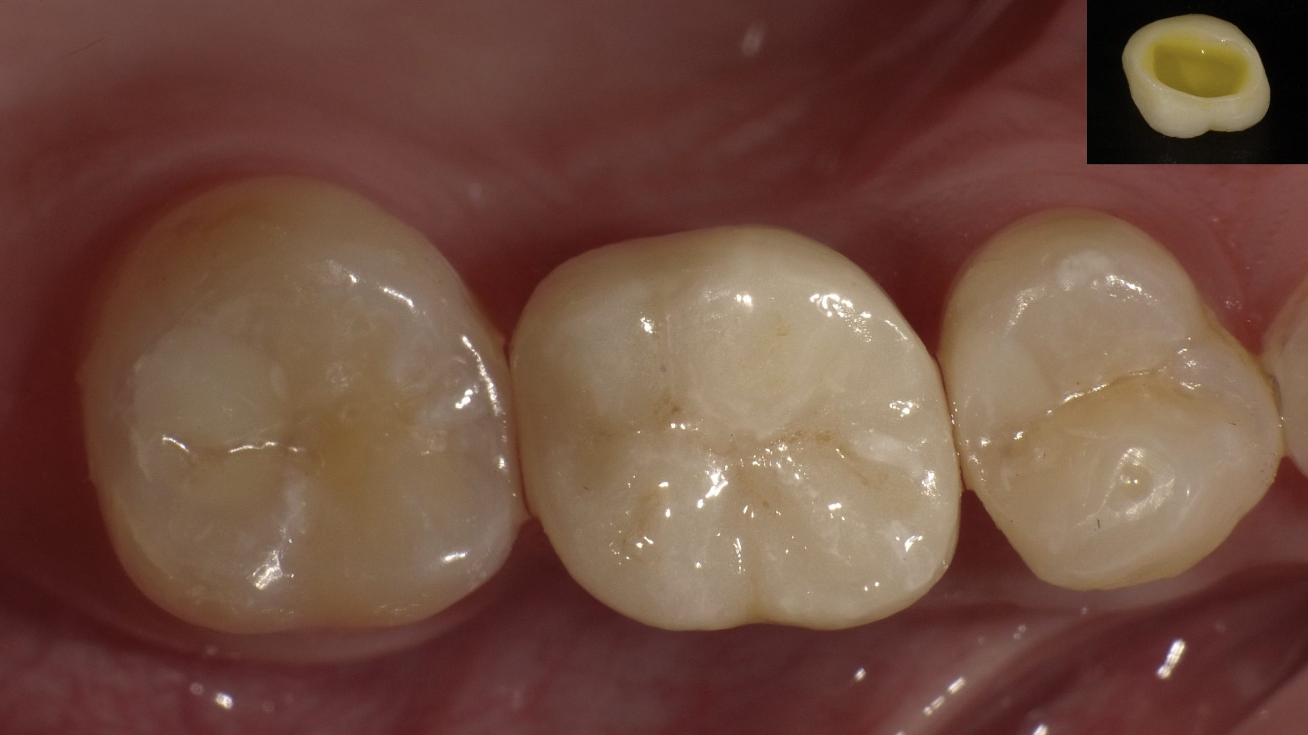
Fig. 14
Molar crown cementation on the premolar tooth.
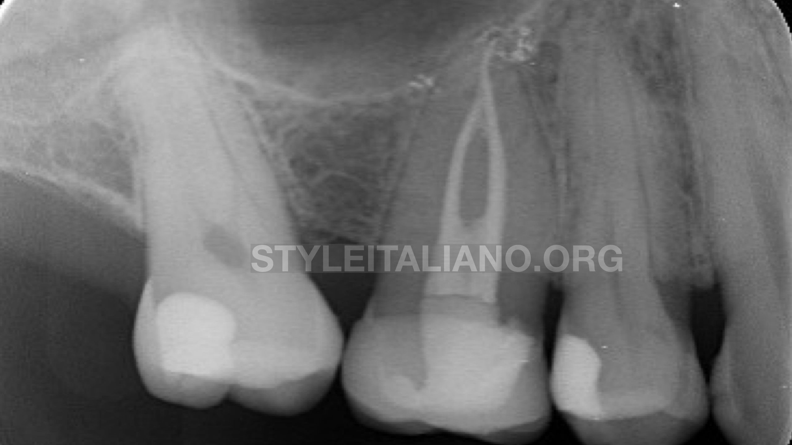
Fig. 15
Post operative X-ray
Conclusions
Pre-treatment case evaluation greatly facilitates our work and avoids any errors that may occur during treatment. The different variations of the restorative part are crucial in these non routine cases as well.
Applying methods for detecting the confluences in this cases are inevitable.
If we have a pre-treatment plane, it will reduce the possibility of mistakes and surprises that may occur during the treatment.
In this case we had two challenges. The first one was the management of the confluent canals and the second one is the restoration solution.
Bibliography
Vertucci Fj. Root canal morphology and its relationship to endodontic procedures, Endodontic topics,2005
Arnaldo Castellucci , Two canals in a single root: clinical and practical considerations, 2003
Furri,M , Differences in the confluence of mesial canals in mandibular molar with three or four root canals, International endodontic journal, 2008
Gianluca Gambarini et AL. Cone-beam computed tomographic analysis on root and canal morphology of mandibular first permamnent molar among multiracial population in Western European population, European Journal of Dentistry, 2018