Mb2 as a first choice
11/07/2020
Riccardo Tonini
Warning: Undefined variable $post in /home/styleendo/htdocs/styleitaliano-endodontics.org/wp-content/plugins/oxygen/component-framework/components/classes/code-block.class.php(133) : eval()'d code on line 2
Warning: Attempt to read property "ID" on null in /home/styleendo/htdocs/styleitaliano-endodontics.org/wp-content/plugins/oxygen/component-framework/components/classes/code-block.class.php(133) : eval()'d code on line 2
This clinical case demonstrates how a strategy is important to approach in a proper way a difficult clinical case. MB1 scouting usually it’s easier and more predictable and only in a second time we dedicate our attention to MB2. Here instead, considering the difficulty of scouting MB1, it was decided to manage the situation in the opposite way.
We know that many studies have reported the presence of two canals in the mesiobuccal (MB) root of the maxillary first and second molars and that the occurrence of second MB canals (MB2) has been reported as high as 95%. Sometimes, clinical situations can move us in using MB2 as a strategy for detecting MB1, in example when it is completely sclerotic as in this clinical case of upper first molar.
After the conservative opening of the access cavity, the Palatal canal and Distal canal were detected, measured and shaped in a proper way. The problem rose with Mb1 scouting. After few minutes of brushing with ultrasounds and Buc1 tip, it still was impossible to detect the canal: for this reason a new strategy was adopted. MB2 was scouted, shaped till protaper S2 and at the end a CBCT was performed in order to check if MB2 was merging with MB1 or it had an independent exit.
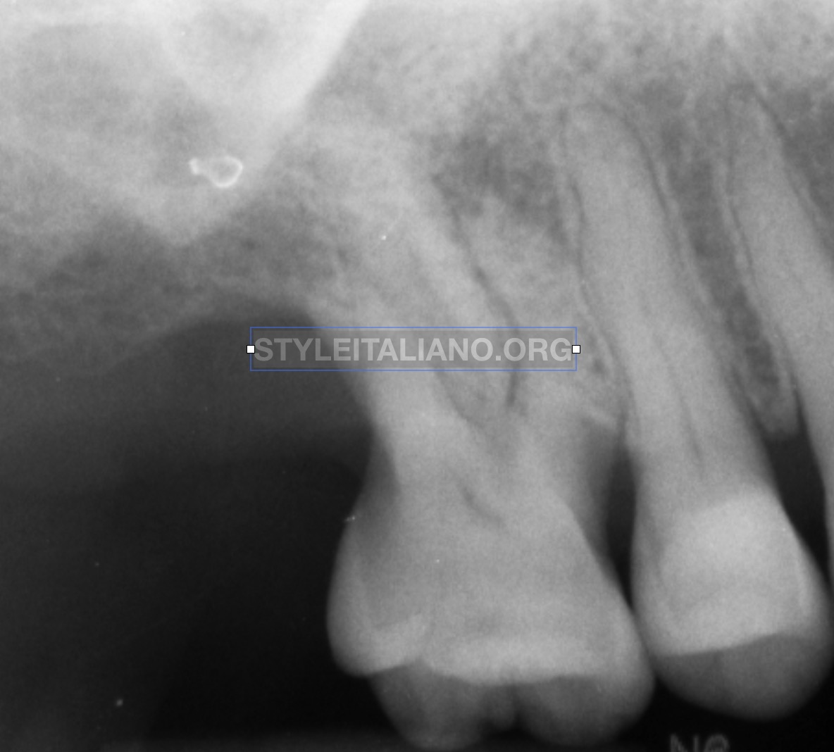
Fig. 1
Necrosis of upper first molar.
From this x Ray is evident that canals are sclerotic and a translucent lesion is present in the apical region of mesial root
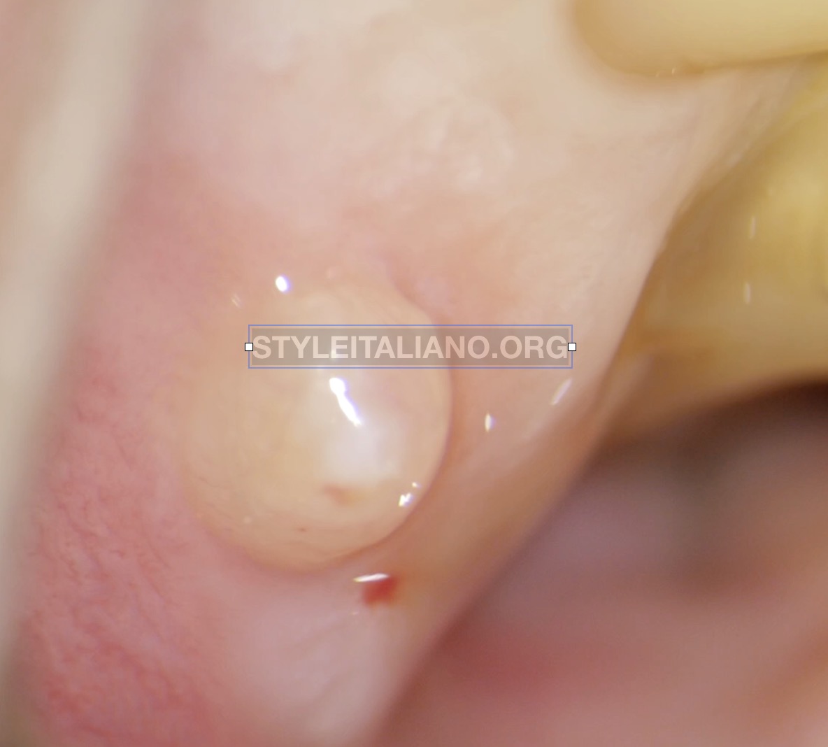
Fig. 2
The sinus tract confirmed necrosis, moreover the cervical abrasion visible on the tooth is a sign for eventual sclerosis of canals
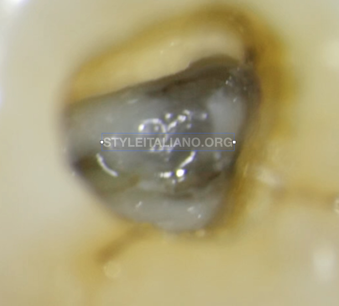
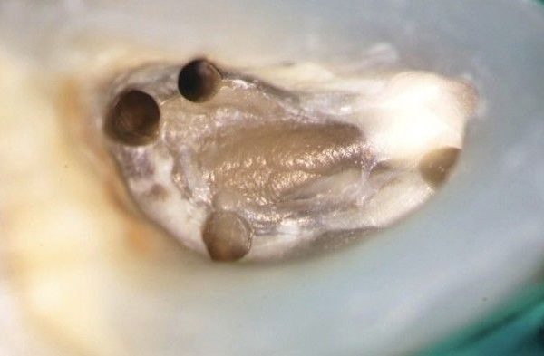
Fig. 3
Access cavity design.
Mb1 and Mb2 seem booth present as distal and palatal
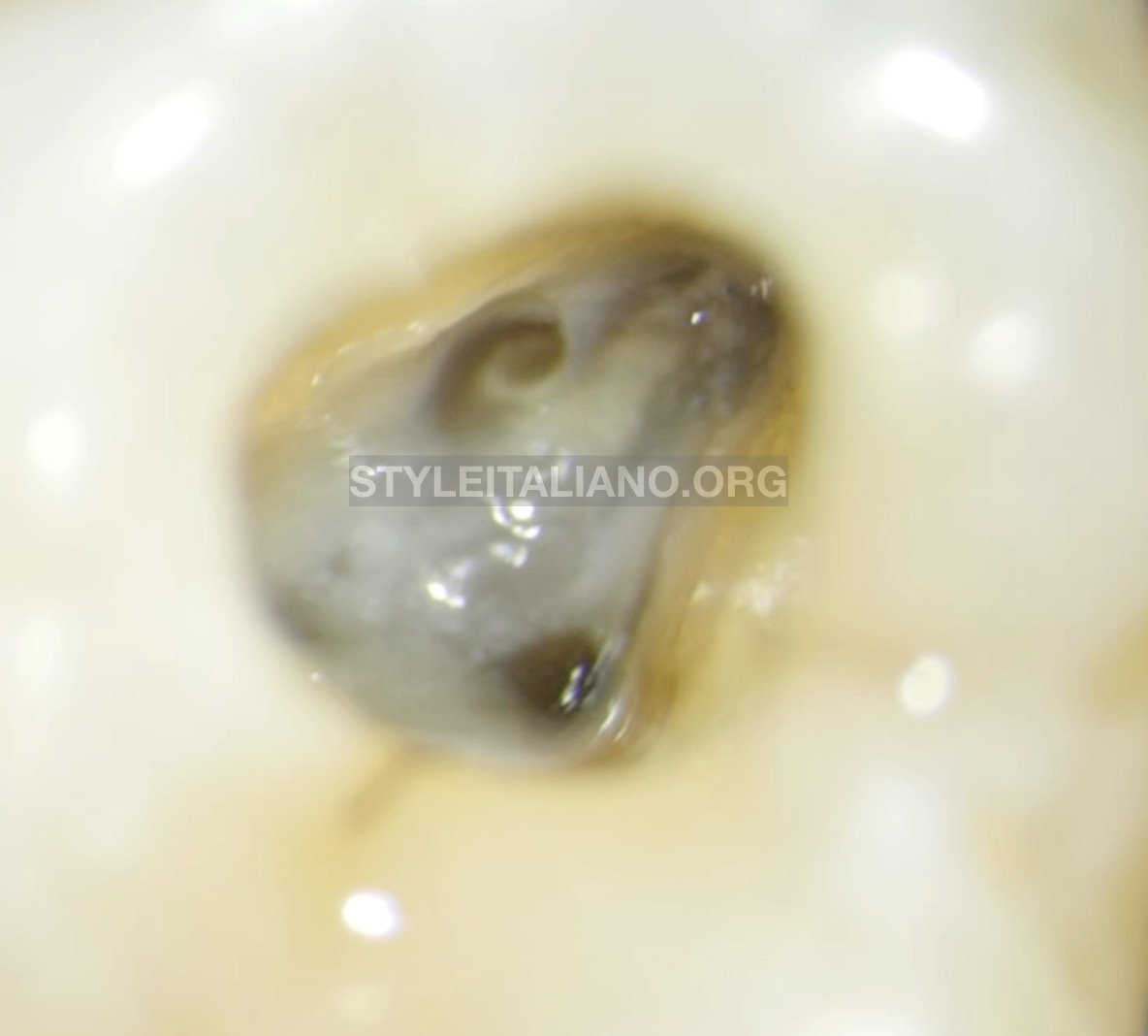
Fig. 4
Mb2 scouting and shaping
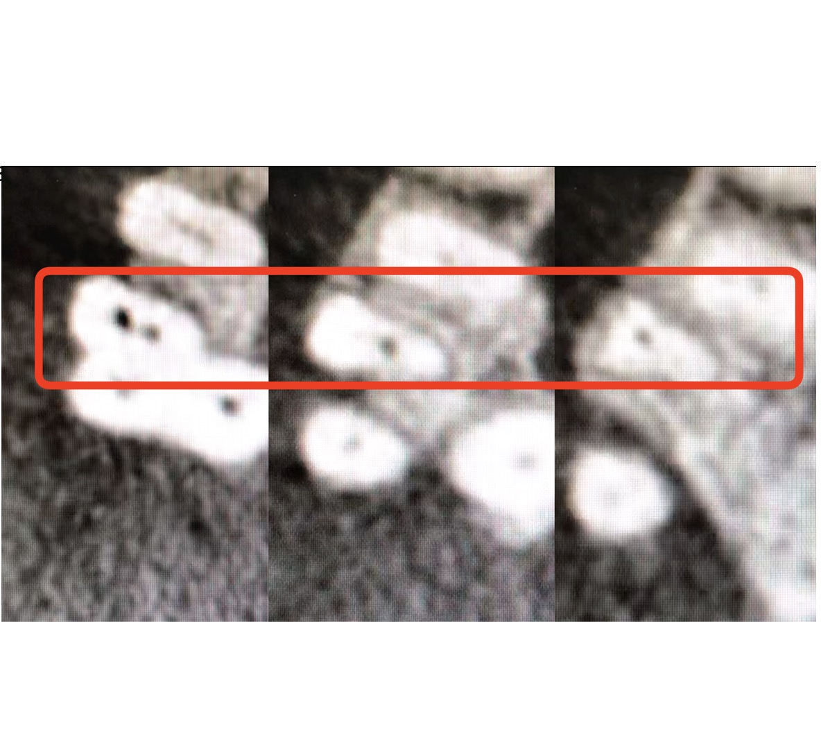
Fig. 5
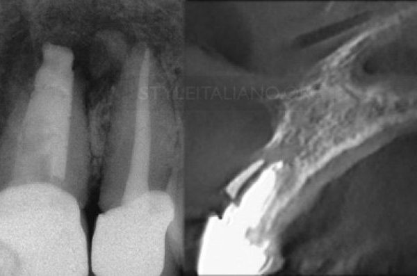
CBCT
Taking a CBCT after shaping is a strategical approach in order to highlight better the anatomy.
From CBCT was evident the MB direction of MB2. CBCT confirmed that MB1 and MB2 were joining and a further MB1 scouting was not necessary anymore.
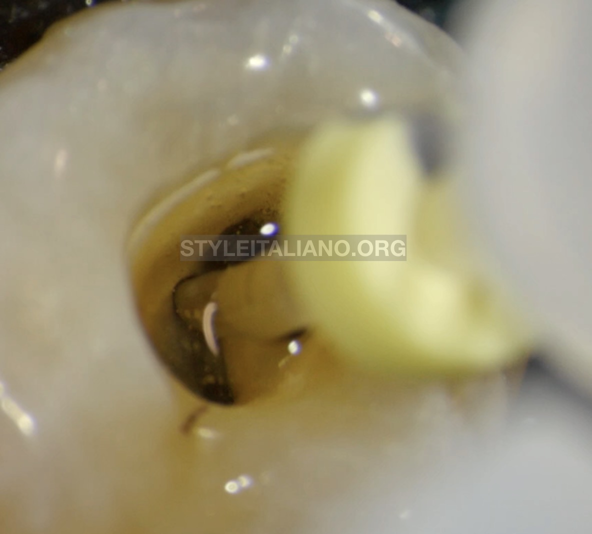
Fig. 6
Cleaning
Irriflex (Produits Dentaires SA) in action for a perfect irrigants delivery
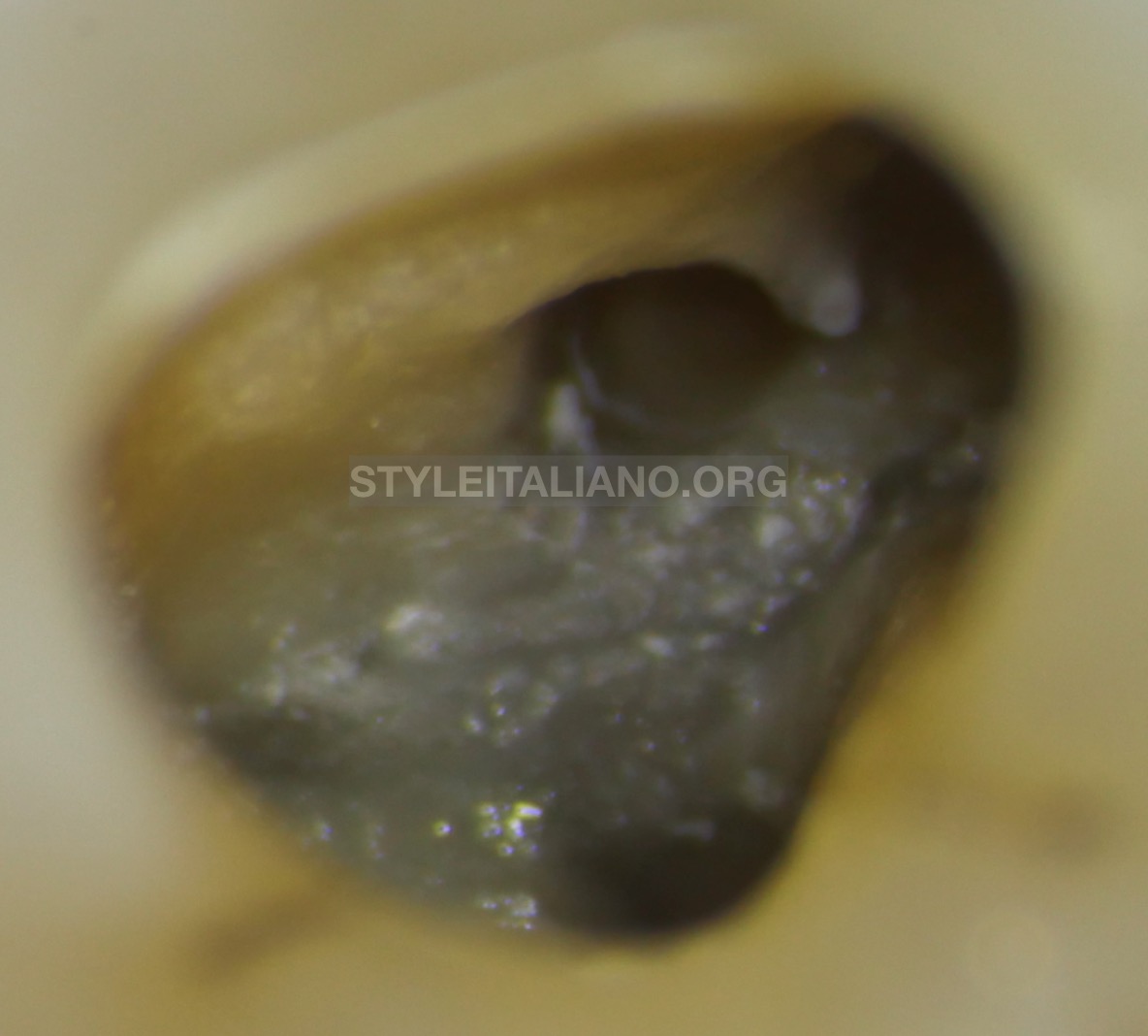
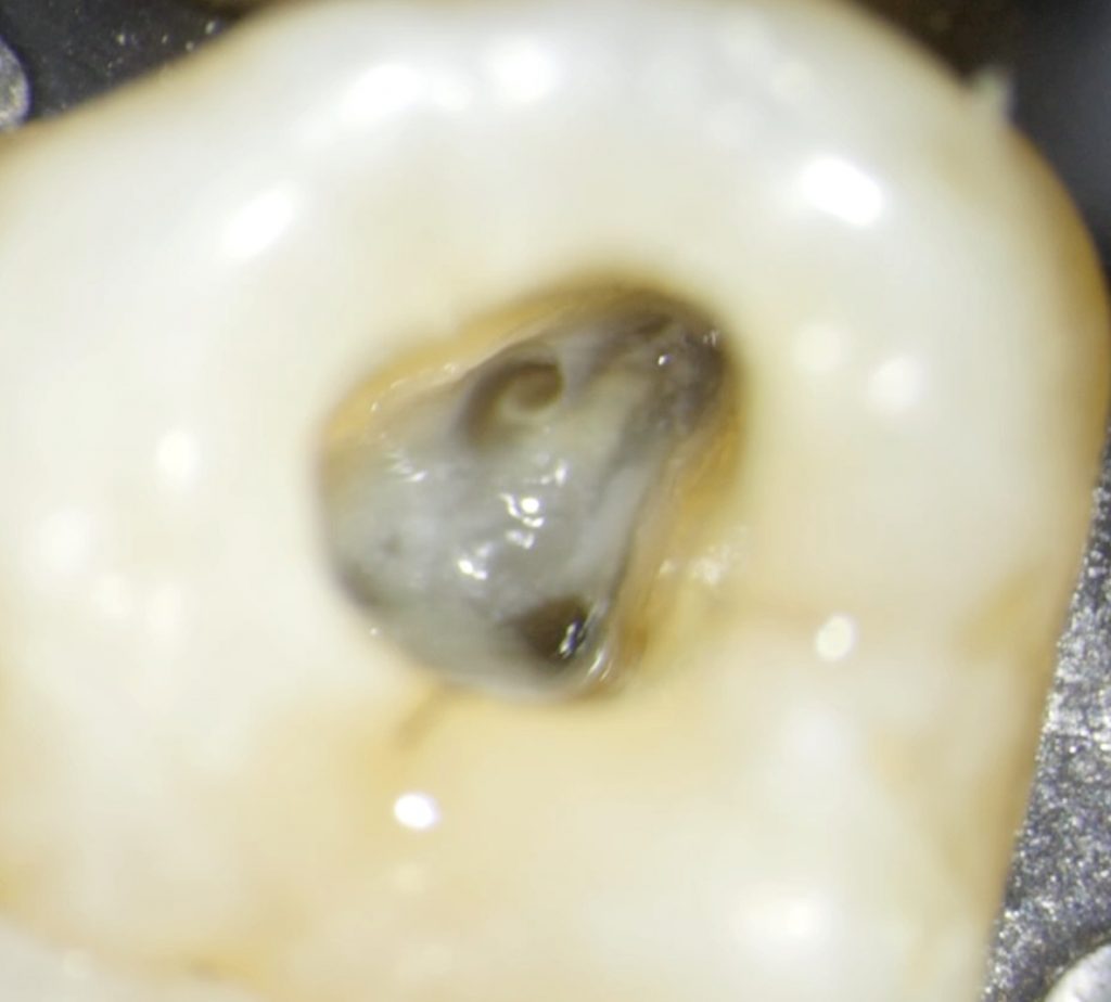
Fig. 7
Mb2 shaped and cleaned before obturation
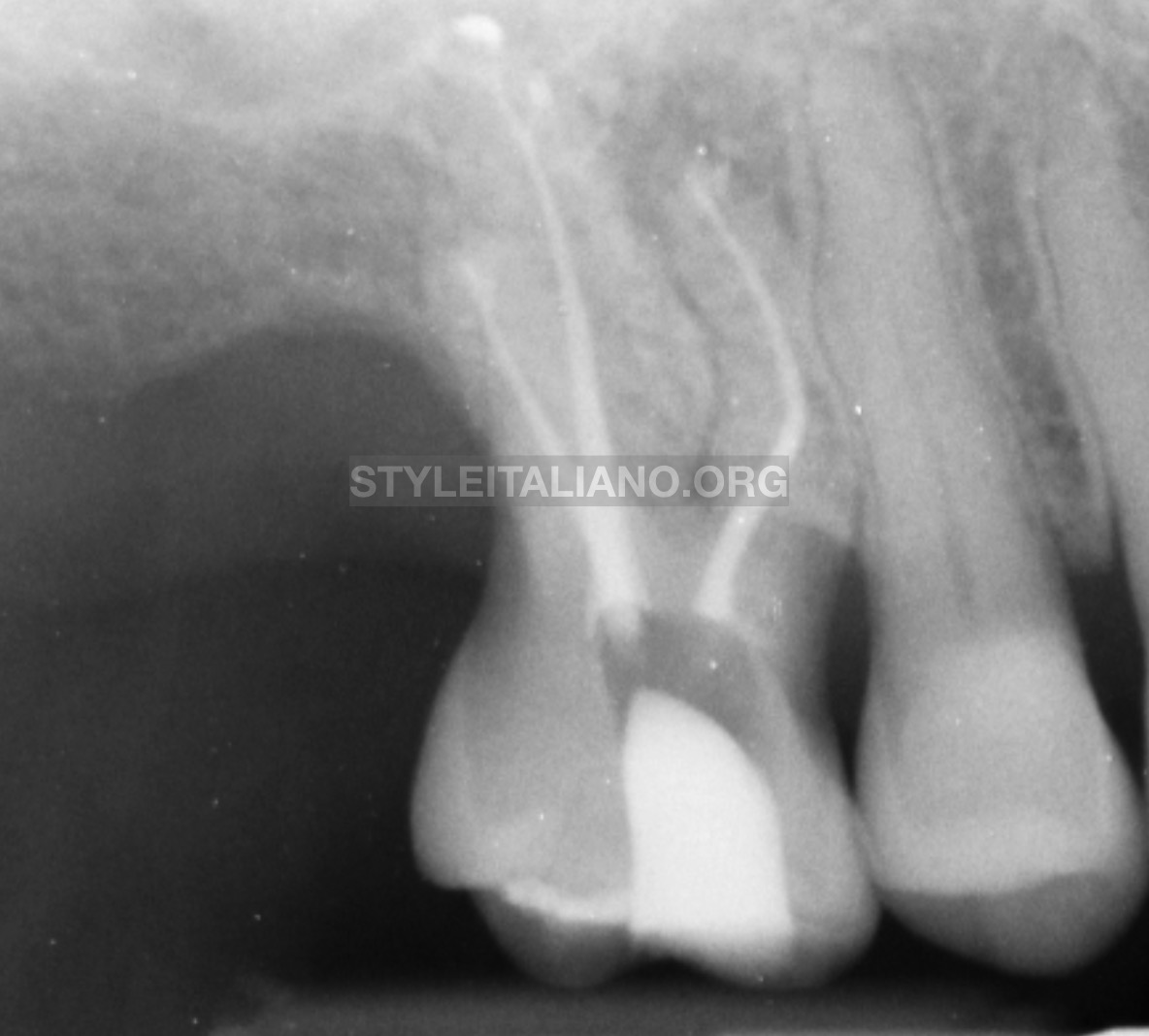
Fig. 8
Post op x ray
The upper first molar was filled with Essenseal and Guttacore.
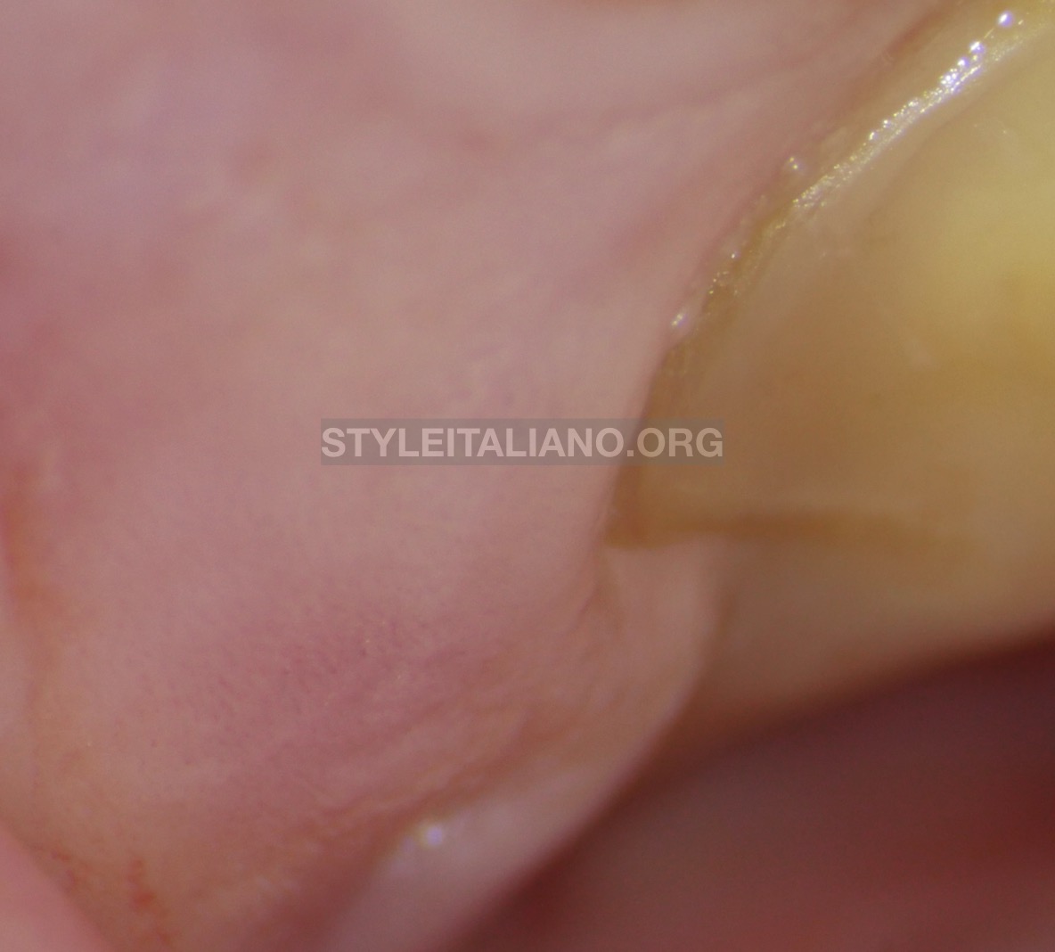
Fig. 9
After 7 days the sinus tract disappeared.
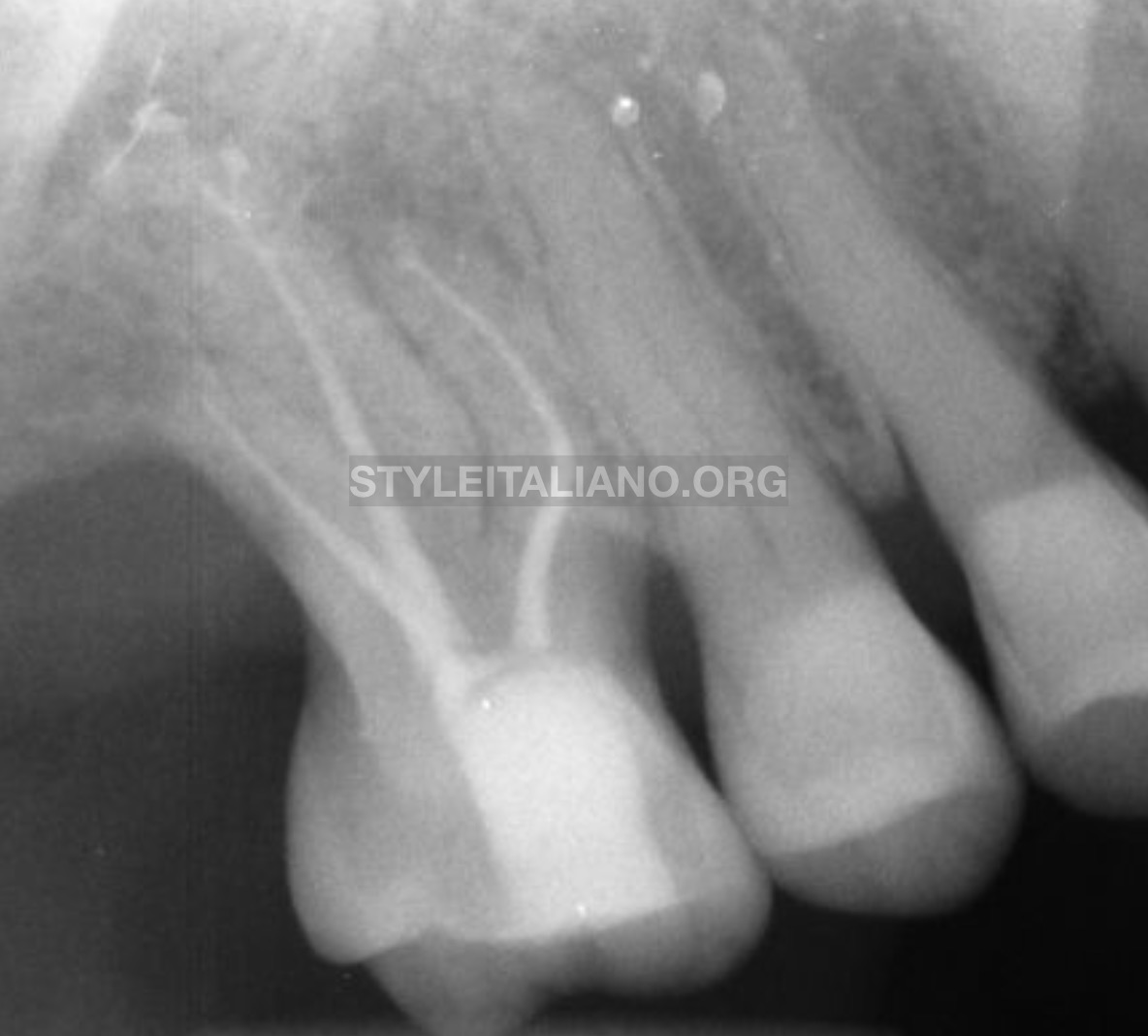
Fig. 10
Direct restoration and first control after 3 months
Conclusions
Endo Strategy is mandatory for a predictable result. In this the case CBCT gave to the operator a great confirmation, but it has to be pointed out that shaping the MB2 before taking the CBCT was strategical in order to observe better the path of the MB2 inside of the mesial root
Bibliography
R. Bauman W. Scarfe S. Clark J. Morelli J. Scheetz A. Farman Ex vivo detection of mesiobuccal canals in maxillary molars using CBCT at four different isotropic voxel dimensions
International Endodontic JournalVolume 44, Issue 8
S. Patel J. Brown T. Pimentel R. D. Kelly F. Abella C. Durack Cone beam computed tomography in Endodontics – a review of the literature
International Endodontic JournalVolume 52, Issue 8
J. Parker A. Mol E. M. Rivera P. Tawil CBCT uses in clinical endodontics: the effect of CBCT on the ability to locate MB2 canals in maxillary molars
International Endodontic JournalVolume 50, Issue 12