Management of separated instruments in different location
27/01/2024
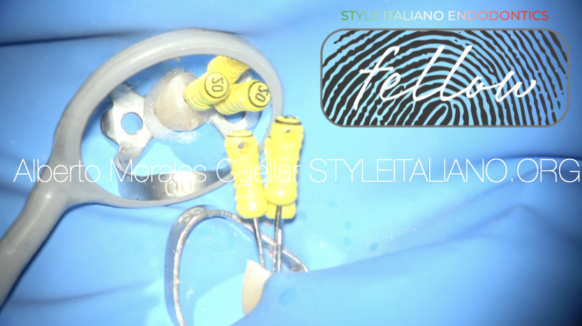
Fellow
Warning: Undefined variable $post in /home/styleendo/htdocs/styleitaliano-endodontics.org/wp-content/plugins/oxygen/component-framework/components/classes/code-block.class.php(133) : eval()'d code on line 2
Warning: Attempt to read property "ID" on null in /home/styleendo/htdocs/styleitaliano-endodontics.org/wp-content/plugins/oxygen/component-framework/components/classes/code-block.class.php(133) : eval()'d code on line 2
Separation of nickel-titanium instrument during root canal treatment can cause serious complications that may lead to treatment failure. It prevents complete cleaning and filling of the entire root canal space. Therefore, retrieval of the instrument should be tried to achieve better outcome for the therapy. There are two ways to manage this problem:
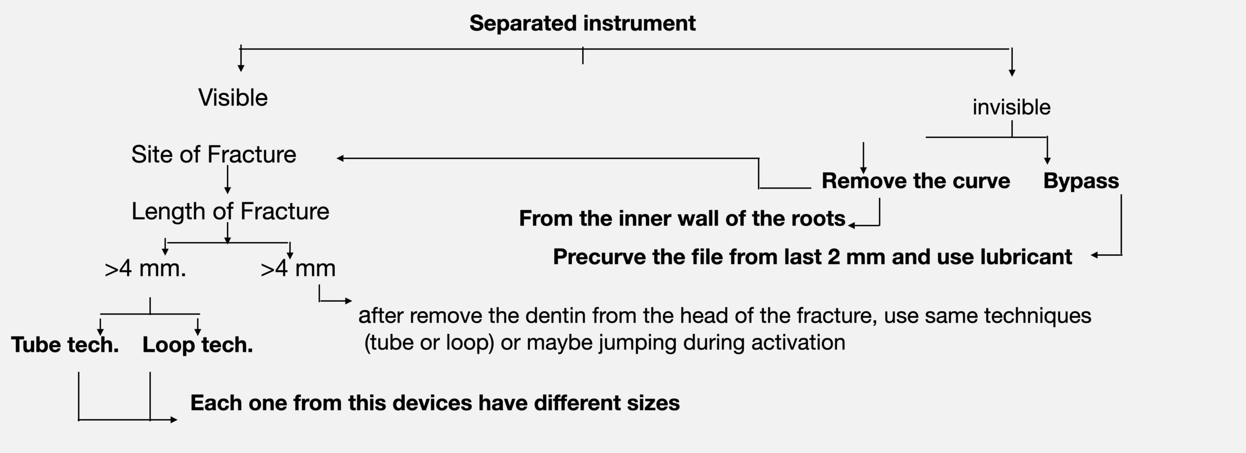
Fig. 1
The first one is bypass which means (Create a path next to the separated instrument). The bypass is the game of hands, this method of management in most cases does not need magnification. It always depend on factors like (patience, time and experience) to pass the separated instrument. So, before I talk about the other ways for management SI we always ask an important question, which method bypass or retrieval should be used. The answer of this question depends on the case data like (visible or invisible and site of fracture). After explaining the first method now we can talk about the second one (retrieval), the important point that must be mentioned here is that this method cannot be done without the presence of magnification and the required tools. The diagram below shows the steps of this method:
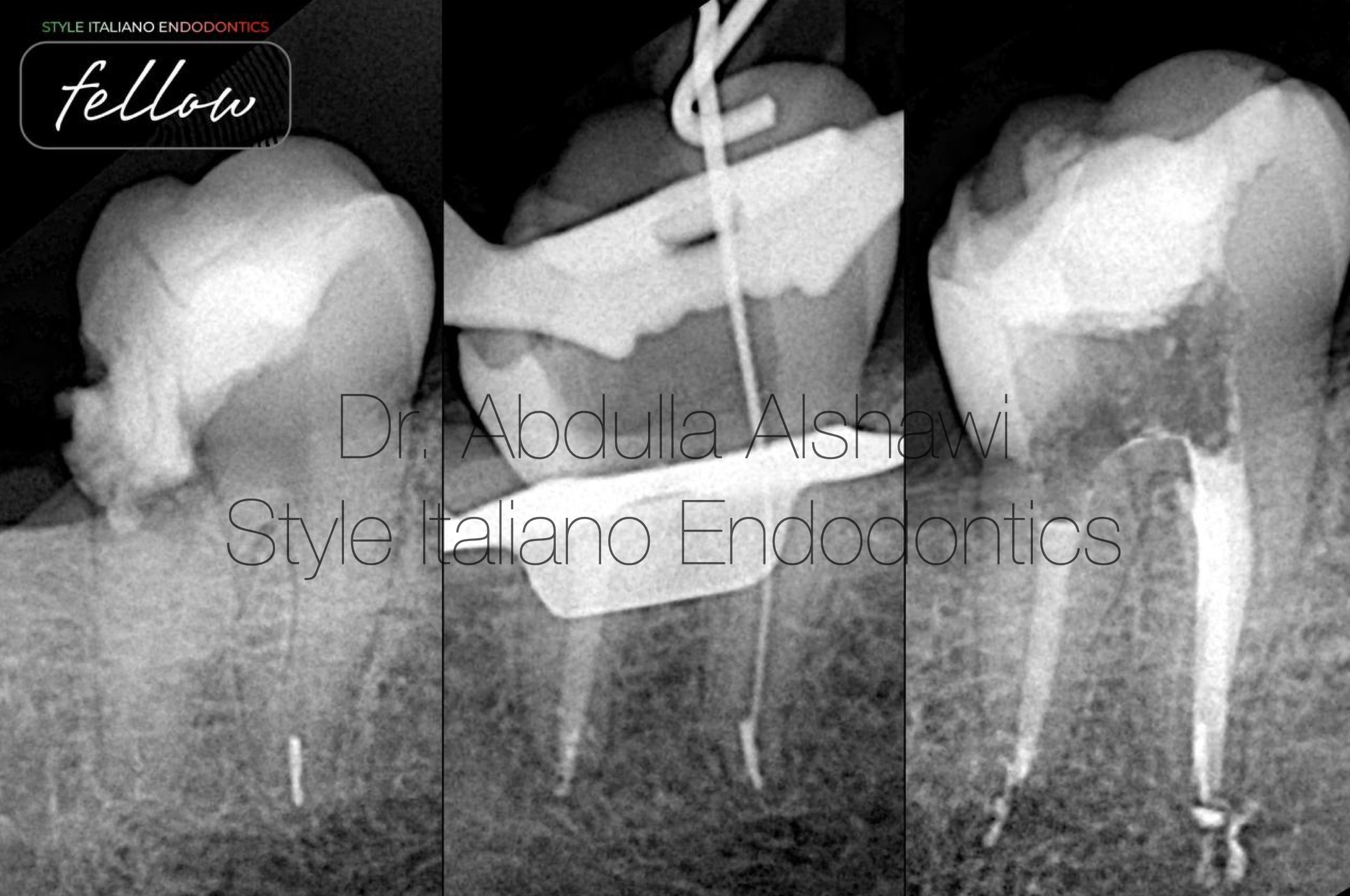
Fig. 2
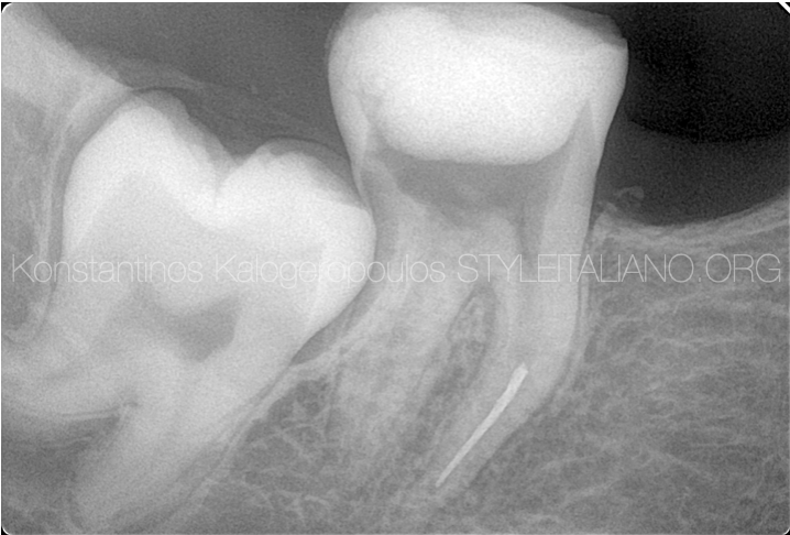
First case. 53-year-old patient was refer to my clinic for management of separated instrument and complete RCT
A pre opertive showed a separated instrument In the mesial canal with the apex so for be conservative with root go for bypass.
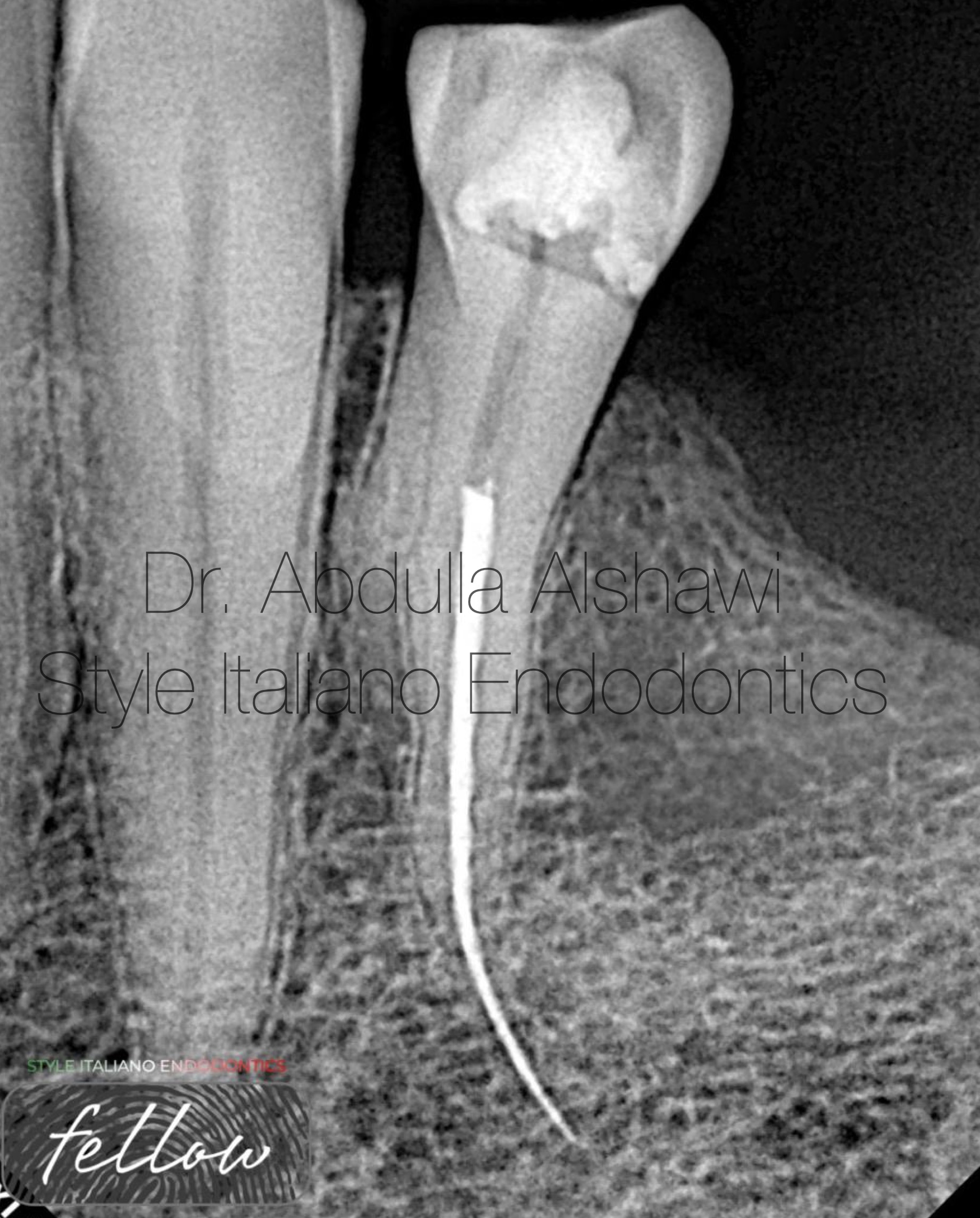
Fig. 3
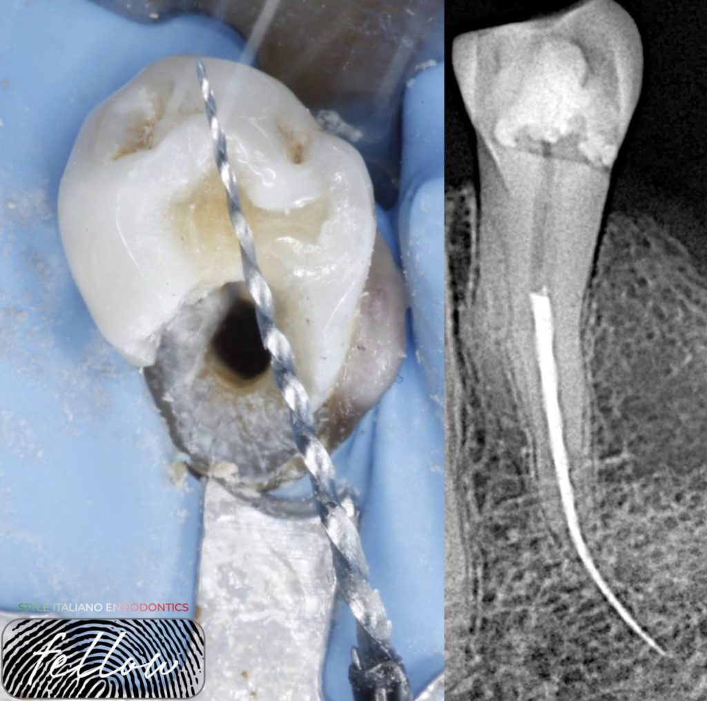
Second case talk about retrieval, x-ray showed long separated instrument extending outside the canal , and the patient refused any surgical intervention.
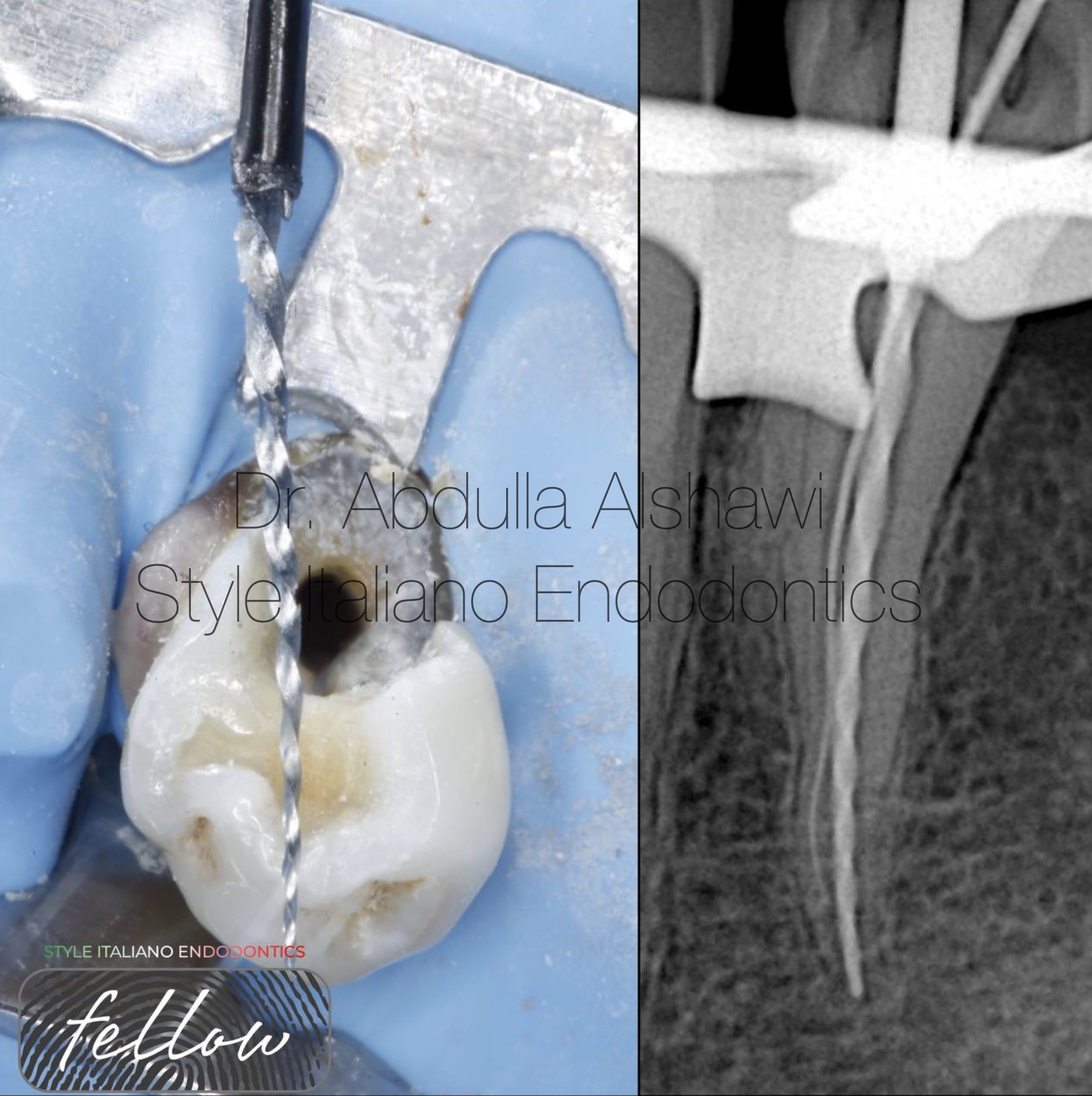
Fig. 4
After removing the dentin around the head of separated instrument then use tube tech. For removing it and check W.L.
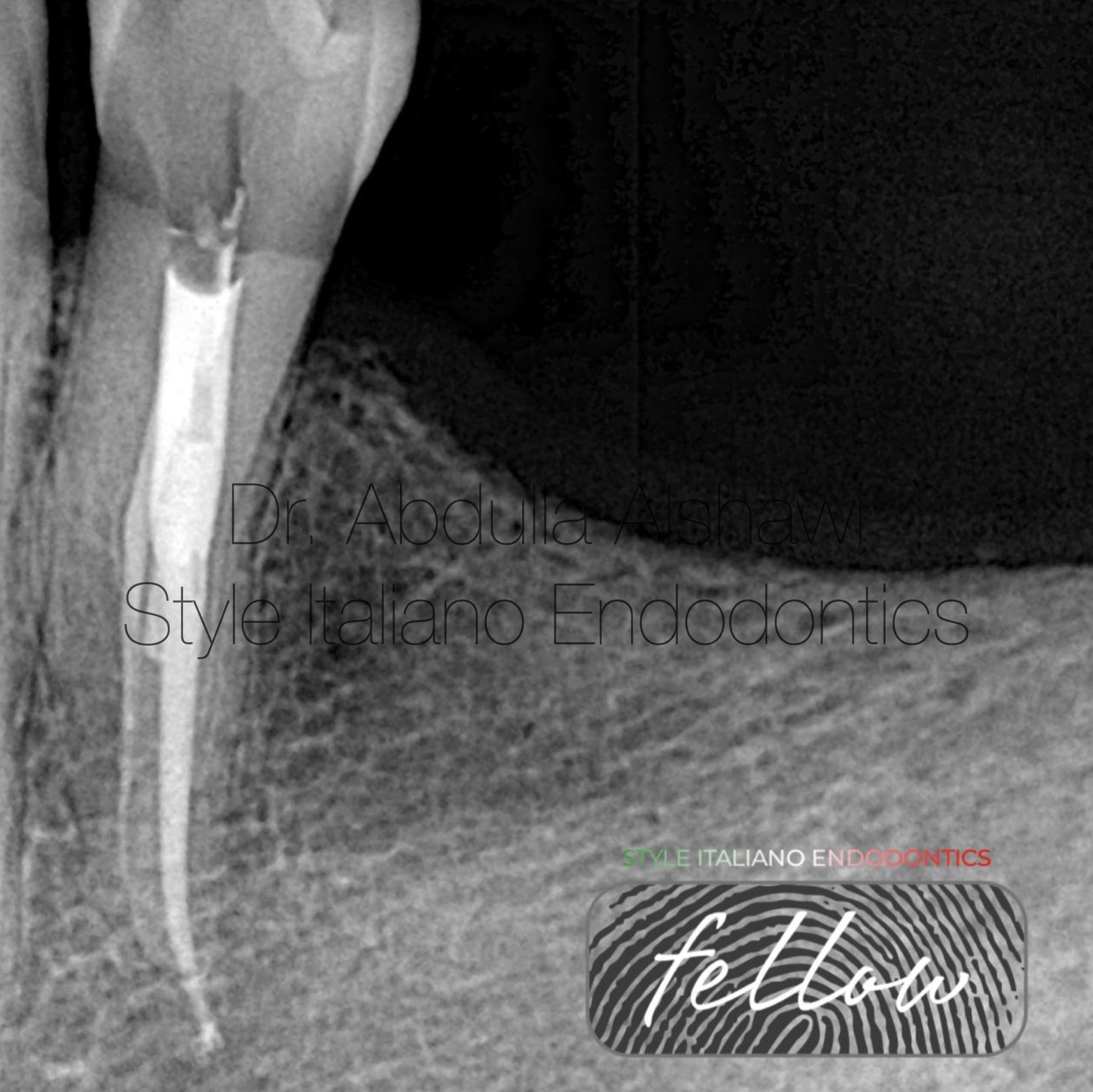
Fig. 5
Final x-ray after obturation
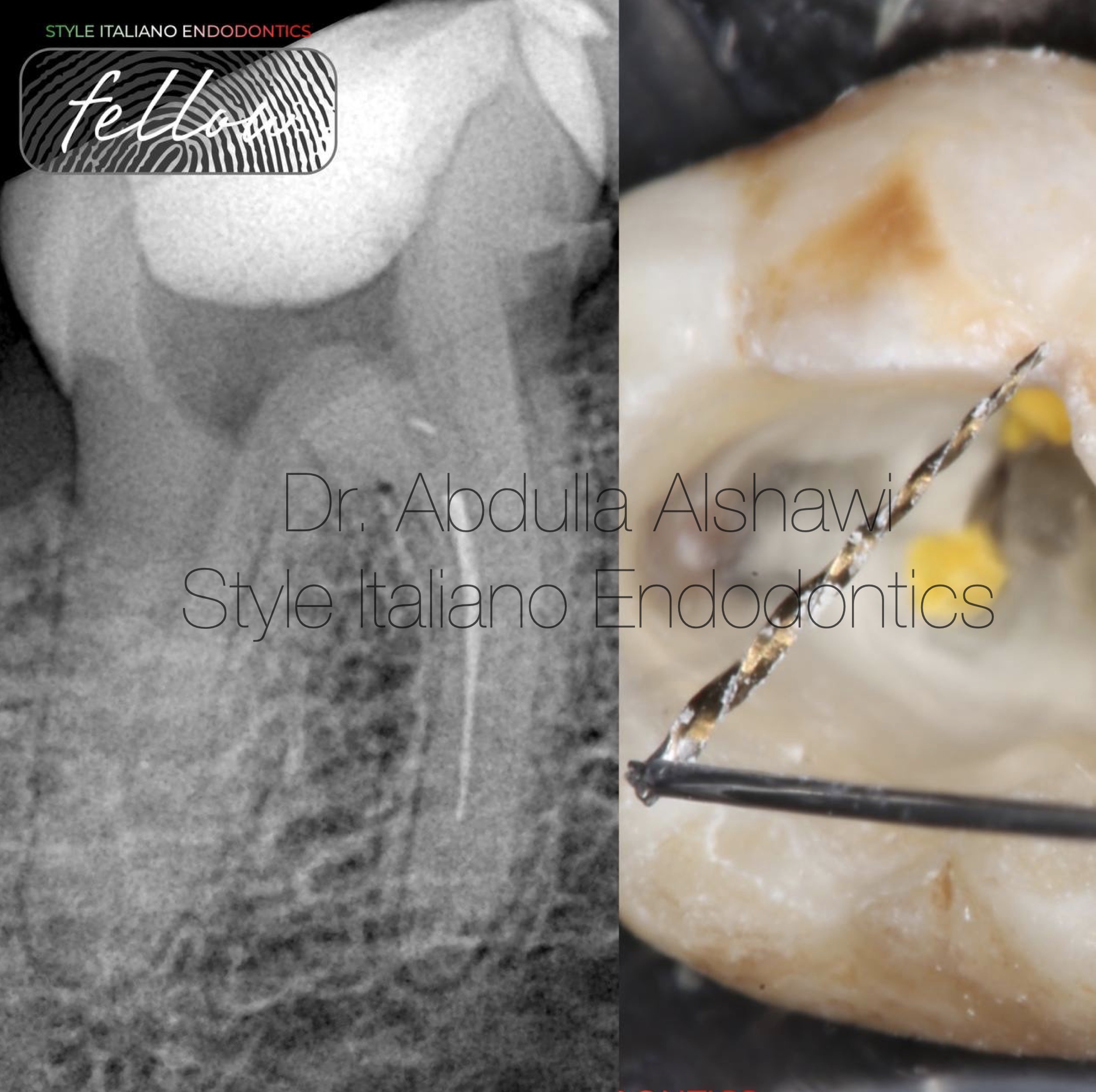
Fig. 6
Another case for retrieval of separated instrument from lower first molar by using loop tech.
This video explains all the steps
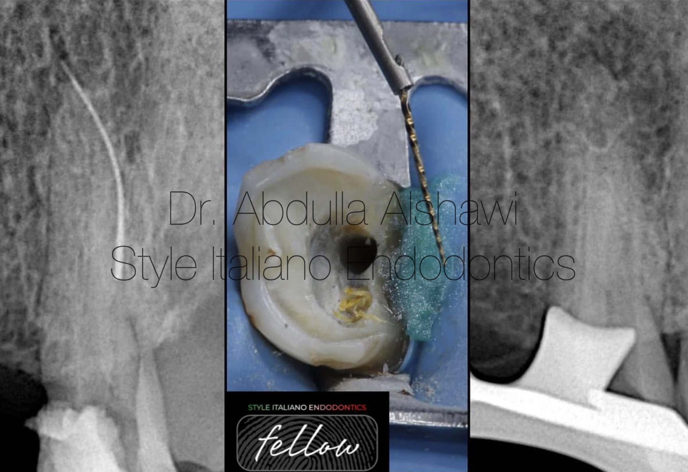
Fig. 7
Fourth case was another fishing for upper first premolar by using tube tech.
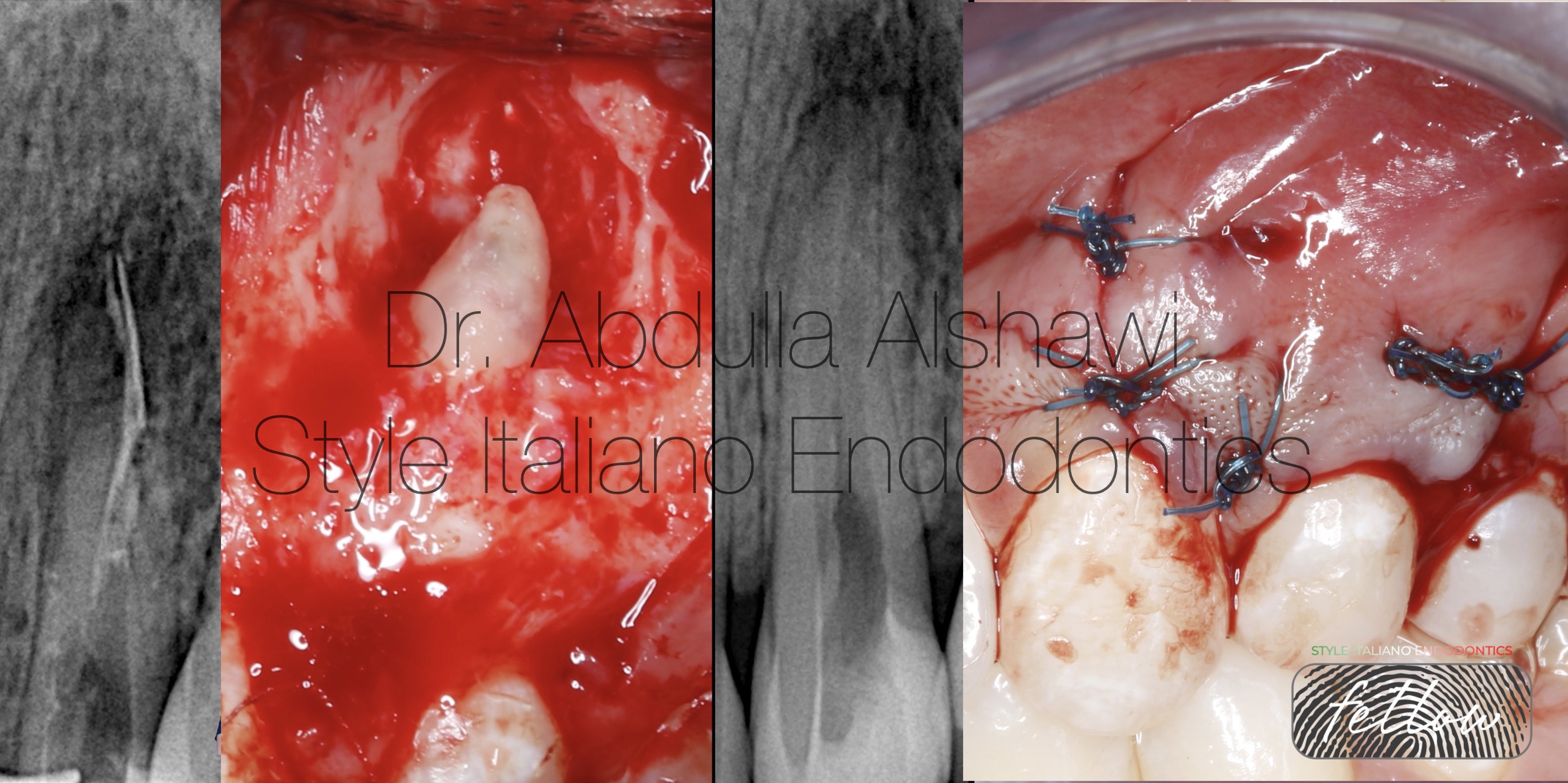
Fig. 8
The fifth case shows surgical intervention for removing the separated instrument beyond the apex.
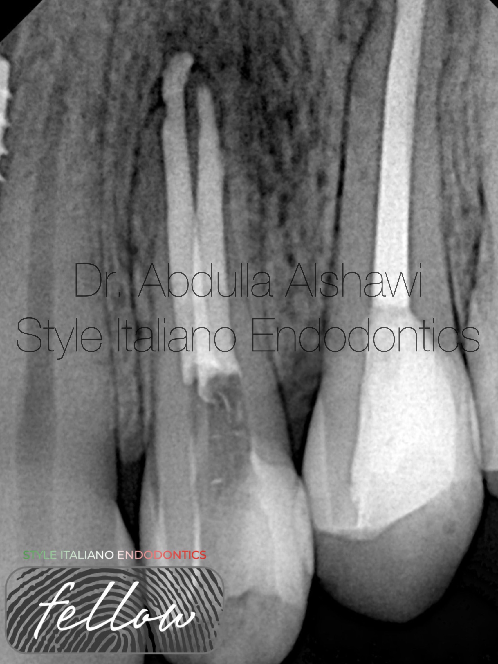
Fig. 9
Final x-ray obturation with MTA after surgical removal for the separated instrument.

Fig. 10
B.D.S
Graduated from Uruk dental collage-Iraq 2018-2019
Conclusions
The most important thing the dentist must take into account when removing any broken instrument inside the root is to know how to deal with the root and to reduce the amount of dentin removal to maintain a longer life of the tooth, in addition to knowing when to stop and do not forget that the availability of the required equipment and experience is a factor very important.
Bibliography
Chew T, Brennan D, Rossi-Fedele G.Comparative longitudinal study on the impact root canal treatment and other dental services have on oral health-related quality of life using self-reported health measures (oral health impact profile-14 and global health measures) J Endod. 2019;45:985–993.
Lea SC, Walmsley AD, Lumley PJ, Landini G. A new insight into the oscillation characteristics of endosonic files used in dentistry Phys Med Biol. 2004;49:2095–102 Factors associated with the removal of fractured NiTi instruments from root canal systems. Shen Y, Peng B, Cheung GS. Oral Surg Oral Med Oral Pathol Oral Radiol Endod. 2004;98:605–610.