Management of anatomy in a first tricky premolar (Deep Split)
06/07/2024
Fellow
Warning: Undefined variable $post in /home/styleendo/htdocs/styleitaliano-endodontics.org/wp-content/plugins/oxygen/component-framework/components/classes/code-block.class.php(133) : eval()'d code on line 2
Warning: Attempt to read property "ID" on null in /home/styleendo/htdocs/styleitaliano-endodontics.org/wp-content/plugins/oxygen/component-framework/components/classes/code-block.class.php(133) : eval()'d code on line 2
The clinician must be aware of the possible anatomical variations of these teeth and their relationship to adjacent anatomical structures when planning and performing endodontic procedures.
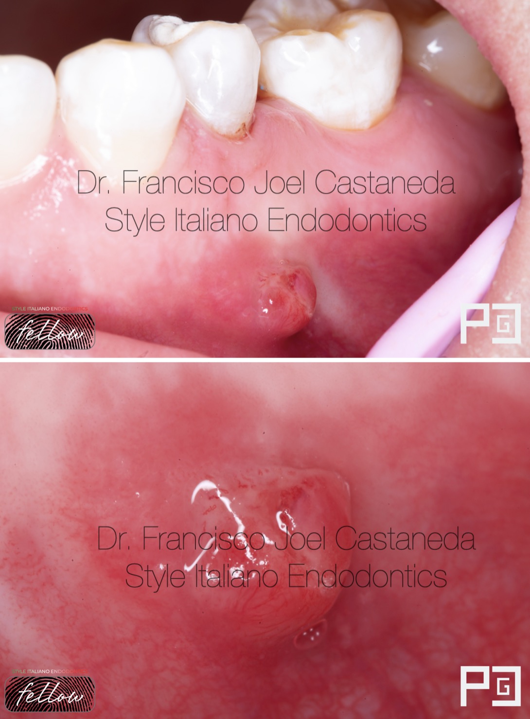
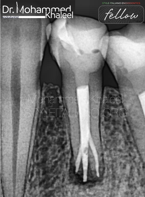
Fig. 1
Initial situation.
Pulp diagnosis: pulp necrosis, chronic apical abscess.
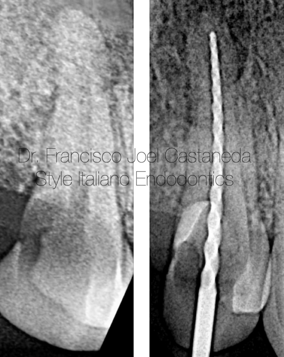
Fig. 2
Pre-op X-Ray
DME and working length and confirmation of electronic apex locator reading.
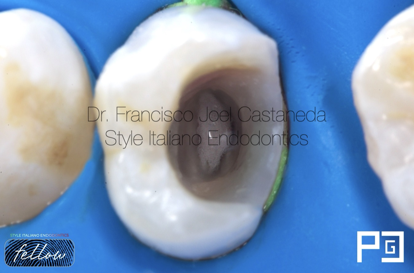
Fig. 3
Sodium Hypoclorite in action
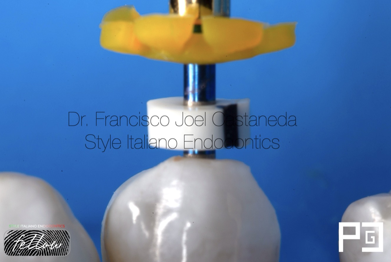
Fig. 4
Shaping up to 45.04, irrigation activation and 5.25% sodium hypochlorite.
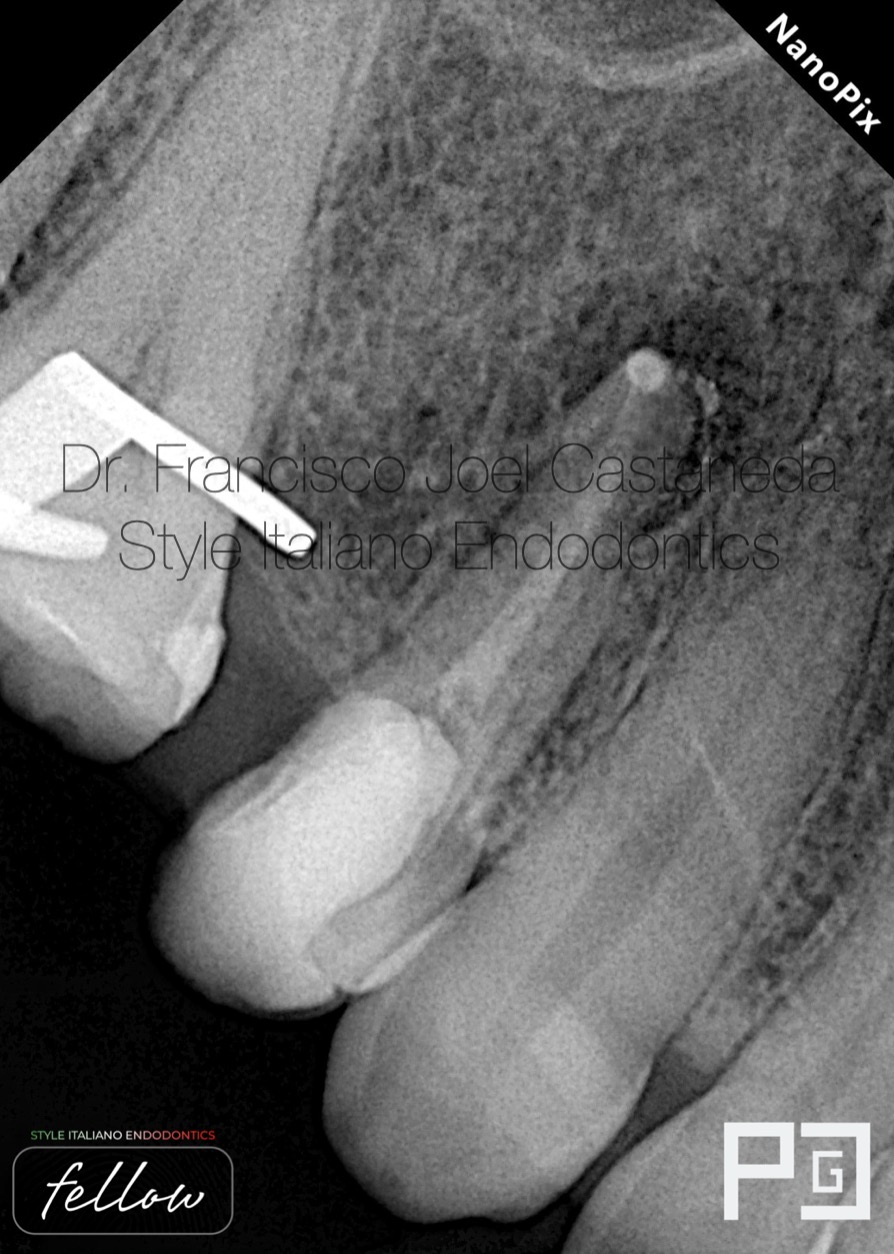
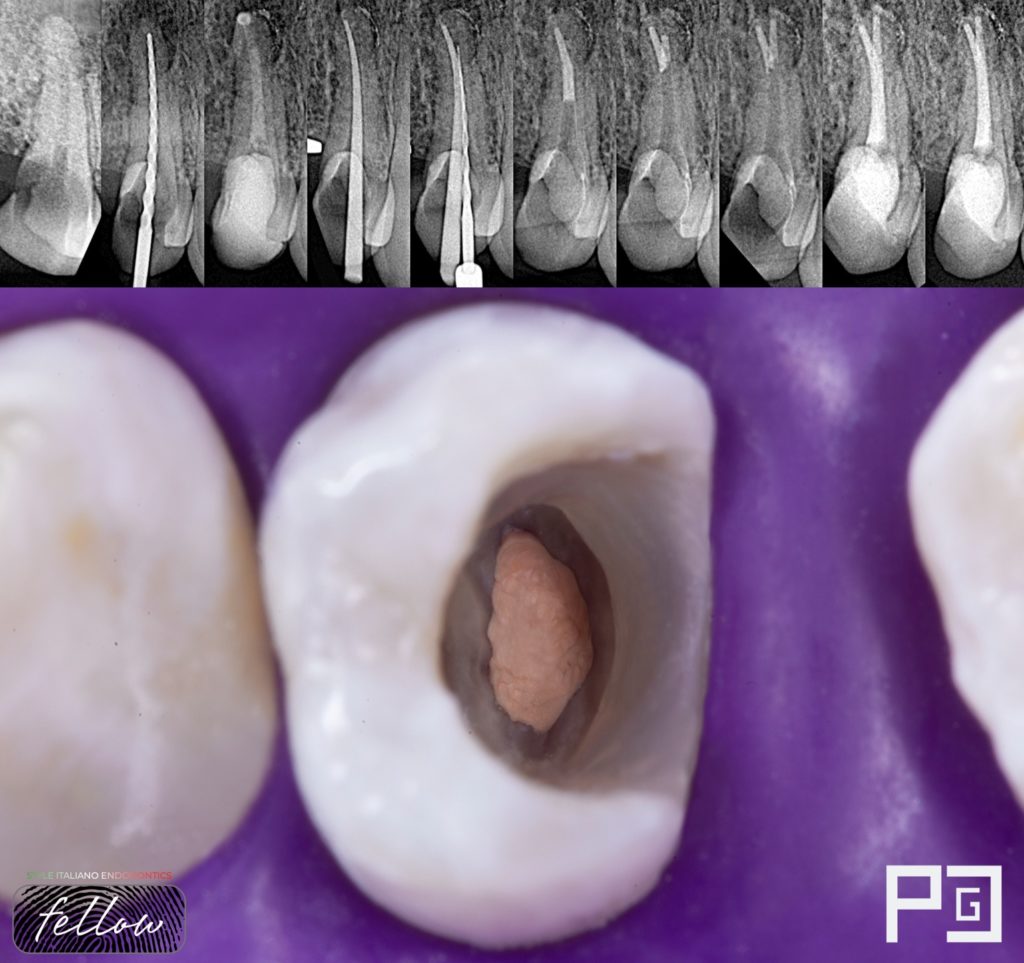
Fig. 5
Placement calcium hydroxide followed by a temporary material by two weeks.
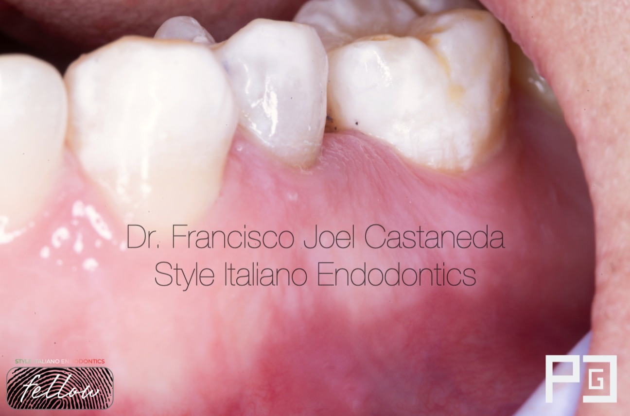
Fig. 6
After two weeks, the sinuous tract disappeared completely, a good sign.
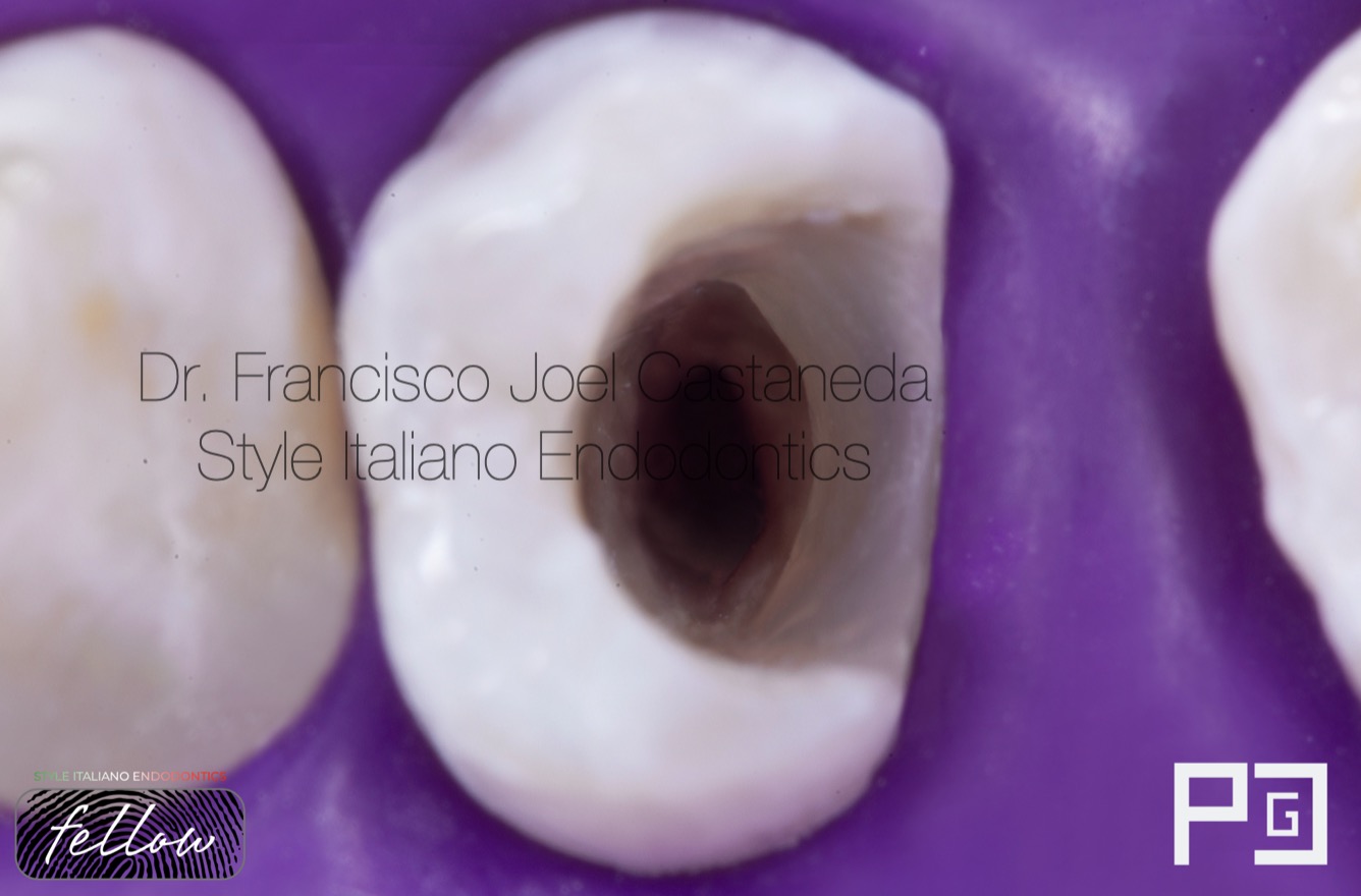
Fig. 7
Placement of rubber dam, elimination of calcium hydroxide by ultrasonic activation and irrigation.
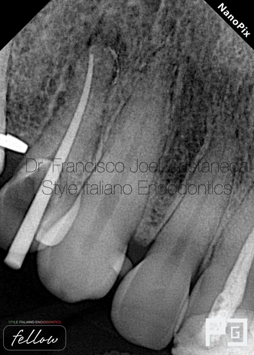
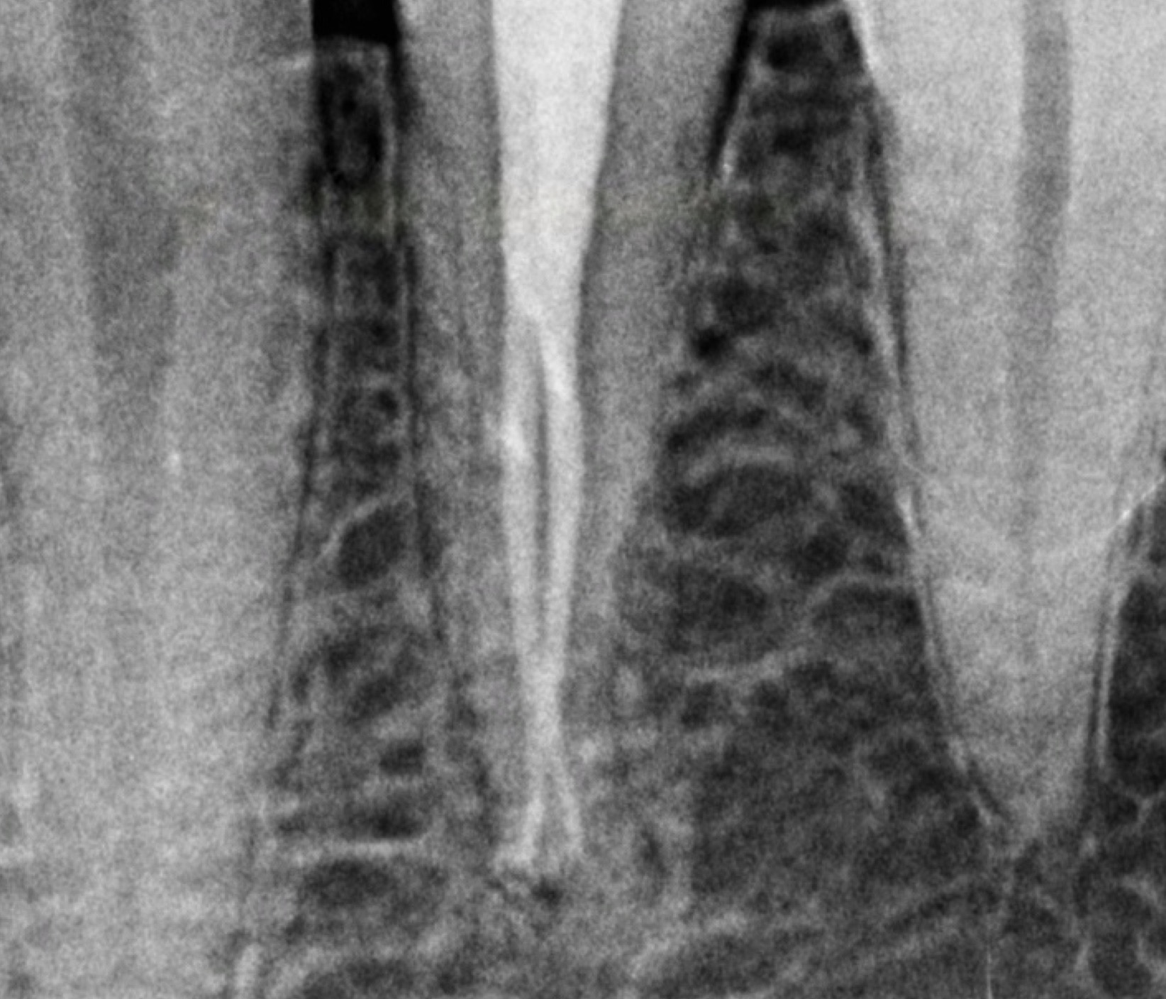
Fig. 8
Cone fit.
Because of the position of the gutta-percha cone, I realized the possibility of an extra canal
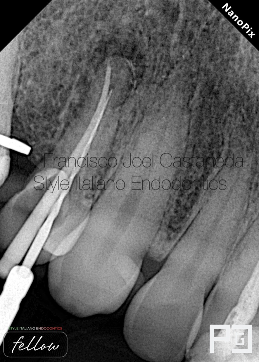
Fig. 9
I placed a rotary file together with the gutta-percha and the entrance of another canal was observed.
We determine the working length with the apex locator and instrument with rotary files.
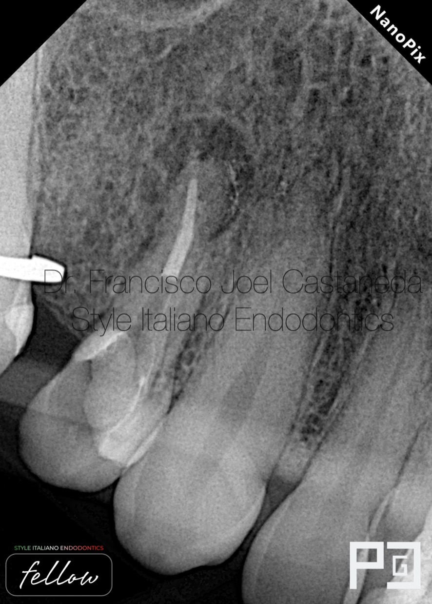
Fig. 10
Down pack, it was impossible to place two gutta-percha points through the very split and deep canal.
AH plus sealer.
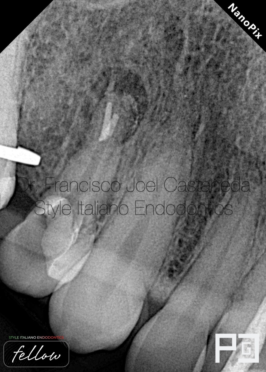
Fig. 11
I made a deeper down pack of approximately 3 mm and with the soft gutta-percha I compacted towards the opposite side of the canal.
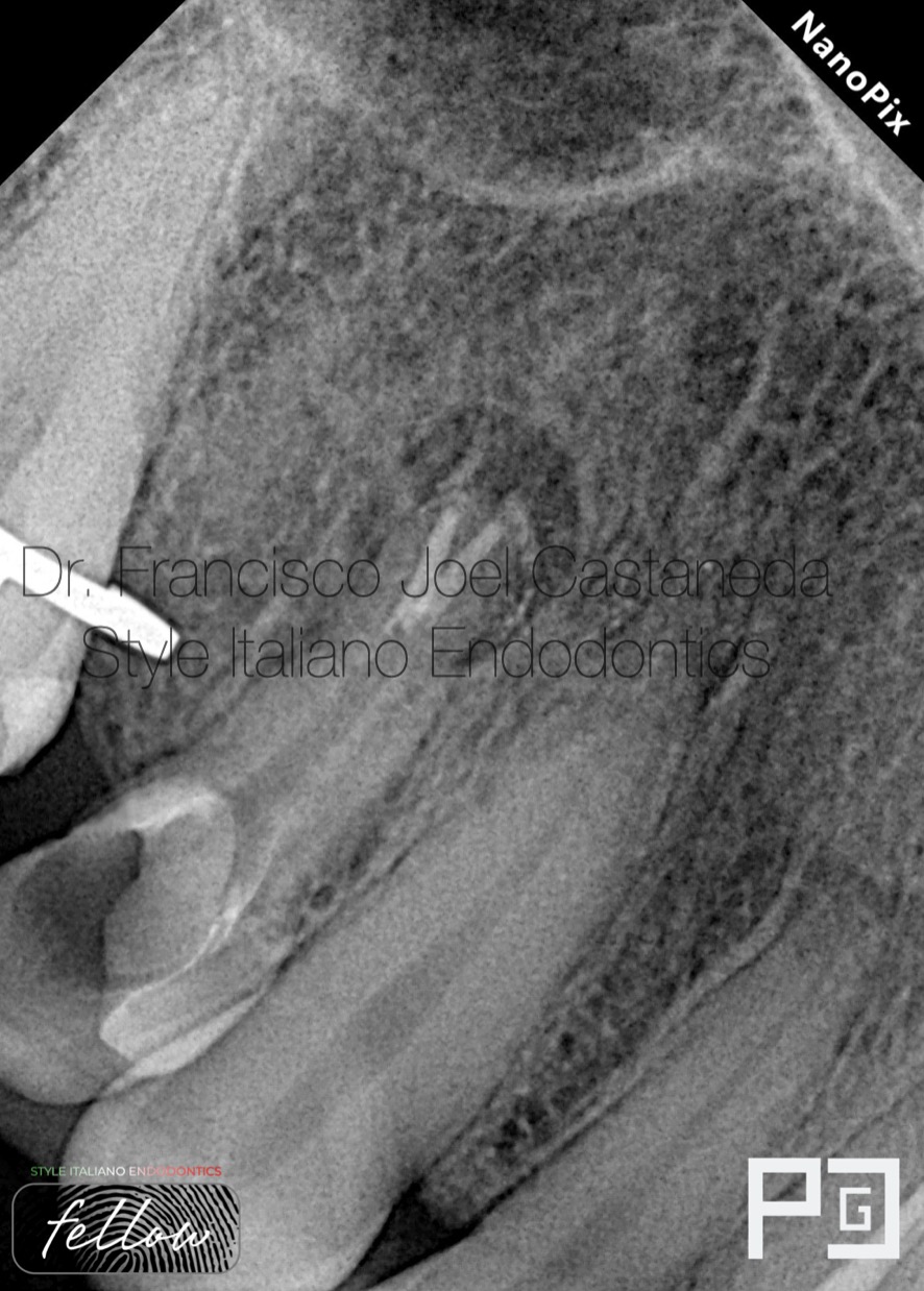
Fig. 12
Once the two canals are sealed, the next step is back filling, with small increments in order to avoid spaces.
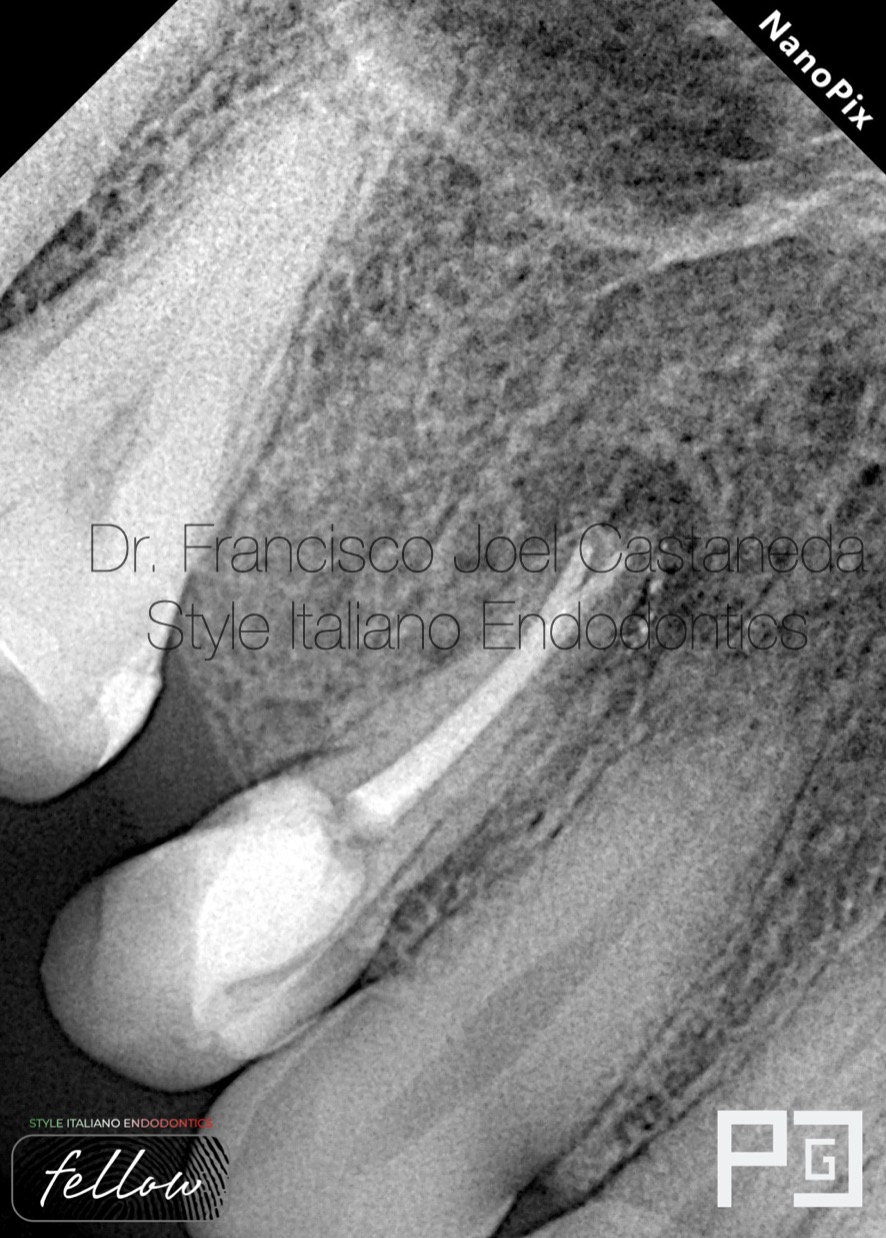
Fig. 13
Final Xray
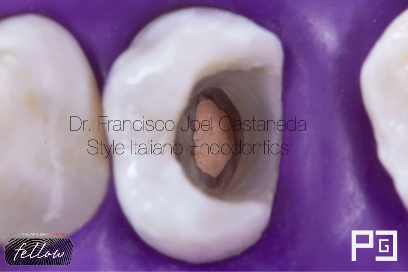
Fig. 14
Obturation done.
Clean the excess of gutapercha and sealer is a very important step in order to do a correct adhesive protocol step.
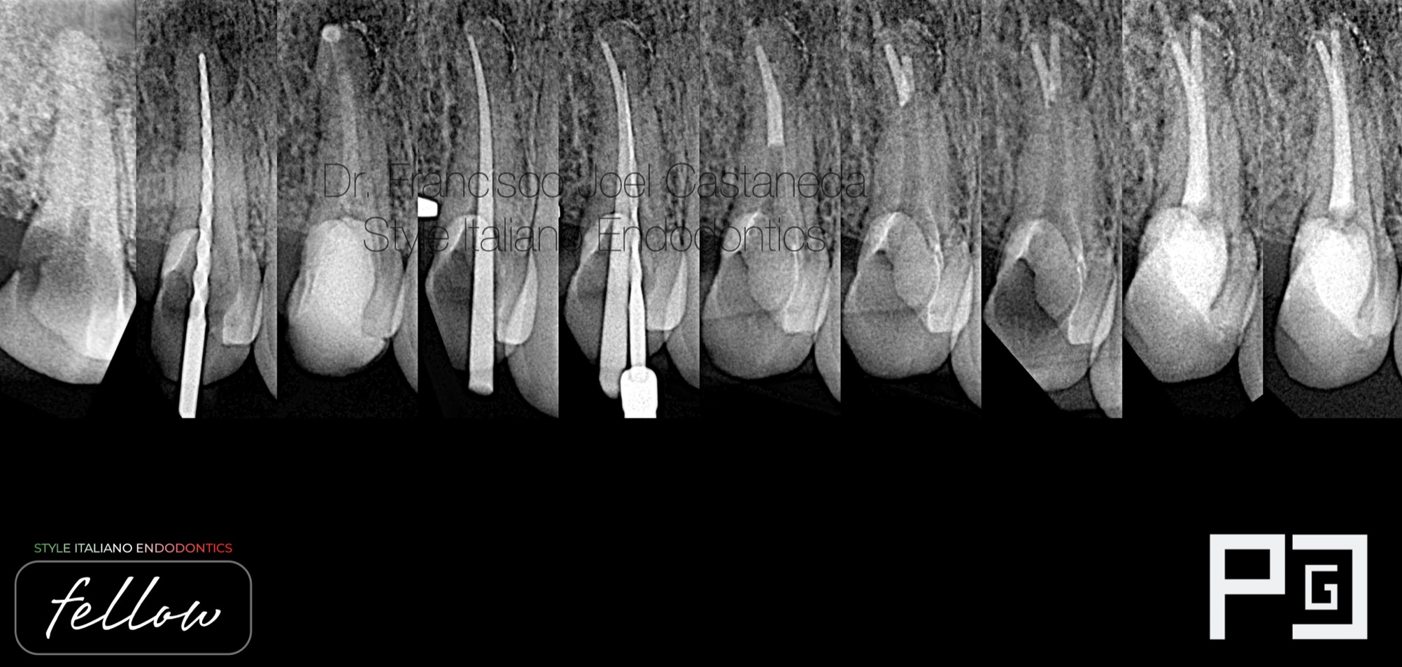
Fig. 15
Endo A-Z

Fig. 16
Dr Francisco Joel Castaneda Guerrero
Guadalajara, Jalisco - Mexico
2014-2018 dental surgeon by UDG (University of Guadalajara)
2021-2023 specialist in endodontics by UAG (University autonomy of Guadalajara)
2021- first place in artistic photography by AME (Mexican Association of Endodontics)
2022- first place in clinical photography, and second place in artistic photography by AME (Mexican Association of Endodontics)
Creator of ENDOGRAPHY and RESTOGRAPGY
ENDOGRAPHY Courses (Endodontics and dental photography)
Conclusions
Premolars undoubtedly have very complicated and complex anatomies, we must respect them, since a day-to-day case could be very difficult if we do not have the planning and ability to face these anatomical challenges.
Bibliography
Ahmad IA, Alenezi MA. Root and Root Canal Morphology of Maxillary First Premolars: A Literature Review and Clinical Considerations. J Endod. 2016 Jun;42(6):861-72. doi: 10.1016/j.joen.2016.02.017. Epub 2016 Apr 20. PMID: 27106718.
Abella F, Teixidó LM, Patel S, Sosa F, Duran-Sindreu F, Roig M. Cone-beam Computed Tomography Analysis of the Root Canal Morphology of Maxillary First and Second Premolars in a Spanish Population. J Endod. 2015 Aug;41(8):1241-7. doi: 10.1016/j.joen.2015.03.026. Epub 2015 May 5. PMID: 25956606.
Xu M, Ren H, Liu C, Zhao X, Li X. Systematic review and meta-analysis of root morphology and canal configuration of permanent premolars using cone-beam computed tomography. BMC Oral Health. 2024 Jun 4;24(1):656. doi: 10.1186/s12903-024-04419-y. PMID: 38835024; PMCID: PMC11149329.